気管支喘息、COPDの薬理学(薬理学II)
著者 柳澤 輝行
呼吸器系に作用する薬
A.気管支喘息とその治療薬
1.気管支喘息
bronchial asthma
2.メチルキサンチン薬
methylxanthine
3.
β2受容体作動薬β
2-Adrenergic agonist
4.吸入抗コリン薬
inhaled anticholinergics (muscarinic
antagonist)
5.コルチコステロイド薬
corticosteroid; inhaled
corticosteroid (ICS)
6.抗アレルギー薬・抗ロイコトリエン
(LTD
4)薬
B.
慢性閉塞性肺疾患
(COPD)とその治療薬;
咳の機序
20110513
1.気管支喘息
bronchial asthma
• アレルギー反応による可逆的な気道閉塞と遅延性炎
症が生ずる病態
• 感作された肥満細胞から抗原によりケミカルメディ
エーターが遊離し,気管支平滑筋収縮を生じる
• 気道の過敏性亢進、気道のリモデリングが基礎
• 気道上皮が剥離した好酸球主体の慢性気管支炎
• 主な治療薬は気管支拡張薬(メチルキサンチン薬・β2
受容体作動薬・抗コリン薬)とステロイド薬・抗アレル
ギー薬
• 吸入式の薬物投与方法
Mechanisms Underlying the Definition of Asthma
http://www.nhlbi.nih.gov/guidelines/asthma/
Coughing, shortness of breath, & wheezing dyspnea
Inverse Relation between the Incidence of Prototypical Infectious Diseases
(Panel A) and the Incidence of Immune Disorders (Panel B) from 1950 to 2000.
NEJM Bach 347 (12): 911, Figure 1 September 19, 2002
Immune Disorders Prototypical Infectious Diseases
0 100 0 200 10 20 年齢 臓 器 別 発 育 (%) 神経系型 リンパ系型 生殖器型 一般型 成長,発達が各臓器で 異なることを示した臓器 別発育パターン
スキャモンの臓器発育類型
気管支喘息治療薬剤
• 気管支喘息の薬剤選択ガイドライン (http://www.nhlbi.nih.gov/guidelines/asthma) • 「発作治療薬reliever」 • 「長期管理薬controller」 • 成人・小児(5歳以下)別に4段階(ステップ)の重症度(1軽症間 欠型,2軽症持続型,3中等度持続型,4重症持続型) • 吸入ステロイド薬が,成人と小児の軽症持続型(ステップ2)以降 の管理薬として,第一選択薬とされた.第二選択治療として,徐 放性テオフィリン(日本では,β2受容体作動薬とともに第一選択 薬である),抗アレルギー薬(クロモグリク酸やロイコトリエン阻害 薬 など)が推奨されている.重症になれば追加薬を用いる. • 治療のゴールを明らかにし,喘息管理を行い,発作を生じないこ とが大切である.Drugs used in asthma
Bronchodilators Beta agonists Muscarinic antagonists Methylxanthines Anti-inflammatory agents Release inhibitors Steroids Slow anti-inflammatory drugs Leukotriene antagonists Lipoxygenase inhibitors Receptor inhibitorsRespiratory system
nerve; muscles; secretory glands; immunological; autacoids
Bronchodilation cAMP Acetylcholine Muscarinic antagonists Adenosine Theophylline Bronchoconstriction ATP β agonists Theophylline AMP − AC PDE
Bronchial tone
+ + + + − − + Leukotrienes − Leukotriene antagonistsAltering
bronchial tone
in Asthma
気管支喘息病態
• 呼吸生理学; FEV
1(%)
– Spirometry; Peak flowmeter
• 閉塞性;拘束性
• 気管支喘息の病理・病態
– Bronchospasm (2 phases)
– Th2 inflammation
– Mast cell; chemical mediators
– Remodeling
予備吸気量(IRV) 2,100-3,100ml 1回換気量(TV) (安静時約500ml) 予備呼気量(ERV) 800-1,200ml 残気量(RV) 1,000-1,200ml 肺活量(VC) 3,400- 4,800ml 全肺気量(TLC) 4,400-6,000ml
呼吸の際に変動する肺気量としては、1回換気量(TV)、
予備吸気量(IRV)、予備呼気量(ERV)、肺活量(VC)が
あげられる。残気量(RV)は、空気を呼出したあとも肺内
に残る空気の量で、呼出後もガス交換が継続して行わ
れ、バファーとしても必要である。
肺機能と肺気量分画
浮ドラム
空気
または
O
2水
生理学テキスト肺活量計(スパイロメーターspirometer)
肺
容
量
努力
肺活量
1秒量
3秒量
1
2
3 sec
0
強制呼出曲線
最大吸息後、最
大呼息を行う。
1秒率FEV
1%
、
3秒率、肺活量
に対して1秒後
および3秒後に
呼出した量の%。
FEV
1/ VC
正常
閉塞性
混合性
拘束性
%
100
70
50
0
0
50
80
100%
肺活量
1
秒率
(肺線維症)
気管支喘息
COPD
慢性気管支炎呼吸機能障害の分類
流量気量曲線
flow-volume curve
気流 (L/s) 呼出 吸入 0 6 10 ピーク(呼気)フローpeak expiratory flow;PEF
1秒量; 1秒率FEV1.0%
FEV1.0%: forced expiratory volume in one second percent
肺活量 vital capacity (%)
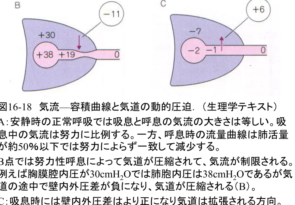
図16-18 気流—容積曲線と気道の動的圧迫. (生理学テキスト) A:安静時の正常呼吸では吸息と呼息の気流の大きさは等しい。吸 息中の気流は努力に比例する。一方、呼息時の流量曲線は肺活量 が約50%以下では努力によらず一致して減尐する。 B点では努力性呼息によって気道が圧縮されて、気流が制限される。 例えば胸膜腔内圧が30cmH2Oでは肺胞内圧は38cmH2Oであるが気 道の途中で壁内外圧差が負になり、気道が圧縮される(B)。 C:吸息時には壁内外圧差はより正になり気道は拡張される方向。
炎症のない気道 炎症細胞の浸潤
狭窄のない気道 狭窄した気道
非発作時の気管支鏡像
(新臨床内科学,第8版 p250) Edema Sputum Plugs of mucusAirway obstruction
(Bronchospasm)
喘息発作時の気道粘膜
気道長径 横断面 安静換気においては、気道内は一般に層流である。 気道の閉塞ならびに粘液の貯留などにより、気流速度が増し、 また高度な乱流が生じる。これが喘鳴として聴取される。
寛
解
期
発
作
時
喘鳴の発現機序
聴診器なしで離れて聞こえる主に呼気時の「ピュー 」 「ヒュー 」 「ゼェー 」 「ギー 」などと、高音性連続性呼吸音。呼吸による気道の気流は気道内腔が狭 窄すると乱流を生じ,笛の原理で笛音を発生する。 呼気性喘鳴expiratory wheeze;末梢気道 吸気時喘鳴inspiratory stridor;中枢気道喘息死患者の気道組織
著明な炎症細胞(好酸球の浸潤が主 体)の浸潤がみられる。(Color Atlas of Respiratory Disease, Vol 2. Allen & Humburys, 1995より。岩手県立中央病院病理センター富地信和先生の提供による)Specimen of Bronchial Mucosa from a Subject without Asthma (Panel A) and a
Patient with Mild Asthma (Panel B) (Hematoxylin and Eosin, x40).
In the subject without asthma, the epithelium is intact; there is no thickening of the sub-basement membrane, and there is no cellular infiltrate.
In contrast, in the patient with mild asthma, there is evidence of goblet-cell hyperplasia in the epithelial-cell lining. The sub-basement
membrane is thickened, with
collagen deposition in the submucosal area, and there is a cellular infiltrate.
Busse and Lemanske: NEJM 344 (5): 350, Figure 1 February 1, 2001
The Importance of Establishing a Balance between Th1-Type and Th2-Type Cytokine Responses. Numerous factors, including alterations in the number or type of infections early in life, the widespread use of
antibiotics, adoption of the Western lifestyle, and repeated exposure to allergens, may affect the balance between Th1-type and Th2-Th1-type cytokine responses and increase the likelihood that the immune response will be dominated by Th2 cells and thus will ultimately lead to the expression of allergic diseases such as asthma.
A Simplified Scheme of the System of Type 1
Helper T (Th1) and Type 2 Helper T (Th2) Cells.
differentiation
regulatory loop
The Role of Eosinophils in Allergic Inflammation. NEJM Busse and Lemanske 344 (5): 350, Figure 3 , 2001
Inhaled antigen activates mast cells and Th2 cells in the airway. They in turn induce the production of mediators of inflammation (such as histamine and leukotrienes) and cytokines including interleukin-4 and
interleukin-5. Interleukin-5 travels to the bone marrow and causes terminal differentiation of eosinophils. Circulating eosinophils enter the area of allergic inflammation and begin migrating to the lung by rolling, through interactions with selectins, and eventually adhering to endothelium through the binding of integrins to members of the immuno-globulin superfamily of adhesion proteins: vascular-cell adhesion molecule 1 (VCAM-1) and intercellular adhesion molecule 1 (ICAM-1). As the eosinophils enter the matrix of the airway through the influence of various chemokines and cytokines, their survival is prolonged by interleukin-5 and granulocyte– macrophage colony-stimulating factor (GM-CSF). On activation, the eosinophil releases inflammatory mediators such as leukotrienes and granule proteins to injure airway tissues. In addition, eosinophils can generate
granulocyte–macrophage colony-stimulating factor to prolong and potentiate their survival and contribution to persistent airway
inflammation. MCP-1 denotes monocyte chemotactic protein, and MIP-1 macrophage inflammatory protein.
Blood vessel FEV1 100 75 0 8 Early
reaction Late reaction
Airway wall Time (h) Histamine Tryptase PGD2 LTC4 PAF IL-4 IL-5 GM-CSF TNF TGF Bronchospasm ECP MBP Proteases PAF Allergen Cell infiltration inflammation
Mast cell Th2 cell Eosinophil Neutrophil IgE
Edema
肥満細胞からのヒスタミンやそ の他のメディエーター遊離 抗原 IgE Lyn Syk PLCγ1 PIP2 DG IP3 PKC IP3 R Ca2+ [Ca2+]i 増加 H 2 受容体 β受容体 A C Gs Gs ATP cAMP 抑制 脱顆粒 Ca2+ Ca2+ IgE受容体
Histamine
PGD2, LTC4, LTD4, PAF プロテアーゼ、ヘパリン、TNFα インターロイキン (特にIL-5:好酸球走化因子)気管支喘息における各種細胞の相互作用と病理所見 外界からの刺激因子(大気汚染物質など) 気道上皮細胞 化学伝達物質遊離刺激 マスト細胞・好塩基球 IL-1 マクロファージ 好中球 Tリンパ球 MBP、ECP、EDN 好酸球 インターロイキン IL-5 慢性気管支炎 好酸球浸潤 上皮剥離 知覚神経過敏
MBP (major basic protein), ECP (eosinophil cationic protein), EDN (eosinophil-derived neurotoxin)は、いずれも好酸球から遊離され、気道上皮細胞を傷害する
The Airway in a Healthy Person (Panel A) and in a Patient
with Asthma (Panel B).
In Panel A, the airway of a healthy person is composed of an epithelium with a thin basement membrane and some relatively
quiescent fibroblasts and smooth-muscle cells. Minimal expression of ADAM-33 may maintain base-line levels of cell proliferation and basement-membrane integrity. In Panel B, the airway of a patient with asthma is characterized by subepithelial fibrosis (with collagen deposition), smooth-muscle hypertrophy or hyperreactivity, and
inflammation. ADAM-33 could participate in these abnormal processes through loss-of-function polymorphisms (by down-regulating the shedding of growth factor receptor) or
gain-of-function mutations (by increasing the shedding of growth factors
with autocrine or paracrine effects on fibroblasts and smooth-muscle cells). ADAM-33 might also be involved in the chronic immune or inflammatory response through the excessive shedding of type 2 helper T cell (Th2) cytokines or by impaired shedding of cytokine receptors (macrophage shown).
2.メチルキサンチン薬
methylxanthines
• テオフィリンtheophylline(徐放性剤型 )
• アミノフィリンaminophylline (喘息発作 i.v.)
• 気道炎症を抑え、気管支平滑筋を弛緩させる.
①ホスホジエステラーゼ
phosphodiesterase
(
PDE)の阻害作用
②アデノシン
A1受容体拮抗作用
③プロスタグランジンなどのケミカルメディエー
ター拮抗作用
④
IgE受容体刺激に共役するCa
2+流入抑制作用
3. Structure of β-Adrenergic Bronchodilators.
Adrenaline
Salbutamol
long-acting β2-agonists (LABA).
β2-Adrenergic agonists are the most effective bronchodilators for the treatment of acute episodes of asthma and for the prevention of exercise-induced
bronchoconstriction. The LABAs prevent nocturnal asthma and provide prolonged protection against exercise-induced bronchoconstriction. LABAs should be
administered only at regular, prescribed intervals. Additional doses should not be given to relieve symptoms; intermediate-acting β2-agonists should be given instead. Despite some concern about these drugs as a class, the adverse effects are probably related more closely to the severity of the asthma than to the drug. Thus,
β2-adrenergic agonists remain the most important class of bronchodilators currently available. When used appropriately, they provide safe and effective relief of the symptoms of airflow obstruction.
臭化イプラトロピウム (アトロピンの四級アミン誘導体;生体膜 biomembraneを通過しにくい)
4. 吸入抗コリン薬
臭化イプラトロピウム
ipratropium bromide
muscarine
受容体
遮断薬
:
副交感神経系亢進による
気管支平滑筋の収縮と粘液の
過剰分泌
抑制
交感神経系のシグナル抑制作
用
遮断
5.(吸入)コルチコステロイド
inhaled corticosteroid (ICS)
プロピオン酸ベクロメタゾンbeclomethasone dipropionate バイオアベイラビリティーの悪い薬物 吸入で用いることにより局所作用のみのため,全身作用ない. 発作予防に最近多用され,喘息死減尐をもたらした 肝臓での 初回通過
プロピオン酸フルチカゾン
fluticasone propionate
ブデソニド
budesonide
消化管
肝
気道 腸から吸収吸入ステロイドの薬物動態
無視できるほど
小さい
吸入噴霧剤の到
達に関するス
Effect of the Interaction between the Intensity of Exercise and the Thermal Environment on the Pulmonary Mechanical Response. Relative Effects of Various Drugs on Airway Obstruction Induced by Thermal Stimuli.
FEV1 denotes forced expiratory volume in one second, VE: minute ventilation. The data were obtained by generating stimulus-response curves during isocapnic hyperventilation with frigid air before and after the administration of placebo and common anti-asthma drugs alone and in combination. This information represents our composite experience with the agents. NEJM 330:1362-1367 May 12, 1994 Exercise-Induced Asthma E.R. McFadden, and Ileen A. Gilbert
6.抗アレルギー薬・
抗ロイコトリエン
(LTD
4)薬
• クロモグリク酸cromoglycate リポキシゲナーゼ活性の阻害,Ca2+流入の阻害(Cl- channel 遮断作用; depolarization),cAMPの増加,膜安定化などをもつ. アレルギー反応によって起こるケミカルメディエーター(ヒスタミ ン,ロイコトリエン,血小板活性化因子PAFなど)の産生・遊離 を抑制,または直接拮抗するので,喘息の予防薬として吸入 により用いられる. • 抗アレルギー薬(抗ヒスタミン作用も有す)(表15-3参照) • Lipoxygenase 阻害薬:ジロイトンzileuton • LTD4受容体拮抗薬:プランルカストpranlukast,モンテルカスト montelukast • トロンボキサンA2(TXA2)合成酵素阻害薬(オザグレル) • スプラタストsuplatast(TH2サイトカイン阻害薬):は IL-4,5と IgE産生抑制作用Biochemical Pathways of the Formation and Action of the Leukotrienes and Sites of Action of Leukotriene-Modifying Drugs. Enzymes are shown in blue, products in yellow, essential cofactor in green, and drugs in red.
Although the synthesis of leukotrienes B4 and C4 probably takes place in close proximity to the nuclear membrane, for clarity they are shown throughout the cytosol. BLT denotes the B leukotriene receptor. An individual cell may produce the cysteinyl leukotrienes, leukotriene B4, or in rare cases both.
肥満細胞 トリプターゼ ロイコトリエン 抗ロイコトリエン Tリンパ球 (Th2細胞) 気管支狭窄と 粘液産生 気道過敏性 ケモタキシス エオタキシン RANTES MCP4 好酸球 増殖と 集積 IL-4,
IL-5
IL-4,
IL-5
抗体
+ + + CC-CKR3 遮断薬喘息の新治療薬の動向
Exposure to antigen (dust, pollen, etc)
Avoidance Antigen and IgE
on mast cells Mediators
(leukotrienes, cytokines, etc) β agonists,
theophylline, muscarnic antagonists, leukotriene
antagonists Early response: bronchoconstriction
Acute symptoms hyperreactivity Bronchial Late response: inflammation Steroids, cromolyn, leukotriene antagonists Cromolyn, steroids, zileuton − − − − 治療戦略の総括
B.慢性閉塞性肺疾患(COPD)とその治療薬
chronic obstructive pulmonary disease
• 気道閉塞を生ずる原因:肺実質の破壊による肺コン
プライアンスの低下と白血球を中心とした気道壁の
病変
• 禁煙が極めて重要
• 気管支拡張薬 (特に、吸入抗コリン薬 )
• チオトロピウムにより、喀痰の減尐や消失、呼吸困難
が改善
• 急性増悪期には,抗菌薬とともに経口ステロイド薬の
短期(
5~7日間)使用
• 良好な水分補給を維持,去痰薬
慢性閉塞性肺疾患(
COPD)対
慢性気管支炎,肺気腫及び喘息の相互関係と病態
(図
6-7)
喘息
慢性
気管支炎
肺気腫
気道閉塞 慢性閉塞性肺疾患 COPD 好酸球増加 CD4:CD8リンパ球の比↑ IL-4・IL-5の遺伝子発現↑ 好中球増加 CD4:CD8リンパ球の比→ IL-4・IL-5の遺伝子発現→ 可逆的気道閉塞• COPD患者の大半が慢性気管支炎と肺気腫の両疾患の特徴を有 する.アレルギーと好酸球増加を基礎に,可逆性気道閉塞の喘息 患者は,COPDと区別する.気道閉塞が完全に寛解しない喘息患 者を部分的に可逆性の気道閉塞と気道過敏症をもつ慢性気管支 炎や肺気腫の患者群から区別することは臨床的には不可能なの で,寛解しない喘息患者は,COPDがあるものとして治療する. • COPDの病態には,①気道抵抗の増加(コリン作動性緊張亢進, 肺弾性低下),②炎症(好中球,マクロファージ中心,IL-8の関与), ③構造的変化(肺胞破壊,気道のリモデリング),④気管支粘膜線 毛機能低下(気道分泌物排出能の低下,細菌の異常付着をもたら す)がある.これらの因子がさらに悪循環を形成する. • 気道閉塞では,肺機能検査上では気道抵抗airway resistanceの増 加,一秒量,一秒率,強制呼出肺活量,peak flowなどの低下をき たす.慢性に繰り返す気道の炎症(主にタンパク質分解酵素の作 用)のために肺胞壁が破壊されてしまう.また,タンパク質分解酵 素の作用を抑制するα1‐アンチトリプシンが先天的に欠損・減尐 している人では,COPDや肺気腫が極めて発症しやすい.タバコは
COPD病態の喀痰と迷走神経
一ノ瀬正和先生の図から COPD増悪予防にはサルメテロールよりチオトロピウム、
POET(Prevention Of Exacerbations with Tiotropium)試験、
COPD増悪予防にはサルメテロールよりチオトロピウム POET(Prevention Of Exacerbations with Tiotropium)
サブスタンスP 気道刺激物、タバコ サブスタンスP ヒスタミン LTs 軸索反射