本邦における下垂体腺腫は原発性脳腫瘍の約 18%を占める.しかしながら,下垂体・視交叉部に 発生する腫瘍では約 78%を占める比較的頻度の高 い疾患である1,2).下垂体腺腫の発生機序の詳細は 明らかにされていないが,遺伝子異常が腫瘍発生の 因子であるとする考え方が一般的である3).下垂体 腺腫の発生機序の詳細が明らかにされない要因とし て,摘出される病巣が小さく,腫瘍組織中の下垂体 腺腫の腫瘍細胞と間質との混在が強く認められるこ とから腫瘍細胞本来の遺伝子解析に必要な RNA 量 が得られず解析が困難であることが考えられる.そ こで Laser Microdissection 法により不均一な組織 中の僅かな腫瘍細胞を取り出し,T7 増幅法によっ て微量な RNA を約 100 倍に増幅して必要な RNA 量を確保し,Microarray による遺伝子解析を行っ た4).
一方,甲状腺刺激ホルモン(以下 TSH)産生下 垂体腺腫は全下垂体腫瘍中の 1 〜 2%を占めるにす ぎない稀な腫瘍であるが5,6)近年は超高感度 TSH 測定技術の向上により TSH 産生下垂体腺腫の報告 数は増加している.しかし,TSH 産生下垂体腺腫
を形成する特異的な遺伝子はいまだ明確にされてい ない7,8).今回われわれは腫瘍形成やホルモン分泌 に関与する遺伝子を明らかにするために,TSH 産 生下垂体腺腫に特異的に発現する遺伝子を検索し た.対照基準として剖検例から得られた正常下垂体 組織とホルモン分泌を伴わない非機能性下垂体腺腫
(null cell adenoma)を用い,遺伝子検索を行った.
研 究 方 法 1.検体
腫瘍組織は昭和大学医学部脳神経外科で経蝶形骨 洞下垂体腫瘍摘出術によって摘出された検体を用い た.
TSH 産生下垂体腺腫 1 例,およびコントロール として正常下垂体 1 例,非機能性下垂体腺腫(null cell adenoma)2 例を対象とした.
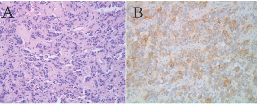
TSH 産生下垂体腺腫は血中値が TSH のみ高値で あり,免疫染色法にて腫瘍組織は TSH 抗体に陽性 を示した.GH,ACTH,FSH,LH は陰性であっ た(Fig. 1).
非機能性下垂体腺腫は臨床的に下垂体ホルモン血
TSH 産生下垂体腺腫における遺伝子解析
昭和大学医学部脳神経外科学教室
中山 禎理 佐々木晶子 桑名 亮輔 桑島 淳氏 阿部 琢巳
昭和大学歯学部口腔病理学教室
山 本 剛 立川 哲彦
要約:ホルモン分泌を伴わない非機能性下垂体腺腫(null cell adenoma)と甲状腺刺激ホルモ ン(以下 TSH)タンパクのみを合成する TSH 産生下垂体腺腫の遺伝子を正常下垂体,null cell adenoma と比較検討し,ホルモン分泌や腫瘍形成に関与する遺伝子を検索した.検索腫 瘍は当施設において手術により摘出した組織を凍結固定し,連続切片を作成した.切片は Laser Microdissection 法を用いて腫瘍細胞だけを採取し,T7 増幅法にて RNA を増幅後,
DNA microarray にて解析をおこなった.その結果,腫瘍形成に関与する遺伝子として MMP と共起する遺伝子 TIMP4,細胞周期 M 期の制御に関わるサイクリン B1,B2 などの高い発現 がみられた.また TSH 産生下垂体腺腫特有の性状である腫瘍硬度を形成する遺伝子として線 維性組織の形成に関与する Transthyretin,Fibroblast growth factor が検出された.さらに ホルモン分泌に関わるタンパク質 CSH1,発生と代謝に関与する転写因子 Forkhead box N4 などが検出され,TSH 産生下垂体腺腫に特有の分化系譜を示唆した.
キーワード:TSH 産生下垂体腺腫,甲状腺刺激ホルモン,laser microdissection 原 著
中値に異常値は認めず,免疫染色においても TSH,
成長ホルモン(以下 GH),副腎刺激ホルモン(以 下 ACTH),卵胞刺激ホルモン(以下 FSH),黄体 刺激ホルモン(以下 LH)は陰性を示した.
正常下垂体組織は,剖検で得られた神経膠芽腫の 患者で下垂体転移のない組織を使用した(Table 1).使用した検体については昭和大学ヒトゲノム・
遺伝子解析倫理審査委員会において承認を得た(申 請番号第 60 号).
全ての検体は OCT compound(Sakura. Torrance,
CA, USA)に入れ,液体窒素を用いて冷却した イソペンタン内で凍結させ,−80℃冷凍庫に保 存 し た. 切 片 は 免 疫 染 色 に は 厚 さ 4μm, Laser Microdissection 用には厚さ 10μm の連続凍結切片 を作成した.
2.Laser Microdissection および RNA 抽出 検体はクリスタルバイオレット染色を行い Laser microdissection(P.A.L.M. Microlaser Technologies AG and Meiwafosis, Osaka, Japan)を用いて 2000 細 胞 以 上 を 取 り 出 し た(Fig. 2).Total RNA を Fig. 1 A: Hematoxylin and eosin staining and immunostaining of a thyrotropic hormone (TSH)-pro-
ducing pituitary adenoma. B: On immunostaining, only TSH is positive. Magnification×400
Table 1 Clinical features in 4 pationts with microarray analysis
patient NO Pathological diagnosis sex Age
1 TSH producing adenoma male 44
2 null cell adenoma male 64
3 null cell adenoma Female 57
4 normal pituitary gland (dissection) male 82
Fig. 2 Tumor cell isolation by laser microdissection.
RNAqueous-Micro Kit(Applied Biosystems/
Ambion, Austin, TX)を用いて抽出した.抽出し た Total RNA は Agilent 2100 Bioanalyzer(Agilent Technologies, Inc., Santa Rosa, CA, U.S.A)を用い て quality check を行った.
3.Total RNA の増幅
Microarray で 分 析 す る た め に 十 分 量 の Total RNA を得るため TargetAmp 2-Round Aminoallyl- aRNA Amplification Kit(Epicentre Biotechnologies, Madison, WI, U.S.A)を用いて RNA の 5 末端に T7 オリゴプライマーを付加し,逆転写によって cDNA を合成した.その後,再度 T7 オリゴプライマーを 付加し Total RNA を増幅して Amminoallyl-aRNA を合成した.
4.Microarray 解析
増幅・合成した Amminoallyl-aRNA を Whole Hu- man Genome Oligo DNA Microarray Kit(Agilent techbologies)を用いて Cyanine3 ラベル化を行い,
Microarray を行った.得られたデータは Genespring GX software(Agilent techbologies)を用いて解析 した.正常下垂体 1 例を対照基準として TSH 産生 下垂体腺腫 1 例,非機能性下垂体腺腫(null cell adenoma)2 例を対照基準としてそれぞれ解析を行 い,相対的に 1000 倍以上増加を認めた遺伝子を有 意な増加と判断し検討を行った.
結 果
TSH 産生下垂体腺腫と正常下垂体を比較し,相 対 的 に 1000 倍 以 上 多 く 発 現 し て い た 遺 伝 子 は Chorionic somatomammotropin hormone1(24,492 倍),Transthyretin(9,689 倍),JEM-1(5,719 倍),
PRKA13(4,465 倍),Forkhead box N4(3,564 倍),
Programmed cell death 6 interacting protein(1,341 倍),Carbonic anhydrase Ⅷ(1,110 倍) 等 の 遺 伝 子であった(Table 2).また,100 倍以上多く発現 していた遺伝子は,細胞骨格因子である中間径フィ ラメントタンパク質 Restin(435.4 倍),細胞接着に 関与し,細胞増殖に関与する Integrin beta4(232.2 倍),線維芽細胞 Fibroblast growth factor(210.2 倍),細胞周期の調節を行う CyclinB2(166.2 倍)
を認めた.
TSH 産生下垂体腺腫と非機能性下垂体腺腫とを比 較し,TSH 産生下垂体腺腫に 1000 倍以上多く発現
していた遺伝子は,Glycoprotein hormones(23,883 倍),Chorionic somatomammotropin hormone1
(15,905 倍),Transthyretin(10,076 倍),Phospho die- sterase 4D interacting protein(8,990 倍),TIMP4
(7,334 倍),JEM-1(5,842 倍),Forkhead box N4
(3,626 倍),PRKA13(3,629 倍),Programmed cell death 6 interacting protein(2,227 倍),Virus- induced signaling adapter(2,055 倍)Pre-B- cell colony enhancing factor1(2,084 倍),Mer- captopyruvate sulfurtransferase(1,471 倍),Cyclin B1(1,455 倍)等の遺伝子を認めた(Table 3).また,
100 倍以上多く発現していた遺伝子は,細胞周期を 調節する CDK6(539.9 倍),細胞骨格を形成するタ ン パ ク Tropomyosin4(563.8 倍), ゴ ル ジ 体 の Trans-golgi network protein2(177.1 倍),細胞骨 格アクチンとチュ−ブリンの転写因子である Zinc and ring finger3(105.6 倍)等の遺伝子発現を認め た.
考 察
本研究では TSH 産生下垂体腺腫の検体を用いて 腺腫形成に関する遺伝子や TSH ホルモン分泌に関 与する遺伝子を検索した.正常下垂体を基準として 増加した遺伝子を検索した結果,Transthyretin
(9,689 倍),Fibroblast growth factor(210.2 倍)な どの線維性組織の形成に関与する遺伝子が多く発現 していたことは,臨床的に TSH 産生下垂体腺腫は 線維化が強く,硬い腫瘍が多いことからも肯定でき る.
Transthyretin はアミロイド病に関連する遺伝子 であり,線維性組織の形成に関与し,他の頭蓋内腫 瘍では Choroid plexus papilloma にて発現すること が報告されている9).
TSH 産生下垂体腺腫は他の下垂体腺腫と比較し て,組織学的にも細胞間に多くの線維形成を認め,
硬い腫瘍が多い.これは線維形成に関与している Transthyretin が影響し,硬い腫瘍の性状を形成し ている可能性が考えられる.
また,Fibroblast growth factor は以前から GH,
PRL,TSH のホルモン分泌に関与するとされてお り,近年では PRL 産生下垂体腺腫の血管形成に関 与すると報告されていると共に,FGF は線維芽細 胞の増殖を促し,線維形成を促進することと関連し
ている10).
非機能性下垂体腺腫を基準として増加した遺伝子 を検索した結果,Glycoprotein hormones(23,883 倍),Chorionic somatomammotropin hormone1
(CSH1)(24,492 倍),TIMP4(7,334 倍),Forkhead box N4(FOXN4)(3,626 倍)の遺伝子が検索された.
Chorionic somatomammotropin hormone1(CSH1)
(24,492 倍)は GH,PRL 等のホルモン分泌に関わ るタンパクの合成制御を行う遺伝子である11)こと から TSH 産生下垂体腺腫は分泌タンパクの合成が 盛んな腫瘍であることが示唆される.
TIMP4(7,334 倍)は細胞外マトリックスを破壊 する酵素マトリックスメタロプロテナーゼ(MMP)
フ ァ ミ リ ー の イ ン ヒ ビ タ ー で あ り,TIMP4 は MMP の活性を阻害させる.MMP の発現調節は TIMP による活性阻害によって調節され,この遺伝
子が下垂体腺腫の浸潤性を評価するインデックスに なる可能性もある12,13).さらに,MMP のもう 1 つ の機能として,MMP はインターロイキンー 1 や腫 瘍壊死因子(TNF)の転写を引き起こす因子とし ても知られている.同時に,EGF の場合では細胞 外マトリックスにアンカーさせる機能がある.以上 のことから TSH 産生下垂体において MMP 遺伝子 の発現が高いことは本腫瘍の細胞増殖や浸潤性発育 に関与していると同時に細胞増殖因子やリンホカイ ンなどで,ホルモン分泌顆粒の生成や分泌に強く関 与している結果であることを示している.
Forkhead box N4(3,626 倍)は,脊髄の 2 つの インターニュロンである V2a と V2b 発生時の共通 の前駆対である DII4 と Mash1 を活性化させ,神経 系の分化と関連している.この分化過程の間には Notch delta シグナルが関与していることが判明し Table 2 List of highly expressed genes in TSH producing adenoma, compared with normal
pituitary gland
Relative amount Gene name
41,296 Guanine nucleotide binding protein (G protein), q polypeptide 32,143 Homo sapiens cDNA FLJ42912 fis, clone BRHIP3024533. [AK124902]
29,610 Sodium channel, voltage-gated, type V, alpha (long QT syndrome 3)
24,492 Chorionic somatomammotropin hormone 1 (placental lactogen)
21,661 Defensin, beta 103A
15,407 Solute carrier family 4, anion exchanger, member 13, Diego blood group)
9,689 Transthyretin (prealbumin, amyloidosis type I)
8,455 Timeless-interacting protein 7,249 Prolactin
5,719 Basic leucine zipper nuclear factor 1 (JEM-1)
5,423 CDNA clone IMAGE:4825318 4,465 A kinase (PRKA) anchor protein 13 4,244 5'-nucleotidase, cytosolic II
3,564 Forkhead box N4 2,949 Sperm associated antigen 9 2,597 Delta-like 1 homolog (Drosophila)
2,132 MRNA from chromosome 5q31-33 region 2,056 Pre-B-cell colony enhancing factor 1
2,020 Homo sapiens cDNA FLJ33266 fis, clone ASTRO2007047. [AK090585]
1,970 PHD finger protein 20
1,772 Delta-like 1 homolog (Drosophila)
1,341 Programmed cell death 6 interacting protein 1,315 PET112-like (yeast)
1,110 Carbonic anhydrase VIII
1,102 Zona pellucida glycoprotein 1(sperm receptor)
1,081 Albumin
1,056 maternally expressed 3
ている14).本腫瘍で Forkhead box N4 の高発現が みられたことは腫瘍細胞の分化に神経系の制御と下 垂体腺腫代謝に関与する転写因子であることが考え られる.この転写因子の増加が認められたことで TSH 産生下垂体腺腫における分化過程が非機能性 下垂体腺腫とは違う系譜を辿ることが示唆された.
Glycoprotein hormones は Gonadotropin,LH,
FSH,TSH ホルモンに共通するαサブユニットで ある.ヒト糖タンパク質ホルモンはαとβサブユ ニットから成る二量体であるがαサブユニットは同 一である.下垂体腺腫の発生起源はαサブユニット
細胞であり,各種転写因子を受けて各ホルモン産生 腺腫に分化することが知られている15).すなわち,
下垂体腺腫の起源となる幹細胞からαサブユニット 細 胞 が 発 生 し,αサ ブ ユ ニ ッ ト か ら Ptx1 と NeuroD1 転写因子が供役して ACTH 産生腺腫に分 化 し,prop1・pit1 の 作 用 で GH 産 生 へ,prop1・
pit1・ER の作用で PRL 産生へ,prop1・GATA2・
SF1 の作用で LH/FSH 産生下垂体腺腫へ,prop1・
pit1 転写因子の作用で TSH 産生へと分化する(Fig.
3).
また,非機能性下垂体腺腫と比較し増加が認めら Table 3 List of highly expressed genes in TSH producing adenoma, compared with null cell adenoma
Relative amount Gene name
42,182 Guanine nucleotide binding protein (G protein), q polypeptide 31,295 Sodium channel, voltage-gated, type V, alpha (long QT syndrome 3)
23,883 Glycoprotein hormones, alpha polypeptide
15,905 Chorionic somatomammotropin hormone 1 (placental lactogen)
15,847 Solute carrier family 4, anion exchanger, member 1 14,653 Proprotein convertase subtilisin/kexin type 5 10,076 Transthyretin (prealbumin, amyloidosis type I)
8,990 Phosphodiesterase 4D interacting protein (myomegalin)
7,334 TIMP metallopeptidase inhibitor 4 6,678 Serine/threonine kinase 11
5,842 Basic leucine zipper nuclear factor 1 (JEM-1)
5,560 CDNA clone IMAGE:4825318 5,009 5'-nucleotidase, cytosolic II
4,881 Zona pellucida glycoprotein 1 (sperm receptor)
4,743 Basic leucine zipper nuclear factor 1 (JEM-1)
4,271 protein kinase C, zeta 4,189 Defensin, beta 103A
3,629 A kinase (PRKA) anchor protein 13 3,626 Forkhead box N4
3,140 Sperm associated antigen 9 2,508 Delta-like 1 homolog (Drosophila)
2,390 MRNA from chromosome 5q31-33 region
2,242 WAP, follistatin/kazal, immunoglobulin, kunitz and netrin domain containing 1 2,227 Programmed cell death 6 interacting protein
2,084 Pre-B-cell colony enhancing factor 1 2,055 Virus-induced signaling adapter
1,706 Albumin
1,471 Mercaptopyruvate sulfurtransferase 1,455 Cyclin B1 interacting protein 1 1,436 PHD finger protein 20 1,285 PET112-like (yeast)
1,106 maternally expressed 3 1,064 Histone deacetylase 4
れた遺伝子 CSH1,TIMP4,FOXN4 は,TSH 産生 細胞への細胞分化に関与している可能性が示唆され た.
非機能性下垂体腺腫と比較して,細胞周期に関す る遺伝子である CyclinB1(1,455 倍)の増加が認め られた.正常下垂体と比べても CyclinB2(166.2 倍)
の発現を認めている.Cyclin B1 遺伝子は M 期にお いて発現し,細胞周期の統御に参画する.Cyclin B2 はゴルジ膜に結合し M 期をピークとして周期的 に変動する16).細胞周期を制御する遺伝子が増加し ていることから,TSH 産生下垂体腺腫は非機能性 下垂体腺腫や正常下垂体よりも細胞分裂が盛んでな おかつ分化が進んでいることが考えられる.
以上のことより TSH 産生下垂体腺腫はホルモン 産生に関わる遺伝子のみでなく細胞周期に関わる遺 伝子の影響を受け,非機能性下垂体腺腫とは違う独 自の分化過程をたどることが示唆された.
文 献
1) Osamura RY, Kajiya H, Takei M, : Patholo- gy of the human pituitary adenomas.
130:495‑507, 2008.
2) Sanno N, Teramoto A and Osamura RY: Long- term surgical outcome in 16 patients with thy-
rotropin pituitary adenoma. 93:
194‑200, 2000.
3) Dworakowska D and Grossman AB: The path- ophysiology of pituitary adenomas.
23:525‑541, 2009.
4) Murakami K, Abe T, Sasaki A, : Gene ex- pression analysis of non-functioning pituitary
adenoma. 20:149‑160,
2008.
5) Beck-Pecoz P, Brucker-Davis F, Persani L, : Thyrotropin-secreting pituitary tumors.
17:610‑638, 1996.
6) Socin HV, Chanson P, Delemer B, : The changing spectrum of TSH-secreting pituitary adenomas: diagnosis and management in 43 pa- tients. 148:433‑442, 2003.
7) Ando S, Sarlis NJ, Oldfield EH, : Somatic mutation of TRbeta can cause a defect in nega- tive regulation of TSH in a TSH-secreting pitu-
itary tumor. 86:5572‑
5576, 2001.
8) Del Barrio MG, Taveira-Marques R, Muroyama Y, : A regulatory network involving Foxn4, Mash1 and delta-like 4/Notch1 generates V2a and V2b spinal interneurons from a common progenitor pool. 134:3427‑3436, 2007.
9) 国塩勝三,白石哲也,三島宣哉,ほか:ヒト正 常脳および脳腫瘍における transthyretin の免疫 Fig. 3 Differentiation process of pituitary anterior lobe cells. FOXN4, CSH1,
and PDCD6IP are thought to be involved at the sites indicated by broken line arrows.
組織学的検索.脳と神経 41:245‑249,1989.
10) de la Torre NG, Turner HE and Wass JA: An- giogenesis in prolactinomas:regulation and rela- tionship with tumour behaviour. 8:
17‑23, 2005.
11) Sedman L, Padhukasahasram B, Kelgo P, : Complex signatures of locus-specific selec- tive pressures and gene conversion on Human Growth Hormone/Chorionic Somatomammotro- pin genes. 29:1181‑1193, 2008.
12) Wang J and Liu YS: Expression of MMPPs and TIMP and invasiveness in pituitary adenomas.
29:
647‑650, 2004.(in Chinese)
13) Páez Pereda M, Ledda MF, Goldberg V, : High levels of matrix metalloproteinases regu- late proliferation and hormone secretion in pitu-
itary cells. 85:263‑
269, 2000.
14) Del Barrio MG, Taveira-Marques R, Muroyama Y, : A regulatory network involving Foxn4, Mush1 and delta-like 4/Notch1 generates V2a and V2b spinal interneurons from a common progenitor pool. 134:3427‑3436, 2007.
15) Aikawa S, Sato T, Ono T, : High level ex- pression of Prop-1 gene in gonadotropic cell lines. 52:195‑201, 2006.
16) Jackman M, Firth M and Pines J : Human cy- clins B1 and B2 are localized to strinkingly dif- ferent structures : B1 to microtubules, B2 pri- marily to the Golgi apparatus. 14:
1646‑1654, 1995.
GENETIC ANALYSIS OF A THYROTROPIC HORMONE-PRODUCING PITUITARY ADENOMA
Sadayoshi NAKAYAMA, Akiko SASAKI, Ryousuke KUWANA, Atsuuji KUWAJIMA and Takumi ABE
Department of Neurosurgery, Showa University School of Medicine
Gou YAMAMOTO and Tetsuhiko TATIKAWA Department of Oral Pathology, Showa University School of Dentistry
Abstract To identify genes involved in hormone secretion and tumorigenesis, we compared genes in a null cell adenoma without hormone secretion and in a pituitary adenoma producing only TSH protein. At surgery, tissue was removed and frozen from two patients with null cell adenomas: two pa- tients with TSH-producing pituitary adenomas, and one patient with a normal pituitary (as a control).
Serial sections of each tissue were prepared, and tumor cells were isolated using laser capture microdis- section. Total RNA was amplified using the T7 promoter and analyzed by DNA microarray. TIMP, co- expressed with MMP and involved in tumor growth, and cyclin Β1, involved in M phase cell cycle regu- lation, were identified as genes involved in tumorigenesis. Additional genes specific for TSH-producing pituitary adenomas were also identified: transthyretin, which plays a role in fibrous tissue formation; fi- broblast growth factor, which is involved in growth hormone, prolactin, and TSH hormone secretion ; CSH1, which regulates proteins involved in growth hormone and prolactin hormone secretion; forkhead box N4, which is a major transcription factor in the development and metabolism; and programmed cell death 6 interacting protein, which is involved in programmed cell death.
Key words: pituitary adenoma, thyrotropic hormone, laser microdissection
〔受付:2 月 15 日,受理:2 月 18 日,2010〕