Structure and biological activities of
calcitonin and procalcitonin amino‑terminal cleavage peptide
著者 加瀬 陽一
著者別表示 Kase Yoichi journal or
publication title
博士論文要旨Abstract 学位授与番号 13301甲第4634号
学位名 博士(理学)
学位授与年月日 2017‑09‑26
URL http://hdl.handle.net/2297/00049557
Creative Commons : 表示 ‑ 非営利 ‑ 改変禁止 http://creativecommons.org/licenses/by‑nc‑nd/3.0/deed.ja
1
学
位
論
文
要 旨
学位請求論文題名:
Structure and biological activities of calcitonin and procalcitonin
amino-terminal cleavage peptide
金沢大学大学院自然科学研究科 自然システム学専攻
加瀬 陽一
(学籍番号:
1424062013
)2 Abstract
The structure and biological activities of calcitonin and the preprocalcitonin amino-terminal cleavage peptide were examined. In brief, full length sequence of the sardine procalcitonin that composed of procalcitonin amino-terminal cleavage peptide (N-proCT) (53 amino acids), calcitonin (CT) (32 amino acids), and procalcitonin carboxyl-terminal cleavage peptide (C-proCT) (18 amino acids) was determined. Using a bioassay with goldfish scales, N-proCT (10-7 M) was shown to activate osteoblastic marker enzyme activity and promote the mRNA expressions of osteoblastic markers.
From Brockmann bodies of the medaka (Oryzias latipes), CT-like substances that reacted to anti-CT antibodies were separated using reversed-phase HPLC. The identified CT-like substance had bioactivity in rats.
Immunoreactive calcitonin-producing cells (iCT) were found in the nervous system of the polychaete with immunohistochemistry. It was found that iCT-producing cells were located bilaterally in each segment of the ventral nerve cord. This suggests that iCT-producing cells have some functional role in the nervous system.
Taking the above results into consideration, CT presents in invertebrates as well as in vertebrates and has some functions. CT has been changed to have a new role in regulating calcium in vertebrates with bone; however, it is thought that CT is originally produced in the nervous system and acts locally as a neurohormone. Ectopic iCT found in Brockmann bodies of medaka is possibly a CT remnant originally produced in the nervous system. In addition, N-proCT was thought to play a physiological role in acting on osteoblasts and regulating bone metabolism, not only in sardines but also in other vertebrates.
1. 研究の背景と目的
脊椎動物において、カルシウム(Ca)は様々な生理現象に関わる重要な元素である。
このため、水生/陸棲に関わらず脊椎動物の血漿中のCa濃度はほぼ一定(10 mg/100 ml)
に保たれている。また、血漿中に比べて細胞内のCa濃度は1万分の1程度であり、この 細胞内外のCa濃度の差が存在することが神経活動において重要な意義を持つ。血漿中の Ca 濃度を調節するために、内分泌系は非常に重要な役割を果たしている。本研究では、
カルシウム濃度を調節するホルモンであるカルシトニン(CT)に注目した。CT は 32 個のアミノ酸残基からなるペプチドホルモンであり、哺乳類では破骨細胞の活動を抑制 しその結果として血中のカルシウム濃度を低下させる。
CTは哺乳類では甲状腺のC細胞で、哺乳類以外の脊椎動物では鰓後腺で産生・分泌 される。CT の生合成では、まずプレプロカルシトニンが産生され、N 末端側のシグナ ル配列が切れてプロカルシトニンになる。プロカルシトニンはさらにプロセッシングを
3
受けて、プロカルシトニンN末端ペプチド(N-proCT)、CT及びプロカルシトニンC末 端ペプチド(C-proCT)の3つのペプチドに分かれることが様々な動物で知られている。
哺乳類のC細胞を培養したところ、細胞抽出物と培地の両方でCTとN-proCTが同等の モル数検出されたことが報告されていることから、CTと同様にN-proCT にも何らかの 生物学的活性があることが示唆される。しかしながら、N-proCT は骨芽細胞の増殖を促 進するという報告と促進しないという報告の両方が存在し、N-proCT に生物学的活性が あるかどうかについては、現在まで一致した見解が得られていない。
このような背景を基盤として本研究では、マイワシ(Sardinops melanostictus)のCT前 駆体の配列を決定し、次に、合成したN-proCTの骨芽細胞及び破骨細胞に対する生物学 的活性の有無をキンギョのウロコを用いたアッセイ系により検討することとした。
また、CT は脊椎動物の腸、生殖腺、肺及び脳など様々な器官で見つかっており、CT 抗体に反応するCT 様物質は、哺乳類、鳥類、爬虫類、両生類及び魚類といったいくつ かの綱の脊椎動物の中枢神経から見つかっている。さらに哺乳類及び魚類ではCT レセ プターも脳で見つかっている。これらの事実はCT がカルシウム代謝を調節するだけで なく神経生理学的な機能も有することを示唆しており、実際にCT は哺乳類で鎮痛作用 を示すことが知られている。さらに無脊椎動物においても、脊索動物の一種のカタユウ レイボヤでCT が見つかっており、幼生では神経複合体で発現していたと報告されてい る。このことから、CTは無脊椎動物の神経系においても何らかの役割を果たしているこ とが示唆される。
これらの事実を踏まえ、本研究では脊椎動物のメダカ(Oryzias latipes)を対象に異所 性のCT 様物質の生化学的特徴や生物学的活性について検討し、また無脊椎動物のアオ
ゴカイ(Perinereis aibuhitensis)を対象に、神経系に存在するCT様物質の体内分布や生
化学的特徴について検討した。
2. 研究内容
2-1) マイワシのN-proCTの一次構造解明と生物学的活性の検討
マイワシの鰓後腺からプレプロカルシトニンmRNAのクローニングを行い、その配列 を決定した。また、合成したN-proCTの生物学的活性をキンギョのウロコを用いたアッ セイ系により検討した。
【実験方法】
マイワシ(Sardinops melanostictus)から採取した鰓後腺からRNAを抽出し、RACE法 でプレプロカルシトニンRNA全長の配列を決定した。この配列に基づいて合成したCT
及びN-proCTを、以下に述べるキンギョのウロコを用いたアッセイに供した。
4
キンギョ(Carassius auratus)からウロコを採取し、CT及びN-proCT(それぞれ10--9
~10--7 M)を添加したL-15培地で6時間培養(曝露)を行った。その後、破骨細胞のマ
ーカー酵素として、酒石酸抵抗性酸性フォスファターゼ(TRAP)活性を、骨芽細胞のマ ーカー酵素として、アルカリ性フォスファターゼ(ALP)活性を測定した。
次に、CT及びN-proCT(いずれも10--7M)を添加したL-15培地で同様にウロコを6
時間培養した後、total RNA isolation kitを用いてTotal RNAを抽出し、RT-PCRにより、骨 芽細胞活性マーカーである1型コラーゲン及びオステオカルシンのmRNA発現量の変化 を調べた。
【実験結果及び考察】
マイワシのプロカルシトニンはN-proCT(53アミノ酸残基)、CT(32アミノ酸残基)
及びC末端ペプチド(18アミノ酸残基)から構成されることが明らかになった。
合成したマイワシのCT及びN-proCTをキンギョのウロコを用いたアッセイで調べた 結果、CTは10--9~10--7 Mの濃度でALP活性を変化させなかったが、N-proCTは10--7 M の濃度でALP活性を統計学的に有意に上昇させた(Fig. 1)。
一方、CTは10--9~10--7 Mの濃度でTRAP活性を濃度依存的に低下させたが、N-proCT は10--9~10--7 Mの濃度でTRAP活性を変化させなかった(Fig. 2)。
また、キンギョのウロコを用いたアッセイに10-7 Mの濃度のCT及びN-proCTを加
えた結果、CTでは変化が見られなかったが、N-proCTでは骨芽細胞活性マーカーである 1型コラーゲン及びオステオカルシンのmRNA発現量が増加した(Fig. 3)。
N-proCT はマイワシのみならず他の脊椎動物においても骨芽細胞に作用して骨代謝を
調節する生理的機能を有している可能性があると考えられる。
Fig. 1. Effects of sardine CT (A) and sardine N-proCT (B) on ALP activities in cultured scales incubated for 6 hours
** indicates a statistically significant difference at p < 0.01 from the values of control scales (n = 8).
5
2-2)メダカのブロックマン小体に存在するCT様物質の生化学的特徴と生物学的活 性
メダカ(Oryzias latipes)のブロックマン小体の抽出物からCT様物質を分離し、分子量
等の生化学的特性やラットアッセイ系における血清Ca濃度低下作用について検討した。
【実験方法】
約500匹のメダカ(Oryzias latipes)から摘出したブロックマン小体を、蒸留水中で煮 沸しプロテアーゼを失活させた後、最終濃度が1Mになるよう酢酸を加えた。これを4°C でホモジナイズした後、4°Cで10分間、25,000 × gで遠心分離し、上清をブロックマン
Fig. 2. Effects of sardine CT (A) and sardine N-proCT (B) on TRAP activities in cultured scales incubated for 6 hours
*, **, and *** indicate statistically significant differences at p < 0.05, p < 0.01, and p < 0.001, respectively, from the values of control scales (n = 8).
Fig. 3. Effects of sardine CT and sardine N-proCT on type 1 collagen (A: CT; B: N-proCT) and osteocalcin (C: CT; D: N-proCT) mRNA expressions in cultured scales incubated for 6 h
* and ** indicate statistically significant differences at p < 0.05 and p < 0.01, respectively.
n = 9 samples; one sample from one fish.
6
小体の粗抽出物とした。ODS-120Tカラム(東ソー)を用いた逆相HPLC(アセトニトリ ル濃度20%~80%のグラディエント溶出法)により、粗抽出物からCT様物質を分離し、
抗サケCT抗体を用いたウエスタンブロッティング法でCT様物質を含むフラクションを 特定した。次にSDS-PAGEによりCT様物質の分子量を推定し、さらにCT様物質をラ ットに投与し、投与前、投与の0.5、1、2及び3 時間後に採血を行い、血清中のCa及び 無機リン濃度を測定した。
【実験結果及び考察】
メダカのブロックマン小体の粗抽出物は逆相HPLCで20 フラクションに分けられた
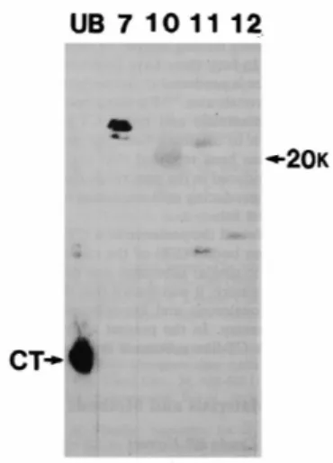
(Fig.4)。ウエスタンブロッティングの結果、アセトニトリル濃度30–33%、39–42%、42–
45%及び45–48%にそれぞれ溶出したフラクション7、10、11及び12 に抗サケCT抗体
に対する免疫陽性反応が認められた。SDS-PAGEの結果から、フラクション7と11に含 まれるそれぞれ2つの免疫反応陽性物質の分子量は、25 kDa と28 kDa、及び10 kDa と
21 kDaとそれぞれ推定された。またフラクション10及び12の免疫反応陽性物質の分子
量はそれぞれ20 kDa及び11 kDaと推定された(Fig. 5)。
ラットバイオアッセイにおける血清中のCa及び無機リン濃度の変化をFig. 6及びFig.
7にそれぞれ示す。フラクション10のみで投与後0.5時間及び1時間の時点で統計学的 に有意な血清Ca濃度の低下が認められた(それぞれp < 0.001及びp < 0.001)。さらにフ ラクション10の投与で投与後3時間まで血清中の無機リン濃度の低下も認められた。
以上の結果を踏まえ、メダカのブロックマン小体のCT様物質は、分子量が約20 kDa の 物質であると結論された。
Fig. 4 Reversed-phase HPLC on an ODS-120T column.
Sample: crude extract of medaka Brockmann bodies; flow rate, 1 ml/min; fraction size, 3 m/tube.
Solvent system: linear-gradient elution from 20% to 80% CH3CN in 0.1% TFA for 60 min.
7
Fig. 5. Molecular weights of immunoreactive calcitonin (CT) in medaka Brockmann body extract.
The positive fractions (Nos. 7, 10, 11, and 12) from Western blotting with anti-calcitonin antiserum and extracts of the ultimobranchial glands of stingrays are compared. The arrows show
immunoreactive CT of 20 kDa in Fraction 10 and stingray CT.
Fig. 6. Changes in serum calcium (Ca) levels in rats after the administration of Fraction 7 (○), 10 (●), 11 (△), 12 (□), or the vehicle (▲).
The vertical bars show the SE. n = 5 for each fraction; n = 6 for the vehicle. *Significantly different from the value for the vehicle only (p < 0.001)
Fig. 7. Changes in serum inorganic phosphorus (Pi) levels in rats after the administration of Fraction 7 (○), 10 (●), 11 (△), 12 (□), or the vehicle (▲).
The vertical bars show the SE. n = 5 for each fraction; n = 6 for the vehicle. Significantly different from the value for the vehicle: * p< 0.05, **p< 0.01, ***p< 0.005, ****p< 0.001
8
2-3) アオゴカイの神経系に存在するCT免疫反応陽性細胞
アオゴカイの神経系に存在するCT免疫反応陽性細胞の分布を観察し、またCT様物質 の分子量を調べた。
【実験方法】
釣り餌として市販されているアオゴカイ(Perinereis aibuhitensis)を購入し、エアレー ションした海水中で3日間飼育して実験に用いた。麻酔したアオゴカイを12分割し酢酸
抜きのBouin液で固定し、一般的な方法で厚さ10μmの連続パラフィン切片を作製した。
脱パラフィン処理した切片を抗サケCT抗体(1/100,000希釈)とインキュベートした 後、ビオチン化抗ウサギ免疫グロブリン抗体(1/2,000希釈)とインキュベートした。そ の後、アビジン-ビオチンkitを用いてCT免疫陽性細胞を検出した。
また、アオゴカイの脳神経節から Fig. 8.に示す方法で粗抽出物を得て、これを
SDS-PAGEにかけた後、抗サケCT抗体を用いたウエスタンブロッティング法でCT様
物質を検出し、その分子量を推定した。
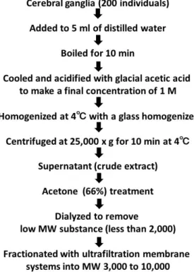
Fig. 8. The procedure for preparing and fractionating crude extracts from cerebral ganglia in the polychaete Perinereis aibuhitensis
Cerebral ganglia were collected from 200 individuals. These ganglia were homogenized and centrifuged. The separated supernatants were treated with 66% acetone and then dialyzed to remove low molecular weight (MW) substances (less than 2,000). Thereafter, the sample was fractionated with an ultrafiltration membrane system into MWs of 3,000 to 10,000.
9
【実験結果及び考察】
アオゴカイの神経系におけるCT免疫陽性細胞の分布をFig.9に示す。
多数のCT免疫陽性細胞(53–70個)が頭部の脳神経節に存在していた(Figs. 10A及び 10B)。他に、食道下神経節には4-6個、腹神経索には体節毎に4個のCT様物質産生細 胞の存在が確認された。CT様物質産生細胞は腹神経索内で体節毎に左右対称に存在して いることから(Fig. 10C)、神経系において何らかの機能的な役割を有することが示唆さ れた。
さらに、SDS-PAGEの結果、その物質の分子量は硬骨魚のCTと同様の約3.5 kDaであ
ると推定された。
Fig. 9. Schematic drawings showing the distribution and number of immunoreactive calcitonin cells (black spots) in the polychaete Perinereis aibuhitensis
The size of the black spots indicates the strength of the positive reaction.
CG: cerebral ganglion; SG: subpharyngeal ganglion; VNC: ventral nerve cord
Fig. 10. Immunoreactive calcitonin (iCT) cells in the polychaete Perinereis aibuhitensis
A: cerebral ganglion (anterior region); B: cerebral ganglion (posterior region); C: ventral nerve cord.
Arrowheads indicate iCT cells.
10 3. まとめ
1. キンギョのウロコを用いたバイオアッセイにより、マイワシのN-proCTは骨形成マ ーカーであるアルカリフォスファターゼ活性を上昇させること及び、骨形成マーカ ーである1型コラーゲン及びオステオカルシンのmRNAの発現を上げる生物学的活 性を有していることを示した。
2. 逆相のHPLCを用いて、メダカのブロックマン小体の抽出物から、抗サケCT抗体 に反応する分子量約20 kDa のCT様物質を分離した。さらにこの物質がラットの血 清Ca濃度を低下させる活性を有することがわかった。この物質はCTの前駆体であ る可能性が高いと考えられた。
3. 抗サケCT抗体を用いた免疫組織化学的手法により、アオゴカイの脳神経節だけで なく、食道下神経節及び腹神経索にCT様物質産生細胞を見出した。CT様物質産生 細胞は腹神経索内で体節毎に左右対称に存在していることから、神経系において何 らかの機能的な役割を有することが示唆された。また、CT様物質の分子量が硬骨魚 のCTと同様の約3.5 kDaであることも判明した。