Acta Med. Nagasaki 30: 186-194
Cancer of the Pancreas in 125 Patients
Shunichi TANABE, Noboru HARADA, Takatoshi NODA, Toshimitsu MIYAMOTO, Tsutomu TOMIOKA, Takashi YAMAGUCHI,
Kensuke YAMAMOTO, Kunihide IZAWA,
Tsukasa TSUNODA, Ryozo YOSHINO,
Toshiya ITO, and Ryoichi TSUCHIYA
The Second Department of Surgery, Nagasaki University School of Medicine
Nagasaki, Japan
Received for publication, June 11, 1985
In this report, we did a retrospective study of 125 patients with cancer of the pan- creas. Seventeen (36.2%) of the 47 patients with cancer of the pancreatic head under- went resection; 11.2% died within 30 days of surgery, 33.3 survived the first year, and 13.3 survived the first three years. The longest survival was for five years. Nine (23%) of the 39 patients with cancer of the pancreatic body and tail underwent resection; 11.1%
died within 30 days, 37.5% survived the first year, and none survived more than three years. The longest survival was for 35 months. Important factors affecting the prognosis were invasion by the cancer of veins or lymph vessels in the surrounding tissues, and metastasis to regional lymph nodes. Lymphatic drainage of small cancers of the pancreatic head were established early. Invasion to the retroperitoneal tissue in cancer of the pan- creatic body and tail was more common than in cancer of the pancreatic head.
For the patient of early cancer of the pancreas, we strongly recommend total pan- createctomy over WHIPPLE's pancreatoduodenectomy, based on results of our analysis of the outcome in our patients following these two operations.
Key words: Cancer of the pancreas, Total pancreatectomy, WHIPPLE's pancreatoduodenec- tomy
INTRODUCTION
The incidence of pancreatic cancer is gradually and steadily increasing, according to the Annals of the Pathological Autopsy in Japan'). The survival rate is very low, and
田辺 篤 一・,原田 昇,野 田剛 稔,宮 本 峻 光,冨 岡 勉,山 口 孝,山 本賢 輔,井 沢 邦 英,角 田 司, 吉 野 奈三,伊 藤 俊 哉,土 屋 涼 一,
this disease is challenging to gastroenterological surgeons. Here, we reviewed a series of patients with cancer of the pancreas seen in our department.
SUBJECTS AND METHODS
At the 2nd Department of Surgery of Nagasaki University Hospital, we have seen 125 patients diagnosed as having pancreatic cancer between November 1969 and Septem- ber 1982. All patients has passed more than three years after their initial treatment.
Seventy-eight patients were male and 47 were female. The mean age was 59.7, with a range from 2 to 80 years.
Each patient's record was reviewed to collect information on early symptoms, loca- tion of the cancer, treatment, factors in survival, and operative management.
The cancers were evaluated using the General Rules for Surgical and Pathological Studies of Cancer of the Pancreas, published by the Japanese Pancreatic Society2).
RESULTS
EARLY SYMPTOMS
In the order of decreasing frequency, the initial symptoms were epigastralgia, ano- rexia, general malaise, jaundice, nausea, vomiting, and weight loss.
Early symptoms are described for each of the four sites of cancer in Table 1.
Upper abdominal pain was the most frequent in all locations. There are no specific early symptoms of cancer of the pancreas, which cause difficulty in its early detection and diagnosis.
Table 1. Early Symptoms by Location of Cancer
Symptoms Patients No. of Head and Tail Body Entire ampullary Peri-
Epigastralgia 78 32 25 14 7
General Malaise 18 10 2 3 3
Anorexia 24 9 6 4 5
Jaundice 12 6 0 1 5
Weight Loss 8 3 4 0 1
Nausea and Vomiting 10 3 4 3 0
LOCATION OF CANCER
The carcinoma was classified as being in one of four sites : the head of the pan- creas, the body and tail of the pancreas, the entire pancreas, or the periampullary region.
Forty-seven patients had carcinoma in the head of the pancreas, 39 in the body and tail
of the pancreas, 20 in the entire pancreas, and 19 in the periampullary region. There was no correlation between age and the location of the carcinoma (Table 2).
Table 2. Sex, Age, and Location of Carcinoma in 125 Patients with Pancreatic Cancer
Location in pancreas Number of Male Female
patients No. Mean age No. Mean age
Head 47 30 57.1 17 57.4
Body and Tail 39 22 56.8 17 54.7
Entire 20 14 59.3 6 58.5
Periampullary 19 12 68.5 7 62.1
Total 125 78 60.4 47 58.2
TREATMENT
We did a radical operation on 26 (20.8%) of our 125 patients (pancreatoduodenec- tomy in 9, total pancreatectomy in 9, and resection of body and tail in 8), a palliative operation on 71 (56.8%), and exploratory laparotomy on 19 (15.2%). Nine patients did not undergo surgery. In only 22.4% of our patients who underwent operation was the cancer resectable. In the palliative operations, biliary enteric bypass operations such as cholecystoduodenostomy, cholecystojejunostomy or external cholecystostomy were done (Ta- ble 3).
Table 3. Treatments in 125 Patients
Operation No. of Patients Incidence
Resection PD 9
TP 9 20.8%
DP 8
Palliative* Operation 71 56.8%
Exploratory Laparotomy 19 15.2%
No Operation 9 7.2%
Total 125 100.0%
PD, Pancreatoduodenectomy
TP, Total pancreatectomy
DP, Distal pancreatectomy
*, Bypass or Drainage operation SURVIVAL
Of the 47 patients with carcinoma of the head of the pancreas, 17 (36.2%) under- went curative operations, 27 (57.4%) a palliative operation, and 3 (6.4%) an exploratory
laparotomy only. Of the 39 patients with carcinoma of the body and tail of the pancreas, 9 patients (23.1%) had curative operation, 11 (28.2%) a palliative operation, and 12
(30.8%) an exploratory laparotomy. By location, the longest mean survival was 13.6 months for the 8 patients who received distal pancreatectomy, and the longest survival among these patient was 35 months. The mean survival for the 9 patients who received pancreatoduodenectomy for carcinoma of the head of the pancreas was 13.1 months ; the longest survival was 4 years. The survival for the 8 patients who received total pancre- atectomy for carcinoma of the head of the pancreas was 6.3 months ; one patient is still alive, 60 months after. No patient with carcinoma of the entire pancreas or the periam- pullary region had a curative operation and all died within 6 months except two of 57 (Table 4).
Table 4. Mean Survivals According to Location of Tumors and Surgical Procedures in 125 Patients
Average Survival Longest Survival
(months) (months)
Laparotomy Only 3( 6.4%) 1.2 1.6
Head Palliative Operation 27(57.4%) 4.9 24.6
(47)
Resection 17(36.2%) 9.9 52.0
PD 9(19.1%) 13.1 48.0
TP 8(17.2%) 6.3 60.0, alive
No Operation 7(17.9%) 1.2 2.4
Body Laparotomy Only 12(30.8%) 3.7 13.0
and
Tail Palliative Operation 11(28.2%) 3.3 12.0
(39)
Resection 9(23.1%) 12.6 35.0
DP 8(20.5%) 13.6 35.0
TP 1( 2.6%) 4.0 4.0
No Operation 1( 5.0%) 0.5 0.5
Entire
(20) Laparotomy Only 4(2.0.%) 3.6 8.0
Paillative Operation 15(75.0%) 3.5 10.0
Peri- No Operation 1( 5.2%) 1.0 1.0
ampullary
(19) Laparotomy Only 18(94.7%) 2.1 5.0
MORTALITY AND SURVIVAL OF RESECTED CASES
Two of the 17 patients with carcinoma of the head of the pancreas who underwent resection, and one of the 9 such patients with carcinoma of the body and tail of the pancreas died within 30 days of the operation, so the mortality rate was 11.8% and 11.1
%, respectively. There was no clear differen'e between operative mortality depending on the procedure or on the site of the tumor. The overall one-year and three-year sur- vival rates of the 17 patients with resectable lesions of the head of the pancreas were
33.3% and 13.3%, respectively. Of these same 17 patients, nine had pancreatoduoden- ectomy, and 50.0% survived for one year. Eight had total pancreatectomy, and 14.3%
survived for one and three years. The one-year survival rate of patients with resectable carcinoma of the body and tail of the pancreas was 37.3%, and the three-year survival rate, 0% (Table 5).
Table 5. Mortality and Survival Rate in Resected Cases
Location Operation PNo. of Operative* Survival for Survival for
atients Death One Year Three Years
Resection 17 2/17(11.2%) 5/15(33.3%) 2/15(13 .3%)
Head PD 9 1/9(11.1%) 4/8(50.0%) 1/8(12.5%)
TP 8 1/802.5%) 1/704.3%) 1/7(14.3%)
Body ody Resection 9 1/ 9(11.9%) 3/ 8(37.5%) 0/ 8( 0 %)
and DP 8 1/8(12.5%) 3/7(42.9%) 0/7( 0 %)
Tail
TP 1 0/1( 0 %) 0/1( 0 %) 0/1( 0 %)
*, within 30days
FACTORS IN SURVIVAL
To evaluate prognostic factors in carcinoma of the pancreas, macroscopic findings about the size of the tumor, the degree of capsular infiltration, the mode of infiltration, the degree of invasion of lymph vessels or veins of the adjacent tissue, and metastasis to regional lymph nodes were examined, and to clarify the relationship between each factor and survival in our 26 patients with resectable cancer, the 21 who lived more than 3 months after the operation were studied. The three factors that had statistical significance
Table 6. Factors in Survival
Factors Patients No. of mean month ± SEM Survival Period
(-) 3 30.0 ± 20.9
Invasion into Veins j P <0.01
(I) 18 10.0 ± 6.2
(-) 2 41.5 ± 9.2
Invasion into Lymph vessels ] P <0.001
(I) 19 10.2 ± 6.1
(-) 6 21.2 ± 17.6
Metastasis to Regional Lymph ] P <0.05
Nodes (-1-) 15 10.0 + 5.6
no invasion or metastasis
(f ), invasion or metastasis
were invasion to veins, invasion to lymph vessels, and metastasis to regional lymph nodes.
Especially, invasion to lymph vessel of the adjacent tissue was evaluated as the most im- portant factor responsible for the prognosis. The mean survival month of resected cases was 41.5±9.2 without invasion to lymph vessels and 10.2±6.1 with invasion to lymph vessels (Table 6) .
On patients having resectable cancer of the head or body and tail of the pancreas
taken separately, we examined histopathologically invasion of the superficial capsule of the
pancreas [positive : S(+), negative : S(-)), invasion of the retroperitoneal tissue of the pancreas (positive : Rp (+) , negative : Rp (-)), and metastasis to lymph nodes (positive : N (+), negative : N (-)'j.
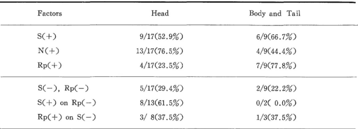
Results are shown in Table 7 . Rp(+) was much more common (77.8%) in cancer of the pancreatic body and tail than in cancer of the pancreatic head (23.5%). The in- cidence of S(+), S(-) and Rp(+), and S(-) and Rp(-) did not showed any signi- ficant difference.
However, metastasis to the lymph nodes was high but 76.5% in cancer of the pan- creatic head, but 44.4% in cancer of the pancreatic body and tail. We were surprised that 80% of the patients who were S(-) and Rp(-) had metastasis to the lymph nodes
Table 7. Incidence of Invasion to Surrounding Tissue By Location
Factors Head Body and Tail
S(+) 9/17(52.9%) 6/9(66.7%)
N(+) 13/17(76.5%) 4/9(44.4%)
Rp(+) 4/17(23.5%) 7/9(77.8%)
S(-), Rp(-) 5/17(29.4%) 2/9(22.2%)
S(+) on Rp(-) 8/13(61.5%) 0/2( 0.0%)
Rp(+) on S(-) 3/ 8(37.5%) 1/3(37.5%)
Table 8. Ratio of Lymph Node Metastasis to Capsular Invasion and/or Retroperitoneal Tissue
Head Body and Tail
N(+) /S(-) 6/ 8(75.0%) 1/3( 33.3%)
/S(+) 7/ 9(77.7%) 3/6( 50.0%)
N(+) /Rp(-) 10/13(76.9%) 0/2( 0.0%)
/Rp(+) 3/ 4(75.0%) 4/7( 57.1%)
N(+) /S(-), Rp(-) 4/ 5(80.0%) 0/2( 0/0%)
/S(-), Rp(+) 2/ 3(66.6%) 1/1(100.0%)
/S(-}-'),f Rp(-) 6/ 8(75.0%) 0/2( 0.0%)
/S(+), Rp(+) 1/1(100.0%) 3/4( 75.0%)
in cancer of the pancreatic head (Table 8) . ASSESSMENT OF OPERATIVE METHODS
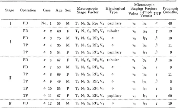
WHIPPLE's pancreatoduodenectomy and total pancreatectomy was used for cancer of the pancreatic head. To evaluate these methods, the resected cases of cancer of the pancreatic head were classified into four stages after the classification 2) and reviewed the operative procedures, histology of the tumor, the prognosis for the four stages (Table 9) . We excluded the patients who died of causes other than recurrence of cancer.
Table 9. Representative List of Patients with Cancer of Head of Pancreas Who died of Recurrence after Resection
(According to Classification of General Rules2))
Microscopic
Stage Operation Case Age Sex Macroscopic St Histological Staging Factors Prognosis age Factor Type Veins Lymph INF (months)
Vessels
j PD No. 1 50 M T1 No So Rpo Vo papillary vo lyo a 48
---
PD ii 2 63 F T2 N1 So RPo Vo tubular v2 IY2 r 19
PD 'i 3 75 M T2 N1 So RPo Vo i, V2 ly A 10
TP 'i 4 35 M T1 N 1 So RPo Vo % v3 1Y3 P 11
TP 'i 5 54 F T2 N1 So RPo Vo papillary v3 ly3 P 9
---
PD i~ 6 67 F T1 No S2 RPo Vo tubular v2 ly2 P 16
PD o 7 53 M T2 N1 So RP2 V2 'i v2 IYi r 9
TP ii 8 69 F T2 N1 S2 RPo Vo % V3 IY3 r 11
1[CI
TP % 9 49 M T3 N1 S1 RPo Vo i, v2 'Y2 P 5
TP 'i 10 55 F T3 N2 So RP2 V2 'i v2 lY2 r 5
TP ii 11 67 F T3 N2 So RPo Vo papillary v2 'Y2 r 60
---
IV PD ii 12 51 M T2 N1 S3 RPo Vo v2 lY2 r 19
Case No. 1 --A 50-year-old male patient who had very small cancer in size 0.2 x 0.2 cm and without any local and distant metastasis to the liver or dissemination to the peritoneum underwent pancreatoduodenectomy. But a recurrence arose from the remaining pancreas, in which a multicentric growth was found. He died of recurrence of cancer four years later. This patient would probably have lived longer with a total pancreatectomy.
These cases lead us to prefer to do total pancreatectomy for cancer of the pancreas.
Case No. 9 --A 49-year-old man in stage III underwent total pancreatectomy. Hi- stological investigation of the resected specimen disclosed a skip lesion confined to the duct of the pancreas 2 cm apart from the main focus pancreatic cancer. Total pancre- atectomy was a fortunate choice for this patient. For patients in stage III, total pancre- atectomy seems not to have any advantage in prognosis if the lymph nodes were already involved.
Of our four patients in stage II with So, Rpo, V0, but with metastasis to lymph
nodes, the survival in the two by pancreatoduodenectomy was 19 and 10 months, and the other two by total pancreatectomy, 11 and 9 months. Histological extension showed more progressive finding in the patients with the total pancreatectomy compared with the pan- creatoduodenectomy. For the patients in this stage, total pancreatectomy failed to show any difference in survival.
DISCUSSION
High diagnostic accuracy for pancreatic cancer is made possible by improvement of imaging diagnostic aids such as endoscopic retrograde cholangio-pancreatography, percu- taneous transhepatic cholangiography, selective celiac arteriography, ultrasound sonography, computed tomography, and aspiration cytology. However, these diagnostic aids are still not able to diagnose early pancreatic cancer. In fact, most of our patients were diagnosed in an advanced stage. Poor prognosis of pancreatic cancer mainly depend on the delay of diagnosis due to vague and non-specific early symptoms and on the tendency of early drainage of cancer into regional lymph nodes even in small pancreatic cancer. To settle these problems of diagnostic delay and poor prognosis, more reliable routine screening tests for early detection of cancer of the pancreas are needed. The usual operation in this disease is WHIPPLE's pancreatoduodenectomy, but prognosis with this operation is poor.
MONGE3) reported 21% operative mortality and 18.2% five-year survival. WARREN') , SMITHS), and others") reported similar or worse results. To increase survival, since 1977 we have utilized total pancreatectomy as a radical operation for cancer of the pancreas.
For the reasons of that we encountered patients with multicentric growths and with metastatic skip lesions in the pancreatic duct, and that possibility of dissemination of free-floating cancer cells7) or direct spread of cancer along the pancreatic ducts'), we have utilized total pancreatectomy as a radical operation for cancer of the pancreas. BROOKS8>
showed the necessity of selection of patients for total pancreatectomy in his results of 19%
five-year survival. REMINE9) reported a 4.7% mortality rate and improvement in the five-year survival rate from 12 to 25% by using total pancreatectomy instead of pancre- atoduodenectomy. LONGMIRE8)1C) found a 12% mortality rate and 20 months average survival with the WHIPPLE procedure, and a_17% mortality and 26 months average survival with total pancreatectomy. IHSE7) reported 21%, five-year survival for sixty-five patients who had undergone total pancreatectomy. However, HERMAN) made comparisons studies of the two operations, and doubted that total pancreatectomy gave longer survival.
In our series, total pancreatectomy has not still achieved improvement in survival in patients in stage II or more advanced stages, especially when they had a metastasis to the lymph nodes. However for the patients in stage I, no metastasis to lymph nodes, total pancreatectomy seem to be a reasonable curative operation to avoid a recurrence from intraductal dissemination or multicentric growth of the cancer. In other attempts to make a more radical dissection of pancreatic cancer, FORTNER11>12)13) has tried regional pan- createctomy. In this recent result, of the 29 patients who underwent this operation, ten
are alive one to 53 months later, and eleven died of recurrences after 5 to 30 months, but follow up period is not enough for evaluation of this operation for the patient of pancreatic cancer.
REFERENCES
1) Annual of the Pathological Autopsy Cases in Japan. edited and published by The Japanese Pathological Society 1979.
2) General Rules for Surgical and Pathological Studies on Cancer of Pancreas edited by Japanese Pancreatic Society 1981.
3) MONGE JJ, JUDD ES, GAGE RP.: Radical Pancreaticoduodenectomy: A. 22-year experience with the complications, mortality rate and survival rate. Ann. Surg. 160:
711-719, 1964
4) WALLEN KW, CHOE DS, PLAZA J, RELIHAM M.: Results of Radical Resection for Periampullary Cancer. Ann. Surg. 181: 534-539, 1975.
5) SMITH R,: Progress in the Surgical Treatment of Pancreatic Disease. Am. J Surg.
125: 143 -153, 1973.
6) SATO T, SAITOH Y, NOTO N, MATSUNO S.: Follow-up Studies of Radical Resection for Periampullary Cancer. Ann. Surg. 186: 581-588, 1977.
7) IHSE I, LILJA P, ARNESJO B, BENGMARK S.: Total Pancreatectomy for Cancer. An appraisal of 65 cases. Ann. Surg. 186: 675-680, 1977.
8) TRYKA AF, BROOKS JR.: Histopathology in the Evaluation of Total Pancreatectomy for Ductal Carcinoma. Ann. Surg. 190: 373-381, 1979.
9) LEVIN B, REMINE WH, HERMAN RE, SCHEIN PS, COHN I Jr.: Panel: Cancer of the Pancreas Am. J. Surg. 135: 185-191, 1978.
10) LONGMITE WPJr, TRAVERSO WL.: The Whipple Procedure and Other Standard Operative Approaches to Pancreatic Cancer. Cancer 47: 1706-1711, 1981.
11) FORTNER JG: Regional Resection of Cancer of the Pancreas. A New Surgical Approach. Surgery 73: 307-320, 1973.
12) FORTNER JG: Cancer of Pancreas. N. Engl. J. Med. 302: 232, 1980.
13) FORTNER JG: Surgical Principles for Pancreatic Cancer, Regional Total and Subtotal Pancreatectomy. Cancer 47: 1712-1718, 1981.