Tropical :Medicine, 13 (,1): 180-192, Dec., 1971 185
Two autopsy cases of a peculiar disease, so called "Pulseless Disease"
Kanehiko YAMASHITA and Yukie KUNITA
Depa1'llnent of Pathology Institute for TrojJical lVIedicine,Nagasaki University
(Director: Froj. Toyosuke WAT ANA HE)
(Received for Publication Kovem ber 27, 1971)
Abstract
T\'v'o autopsy cases of "Pulseless Disease", a peculiar dsease of the aorta and its branches, are reported in this paper. In "Pulseless Disease" autopsy cases of old persons are relatively rare. The two cases are both of old persons, and show not only stenosis or obstruction as indicated by the name, "Pulseless Disease" , but also marked dilatation of arterial lumen. There are no histological differences between stenosed and dilated portions. One of the two cases demonstrates so· called \\Atypical coarctation of the aorta
N ,and the other is combined with severe atheroscleros's with marked calcification.
Introduction The disease known by the name, "Pulse-
less Disease" , was first reported by a Japanese ophthalmologist, Takayasu
D .In
1908he made a report on "a case of peculiar changes of the central retinal vessels
ll ,in which he pointed out a wreath-like arteriovenous anastomosis surrounding the optic disc, but no refe- rence was made by him of pulselessness.
A t the same time Oonishi and Kagoshima supplemented each a similar case with abscent radial pulses. Tn
1948Shimizu and San0
2 )described the symptomatology of this disease, and first used the Ina me ,
\\Pulseless Disease\\. They mentioned
clinically three cardiac symptoms of this disease:
(1)abscent pulse of upper limbs,
(2)arteriovenous anastomosis surrounding the optic disc, and
(3)an acceleration of carotid sinus reflex. The "Pulseless Disease
llwas reported under various names; Takayasu's disease, Takayasu's arteritis, aortic arch syndrome and aorti- tis syndrome etc, and pathologically named as truncoarteritis productiva obliterans by a Japanese patholog:st,
~asu'~).
This d:sease was found predomi- nantly in Asia, however infrequent in other continents, and seems to have a predilection for young female. Some
Contribution No.6?5 from the Institute for Tropical Medicine, Nagasaki University.
186 Kanehiko YAMASHITA and Yukie KUNITA
cases of so called "Atypical coarctation
of the aorta
N ,noticed by Inada et a1
4 ) ,was considered to belong in the same categoly of this d:sease, and recently frequent association of hypertension with this disease has been notced. The etio- logy of this disease is still unknown.
Clinically this disease was diagnosed mainly as follows; pulseless disease, val- vular disease, coarctation of the aorta, aortic arch syndrome and renovascular disturbance. Causes of death were chiefly heart failure, death under operation,
sudden death and uremia.
Pathologically the main lesions of
"Pulseless Disease" are mainly localized to the arteries of elastic type including pulmonary artery. The chief canges in the initial stage are supposed to be limit·
ed to adventitia and outer portion of media, based on a inflammatory reaction, which in turn brings about several follo- wing changes in arterial wall; marked fibrous thickening of adventitia, destruc- tion of media and intimal thickening.
Case reports
Case 1: The patient is a 54 year·old Japanese female, who has noticed a pulsation of neck at the age of 34 years but did not seek medical attention. At the age of 45 years she saw a doctor with the complaints, sense of pressure at chest and angina-like attacks. At that time hypertension and cardiomegaly were pointed out, so she was admitted to the Nagasaki University Hospital and was diagnosed as aortic insufficiency and aortic arch syndrome. The angiocardiog- raphy revealed obstruction of the left common carotid artery, aneurysm of the right subclavian artery, stenosis of the descending aorta and kinking of the right common carotid artery, and thrill and pulsation were noticed in all portions mentioned above. After treatment in the hospital for two months she had been followed up as outpatient for eight years.
One day prior to her death anorexia and orthopnea appeared and suddenly she fell on her back and was dead. The entire clinical course is approximately 18 years.
Case 2: The patient is a 66 year-old
Japanese male. About two years ago he noticed a dyspnea at the rest and a strangulated sense of chest, and visited a doctor to be diagnosed as hypertension and cardiac asthma. Three months after he was admitted to the Nagasaki Univer- sity Hospital and was diagnosed as severe atherosclerosis, aortic arch syndrome, coronary insufficiency with ventricular hypertrophy on both sides and gastric ulcer etc. The left radial pulse was difficult to palpate, and the variation of blood pressure between left and right arm was noticed. The angiocardiography revealed aneurysmatic dilatation of the ascending aorta, narrowing of the left common carotid artery and stenosis of the left subclavian artery. After medi- cation for about one month the symptoms were improved, but the sense of pressure at chest, dyspnea and stridor appeared and he died of increased heart failure.
The both cases are negative for Was-
sermann reaction and positive for tuber-
culine reaction. The blood pressure of
Case 1 is 186/30-0 mmHg, and that of
Two autopsy caoes of a peculiar ciiseaO'e, so called "Pu!seless Disease" IS7
Case 2 is 170(90 mmHg in the right arm, 110(80 mmHg in the left arm and 230(90 mmHg in the lower extremities. The erythrocyte·sedimentation rate moderately accelerates in both cases, but the other
examinations are within normal limits.
Case 1 has a history of pulmonary tuber- culosis at the age of 43 years, and Case 2 has that of pleurisy at the age of 28 years.
Autopsy findings Case 1: The main lesions correspond
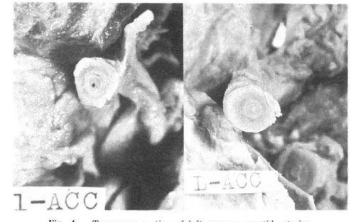
to the artery of elastic type including pulmonary artery (Fig. 1). The wall of affected arteries are diffusely thickened and the inner surface reveals relatively smooth appearance with occasional irregu- lar white plaques (Fig. 2). But neither atheroma nor thrombi are noticed. The wall of the left common carotid artery is strongly thickened and markedly contrac- ted in the entire length, so that the lumen is stenosed or obstructed (Fig. 4, Fig. 5). The lumen of the descending aorta is narrowed to the diaphragmatic portion as described as "Atypical COal'cta- tion of the aorta
ll ,\vhile that of the abdominal aorta is not remarkably chan- ged. On the contrary the aortic arch, brachiocephalic artery, right common carotid artery and right subclavian artery are markedly dilated and partially aneu·
rysmatic. Findings of other organs are as follows; cardiomegaly (600 g) with moderately thickened and contracted aortic val ves, occasional pul monary thromboarteritis with infarction in both lungs, partial atelectasis of the left lower lobe, two calcified nodules in the left
lower lobe. eloudy swelling of the liver, moderate nephrosclerosis, cholelithiasis and capillary hemorrhage of the stomach and intestine.
Case 2: The distribution of "Pulseless Disease
llis similar to that of Case 1 (Fig. 1). The wall of the affected arteries are diffusely thickened. In this case, there is severe atherosclerosis with marked calcification of the aorta and its large branches. especially of the thoracic aorta, brachiocephalic artery, right sub·
clavian artery and right common carotid artery, the inner surface of them being rough and irregular (Fig. 3). The lumen of the left common carotid artery and that of the left subclavian artery are almost obstructed (Fig. 4, Fig. 6). The thoracic aorta, especially of the descen- ding aorta, brachiocephalic artery, left subclavian artery and proximal portion of the rigth common carotid artery are markedly dilated. Findings of other organs are as follows; cardiomegaly (680 g) ,'lith fibrinofibrotic pericarditis, subacute splenitis, severe fatty liver, pyelocystitis and moderate pulmonary edema.
Histological findings of arteries In the adventitia, fibrosis is more
remarkable. Diffuse and marked prolife·
ration of collagenous fibers with partial
hyalinization are arranged in various pattern; a nodular, fascicular and stra"
tionform ones. The adventitia is strongly
188 Kanehiko Y AMASHIT A and Yukie KUNITA
thickened and occupies the almost half or
more width of the arterial wall. Several granulomas consisting of lymphocytes also plasma cells, a small number of histiocytes and some giant cells of Lan- ghans type, are found in the adventitia and also in the media at the proximal portions of the pulmonary artery and ascending aorta in Case 1 (Fig _ 8, Fig_
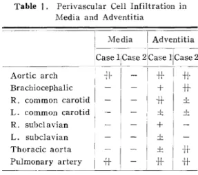
9). Beside these changes, there are perivascular cell infiltrations of various degree chiefly composed of lymphocytes in the adventitia and partially in the media of all the affected arteries, which sometimes show lymph-follicle like appea- rance (Table 1). In Case 2, the perivas- cular cell infiltrations are almost equal to Case 1, but granuloma formation is not seen. The granuloma seems to be prone fibrosis.
In the media, the changes are found chiefly at the outer one third to half portion, and are summarized as follows;
newly proliferated capillaries and arterio- les. deposition of mucoid substance.
partial fibrosis and interruption and coagulation of elastic fibers. At some
parts the media
ISmore thin oppressed by the nodular fibrosis of the adventitia.
In the intima, fibrocellular prolifera- tion is remarkable with newly produced elastic fibers and deposition of mucoid substance.
The wall of vasa vasorum at the affec- ted portions are markedly thickened with fibrocellular proliferation of the intima and deposition of mucoid substance, so that the lumen is stenosed or obstructed.
There are no histological differences between the stenosed and dilated portions (Fig_ 5, Fig. 6, Fig. 7).
Table 1. Perivascular Cell Infiltration in J\'Tedia and Adventitia
, Media I Adventitia
i---- -1--- -_.
Case lCase 21Case llCase 2 Aortic arch
-:+
• • I
BrachlOcephahc I' R. common carotid L. common carotid R. subclavian L. subclavian Thoracic aorta
Pulmonary artery
-it
T*I -11-
I~ ;
I _L
+
±
± I~
-H- +t
Summary Autopsy cases of "Pulseless Disease" in
old persons are relatively rare. The two cases reported in this paper are both of old persons. In "Pulseless Disease
ll ,the lumen of the affected artery tends to be stenosed or obstructed, but also can be dilated and even partially aneurysma- tic as in these two cases. The stenosis of the lumen is chiefly caused by the contraction of the adventitial fibrosis overcoming the pressure on the arterial wall, promoted with the intimal thicke-
ning. Dilatation of that may be caused
by increased arterial pressure, which
exceeds the contracting effect of wall,
and also influenced by the duration of
the disease. There are no histological
differences between stenosed and dilated
portions. One of the two cases demonst-
rates so-called "Atypical coarctation of
the aorta/1 , and the other is combined
with severe atherosclerosis with marked
ca1cifica tion.
Two autopsy cases of a peculiar disease, so called "Pulseless Diseao:eio 189 The Pulseless Disease has been reported during the last 60 years in Asia, chiefly in Japan. but might be found more frequently in tropical and subtropical countries of Asia, as more attension will he paid to this conditiun.
References
1. Takayasu, M. : A case of peculiar chan·
ges of the central retinal vessels. Acta Soc.
Ophtha!. Jap. 12 ; 554-555, 1908 (in Japanese).
2. Shimizu, K. and Sano, K.: Pulseless disease. Rinsho·geka 3 : 377--396, 1948 (in Japanese) .
3. Nasu, T. and Mamiya,:\!. : Truncoar·
teriti3 productiva obEterans. Nis,hin·igaku 50 :
435~-451. 1963 (in Japanese;.
4. Inada, K., Shimizu. H. and Yokoyama, T.: Pulse less disease and atypical coarctation of the aorta with special reference to their genesis. Surgery 52 : 433-443, 1962.
5. Ueda. H. et aL : Clinical and patholo' gical studies of aortitis syndrome. Saishin·igaku 23 : 181-188, 1968 (in Japanese).
I, ·:iJvp6 iWrd" U~J O);f;cilf-fih.
;t::11J1JIR
t.1:"OJ±:~5t.tZ)~+WlJ£~~-t 6J*.~,(/:Ci'ilj~fjIJ'kf.,f!i6'i L t:.O)c'¥R"l'r Lt:.. 1~I~t" LWlJ 0) ,~j~~O)trlj~{9IJO)*fi,;:JtijijH!']'Yt" \,·tJ~. 4:jj£{JUi;,=-j).ttt1~1.,;;;~0)!rti9~-(i/i)':;'.
3(. *~IT?c'I:;,1:"O)::g;h;*-t!::: J-; i) jJ!..b,~r:9}!'1'0)~~i/i) Q HiFof]'Wi'k*t:.-j ~ UJ;19'pitc';ji,.:;, D;. .~ '5 Lt:.ffl~
v;::,
tm;t, *
Rjj~fJlj -r,:-,:t-i¥l1
~=.if) Jjiil!'iIltiR t. ts:.
"'0 t:.#11:;r 71
dyjilf il)J tdl)tl~ V~Il'i
0)Dl;'iEf;HDj Il#
v=. 6~'g6':>G h
t:.. - }j.Hl.
~+i¥Jv='fi, ~%!'fffil, tI!:;5.Rg1\O)rI31f~i¥:j£i;,~rr;6':> Ght"D'·) t:.. ~'.f5'JO) rf1-fJIH i~'1jiI;t::ilJ~~m!'j"'JlEO)ffl Rj);;li) • ftlJ(i"ilJllt,,:fi!J({tHl'~) t:.1i!i1lit0)1lW!lI~@!:{U1E~I+
'"
-Cv·:t!.190
.
-.
.~ -, -
--: .
,-
~'-::
. -
Cast· 1.
Kanehiko YAMASHITA and Ylikie KL;NITA
Fig. 1.
Ca!lC 2.
Affl't'lt'd Portion of Arlcrirtl Wall
Schemas presenting the arterial lesions.
.- ,-
, , .-
• , .- .- -
.-.- .-. -
Fig. 2. Aorta an:! its large branches (Case I). Fig. 3. Aorta andiitsllarge branches (ease 2).
Two autopsy cases of a peculiar di£ease, so called "Pulseless Di£easell 191
Fig. 4. Transverse section of Jeft common carotid arteries (right: Case 1, left: Case 2).
:
': - . .
Fig. 5. Typical arterial lesion showing the narrowing of lumen (the left common carotid artery of Case J).
Fig. 6. Typical arterial lesion showing the obstructiGn of lumen (the left common carotid artery of Case 2).
192 Kanebiko YAMASHITA and Yukie ICJNITA
Fig. 7. Typical arterial lesion at the portion of dilated lumen (the right common carotid artery of Case 1).
Fig. 8. Granuloma in the media of the pulmo- nary artery of Case I.
Fig. 9. Granuloma in the media.