A novel role of eIF5A in HIF-1α-mediated hypoxic
response
(HIF-1αを介した低酸素応答における翻訳制御因子 eIF5A の
新しい機能に関する研究)
2016
年 3 月
埼玉大学大学院理工学研究科(博士後期課程)
理工学専攻(主指導教員 吉田 稔)
TARIQ MOHAMMAD
Mohammad Tariq
Graduate School of Science and Engineering
Saitama University
Supervisor: Professor Minoru Yoshida
Abstract
Eukaryotic translation initiation factor 5A (eIF5A) is an important cellular protein and is highly conserved from bacteria to humans. eIF5A is essential for cell growth and survival. Initially eIF5A was identified to be a translation initiation factor. However recent findings suggested that eIF5A is not involved in translation initiation but has a role in translation elongation and other cellular processes such as mRNA transport. eIF5A is quite unique as it is the only cellular protein containing an unusual, polyamine-derived amino acid, hypusine [Nε- (4-amino 2-hydroxybutyl) lysine]. A conserved lysine residue of eIF5A i.e., lysine 50 of human eIF5A, is modified to hypusine. eIF5A undergoes acetylation, at lysine 47. Our lab previously reported that acetylation regulates subcellular distribution of eIF5A and that acetylated eIF5A localizes to the nucleus. Regulation of eIF5A acetylation by cellular environment is unknown. In the current study I focused to reveal a novel function of eIF5A, under different cellular environments.
In order to unravel the role of eIF5A, I studied physiological conditions promoting eIF5A acetylation. I found glucose and hypoxia as the cellular cues of eIF5A acetylation induction. eIF5A was found to be an important factor for cell growth and survival in hypoxia. Furthermore, I found that hypusinated eIF5A is essential for the expression of HIF-1α in hypoxia. In the absence of eIF5A HIF-1α is degraded by the proteasome. My study revealed the importance of eIF5A in tumor growth and survival. On the basis of my observations, it is suggested that hypusination of eIF5A may be a potential target for therapeutic intervention.
ACKNOWLEDGEMENTS
I would like to express my deepest gratitude to my supervisors Prof. Minoru Yoshida and Dr. Akihiro Ito, for their guidance, support, encouragement and providing me with an excellent atmosphere for doing research. Without their continuous support, the successful completion of my research work would not have been possible.
I am thankful to all members of the Chemical Genetics Laboratory at RIKEN especially Ms. Satoko Maeda, Ms. Kazuko Uchida, Ms. Naomi Hayashi and Ms. Megumi Takase for their support throughout my stay.
I would like to thank all my friends especially Dr. Muhammad Ishfaq, Dr. Khalid khan and Mr. Asad Ali Shah for their support.
I am thankful to my parents and family members for their love, care, support and encouragement throughout my life.
Abbreviations
eIF5A Eukaryotic Initiation Factor 5A Ac-eIF5A acetylated eIF5A
Hyp-eIF5A hypusinated eIF5A
Dhp-eIF5A Deoxyhypusinated eIF5A
HIF-1α hypoxia inducible factor 1 alpha DHS Deoxyhypusine synthase
DOHH Deoxyhypusine hydroxylase PHD Prolyl hydroxylase
HRE Hypoxia response element HDAC Histone deacetylase
NAD Nicotinamide adenine dinucleotide TSA Trichostatin A
NA Nicotinamide
DFX Deferoxamine mesylate SIRT Sirtuin
PCR Polymerase chain reaction
qPCR Quantitative polymerase chain reaction RNA Ribonucleic acid
mRNA Messenger ribonucleic acid siRNA Small interfering RNA RNAi RNA interference
SDS-PAGE Sodium dodecyle sulfate polyacrylamide gel electrophoresis
VHL Von Hippel-Lindau
HAF Hypoxia associated factor
RACK1 Receptor of activated protein kinase C VEGFA Vascular endothelial growth factor A GLUT1 Glucose transporter 1
PGK1 Phosphoglycerate kinase 1 LDHA Lactate dehydrogenase A
BNIP3 BCL2/adenovirus E1B 19kDa interacting protein 3 PDK1 Pyruvate dehydrogenase kinase isozyme 1
Contents
Chapter 1: Introduction
1.1 Eukaryotic initiation factor 5A (eIF5A)………...1
1.2 Post-translational modifications of eIF5A……….1
1.3 Role of eIF5A in translation………....2
1.4 Role of eIF5A in cancer………...3
Chapter 2: Regulation of eIF5A acetylation
2.1 Introduction……….072.2 Materials and methods……….09
2.2.1 Compounds and cell culture 2.2.2 Antibodies and immunoblotting 2.2.3 Hypoxic conditions 2.3 Results and discussion……….10
2.3.1 Increasing glucose concentration induced acetylation of eIF5A 2.3.2 acetylation of eIF5A is induced by hypoxia 2.3.3 Effect of HDAC inhibitors and hypoxia on eIF5A acetylation Summary………11
Chapter 3: eIF5A is essential for HIF-1α expression in hypoxia
3.1 Introduction……….143.1.1 Hypoxia inducible factor 1 alpha (HIF-1α) 3.1.2 Regulation of HIF-1α 3.1.3 HIF-1α and cancer progression 3.2 Materials and methods……….18
3.2.1 Compounds and cell culture 3.2.2 Antibodies and immunoblotting 3.2.3 RNAi and hypoxia
3.3 Results and discussion………19 3.3.1 Time course of eIF5A acetylation and HIF-1α expression is different
3.3.2 eIF5A is essential for HIF-1α expression in hypoxia
3.3.3 Functional eIF5A is important for HIF-1α expression in hypoxia 3.3.4 GC7 treatment or eIF5A knockdown-mediated suppression of HIF-1α is independent of prolyl hydroxylase (PHD)
3.3.5 Mechanism of HIF-1α suppression by GC7 treatment or eIF5A knockdown
Summary………...22
Chapter 4: eIF5A is required HIF-1α-mediated transcriptional
activation in hypoxia
4.1 Introduction………29 4.2 Materials and methods………30
4.2.1 Compounds and cell culture 4.2.2 Antibodies and immunoblotting
4.2.3 RNA interference, GC7 treatment and hypoxia 4.2.4 RNA extraction and qPCR
4.3 Results and discussion………32 Summary………..33
Chapter 5: eIF5A is important for growth of tumor spheroid
5.1 Introduction……….37 5.2 Materials and methods………..38
5.2.1 Compounds and cell culture 5.2.2 Antibodies and immunoblotting 5.2.3 RNA interference and GC7 treatment
5.3 Results and discussion………..39 5.3.1 HIF-1α knockdown leads to reduction in the size of spheroid 5.3.2 eIF5A knockdown has negative effect on spheroid growth
5.3.3 Functional eIF5A is essential for spheroid growth
Summary……….40
Chapter 6: Conclusions……… 44
References
Chapter 1:
Introduction
1.1 Eukaryotic initiation factor 5A (eIF5A)
Eukaryotic Initiation Factor 5A (eIF5A) is a small (~17 kDa) essential protein and highly conserved from bacteria (EF-P) to humans [1-3]. eIF5A was initially identified in reticulocyte lysate along with other translation initiation factors, as a protein which could stimulate methionyl puromycin synthesis. eIF5A (formerly called eIF4D) was then considered to be a translation initiation factor [4] but further studies did not validate this observation and in an in vitro translation assay, the absence of eIF5A had no negative effect on translation initiation and thus making the role of eIF5A in translation initiation controversial [1,5]. Further studies suggested that eIF5A is involved in translation elongation instead of initiation [6]. eIF5A has two isoforms, eIF5A1 and eIF5A2. eIF5A1 is constitutively expressed in all tissues and shows higher expression in caner and proliferating cells. eIF5A2 shows tissue specific expression being expressed in brain, testes and certain cancer cell lines [7-9]. Both isoforms contain conserved lysine residue (K50), which is post-translationally modified to hypusine [10,11].
1.2 Post-translational modifications of eIF5A
eIF5A undergoes several post-translational modifications, including hypusination and acetylation. It is a quite unique protein as it is the only known cellular protein containing a modified lysine residue called hypusine [Nε- (4-amino 2-hydroxybutyl) lysine] [12]. Post-translational conversion of a specific lysine residue (Lys 50 in Human eIF5A) to hypusine is carried out in two steps. In the first step, an amino butyl moiety of polyamine spermidine is transferred to the ε-amino group of lysine residue converting it into deoxyhypusine. This step is catalyzed by deoxyhypusine synthase (DHS). Deoxyhypusine is an intermediate (precursor) form and is soon converted to the mature (hypusine) form in the second step by deoxyhypusine hydroxylase (DOHH) [13]. It was shown previously that post-translational hypusination of
eIF5A is essential for its activity and that unhypusinated or deoxyhypusinated forms of eIF5A are inactive in the protein function [11,14].
Hypusination is an irreversible modification and eIF5A is hypusinated immediately after translation and retain in the cytoplasm. It was tempting to speculate that eIF5A may possess another reversible post-translational modification that may mediate biological activity of this protein. In search of such reversible post-translational modifications of eIF5A, it was observed that this protein undergoes acetylation at conserved lysine residues (K47, K68) [15,16]. Furthermore, it was also reported that acetylation of the hypusine residue of eIF5A by spermidine/spermine acetyl-transferase 1(SSAT1) impairs eIF5A activity in methionyl-puromycin synthesis suggesting that reversible acetylation of the hypusine residue may be important for regulating eIF5A activity [17]. Although the basic charge at lysine 47 is essential for its activity but the physiological importance of acetylation at this site was not shown. In order to reveal the physiological significance of eIF5A acetylation at lysine 47, our lab reported the enzymes responsible for acetylation and deacetylation at K47 as well as it was shown that acetylation of eIF5A at lysine 47 dictates its sub-cellular location and that acetylated eIF5A is primarily localized in the nucleus. It was also found that hypusination of eIF5A inhibits its acetylation [18]. Both eIF5A isoforms have the conserved lysine residues (K47, K50), but acetylation of eIF5A2 was never reported. Our lab research found that lysine 47 of eIF5A2 is acetylated and like eIF5A1 acetylation, eIF5A2 acetylation at lysine 47 accumulates it in the nucleus [19]. Localization of eIF5A in the nucleus might be due to regulation of its biological activity by reversible acetylation. However, the physiological role of eIF5A nuclear accumulation is yet unknown.
1.3 Role of eIF5A in translation
Precise cellular function of eIF5A has remained controversial for decades. Based on studies in the past, eIF5A was shown to be involved in nucleocytoplamic transport [20], nonsense-mediated mRNA decay (NMD) [21] and brain aging [22].
The role of eIF5A in translation is well documented but depletion of eIF5A in yeast had only a slight effect on protein synthesis suggesting that eIF5A is not
a general translation factor but may be involved in translation of a subset of mRNAs [23]. A bacterial ortholog of eIF5A, elongation factor P (EF-P), attracted the attention of scientific community in the past few years because of structural similarity between the two proteins. EF-P was reported to be involved in stimulating formyl-methionyl puromycin synthesis[24]. It was reported that a conserved lysine residue (K35) in bacterial EF-P is β-lysinylated, a post-translational modification similar to hypusination of eIF5A [25] and β-lysyl-lysine is important for the function of EF-P in translation elongation [26]. A study on Drosophila cells showed that eIF5A knockdown results in polysome retention indicating the involvement of eIF5A in translation elongation[27]. In yeast, inactivation of eIF5A leads to accumulation of polysomes and an increase in transit time of ribosome. The inactivation of eIF5A had the same effects as inhibition of eEF2 (eukaryotic elongation factor 2) [28] as well as a functional interaction of eIF5A and eEF2 was shown which further supported the idea that eIF5A is a translation elongation factor [29]. Recently, it was reported that bacterial EF-P enhanced synthesis of proteins containing consecutive proline residues. In vitro translation assays revealed that translation of peptides containing consecutive proline stretches was strongly dependent on EF-P and its modifying enzymes while peptides having no proline stretches were not dependent on EF-P and its β-lysyl-lysine modification for translation [30,31]. As EF-P has structural and functional homology with eIF5A, studies were carried out to find out whether eIF5A like its bacterial ortholog EF-P is also involved in stimulating the synthesis of proteins containing proline stretches. It was found that eIF5A is indeed required for translation of proteins containing consecutive proline residues in yeast. eIF5A relieved stalled ribosome at consecutive proline residues and to perform this role, hypusine modification of eIF5A was essential [32]. All these studies reiterate that eIF5A is involved in translation as was suggested in the past.
1.4 Role of eIF5A in cancer
Although eIF5A and its hypusine modification is essential for cell proliferation but the role of this protein in such processes is not yet fully understood. eIF5A and the enzymes of hypusine modification are essential for survival of
organisms and it has been shown that knockout of the eIF5A gene or its modification enzymes leads to embryonic lethality in mammals [33,34]. eIF5A has been shown to play a vital role in cell growth and proliferation [10]. eIF5A isoforms have 84% identity and both isoforms have growth promoting activity but it is not well understood whether both isoforms have a similar role in cancer cell survival and proliferation. It was found that tissue restricted expression of eIF5A2 is due to inefficient translation although its mRNA expressed in various cells [8], however, overexpression of eIF5A2 in certain cancer cells is associated with gene amplification [11,35,36]. Based on these studies eIF5A2 was proposed to be an oncogene. As hypusine modification is important for eIF5A activity, it has been shown that inhibition of deoxyhypusine synthase or deoxyhypusine hydroxylase has anti-proliferative activity in various human cancer cell lines [37-39]. Mémin et al. reported that eIF5A supports cancer cell growth by controlling expression of cancer driving genes [40]. Hypusine modification being unique, offer the possibility of selective intervention. Studies show that blocking hypusine modification or overexpressing a form of eIF5A that cannot be hypusinated, promote apoptotic cell death [41-43].
Figures chapter 1:
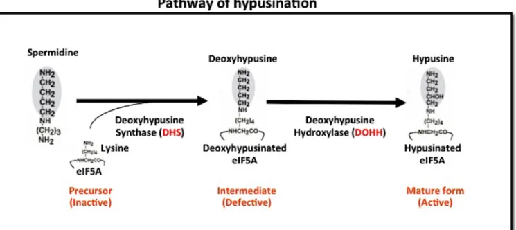
Figure 1.1: Pathway of eIF5A hypusination
.After translation of eIF5A, a conserved lysine residue (K47) of eIF5A is modified to hypusine. This pathway has two steps catalyzed by two different enzymes. In the first step of hypusine modification, a butylamine moiety from polyamine (Spermidine) is transferred to a lysine residue of eIF5A converting it to deoxyhypusine. This step is catalyzed by deoxyhypusine synthase (DHS). In the second step deoxyhypusine is hydroxylated by deoxyhypusine hydroxylase (DOHH) that converts deoxyhypusine to hypusine.
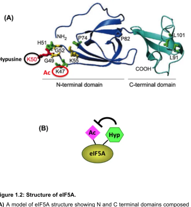
Figure 1.2: Structure of eIF5A.
(A) A model of eIF5A structure showing N and C terminal domains composed
of β-sheet core structures. Lysine 47 and 50 undergoes acetylation and hypusination respectively. (B) Hypusination of eIF5A inhibits its acetylation.
Chapter 2:
Regulation of eIF5A acetylation
2.1 Introduction
In order to unveil the physiological significance of eIF5A post-translational modifications, our lab previously explored the enzymes responsible for acetylation and deacetylation of eIF5A as well as it was shown that hypusination of eIF5A inhibits acetylation [18,44]. A member of NAD dependent class III HDACs, SIRT2 was revealed to be one of the two deacteylases of eIF5A in mammals. The second deacetylase of eIF5A was shown to be HDAC6. Requirement of NAD as a cofactor by SIRT2 shows that the activity of this enzyme is dependent on cellular energy status, therefore physiological function of eIF5A might be modulated with the change in metabolic state of the cell. Under energy rich conditions, NAD is required for glycolysis to proceed and is not available to serve as a cofactor of SIRT2 so the activity of SIRT2 might be inhibited under such conditions leading to hyper-acetylation of eIF5A. On the other hand under energy-deprived conditions, sufficient amount of NAD may be available to stimulate its deacetylation activity and eIF5A might be deacetylated. As our lab showed previously, hypusination of eIF5A blocks its acetylation, it was also important to monitor cellular states in which hypusination is affected. Hypusination is controlled by the activity of two enzymes, deoxyhypusine synthase (DHS) and deoxyhypusine hydroxylase (DOHH). These two enzymes also are under tight control and are affected by cellular environment. DHS is an NAD-dependent enzyme and might be affected by energy state of the cell. Like SIRT2, the activity of DHS might also be lower in energy-rich conditions while high in low energy state. On one hand SIRT2 inactivation will lead to hyperacetylation of eIF5A and on the other hand inactivation of DHS will lead to decreased hypusination, which in turn will increase acetylation of eIF5A. The second enzyme in the hypusine pathway also is under tight control. As the activity of DOHH is dependent on the availability of molecular oxygen, hydroxylation of
deoxyhypuisnated (Dhp) eIF5A to hypuisnated eIF5A might be affected by the availability of molecular oxygen. In high oxygen environment, DOHH will actively hydroxylate deoxyhypusinated eIF5A to hypuisnated eIF5A while in low availability of oxygen the activity of DOHH might be limited leading to accumulation of deoxyhypusinated eIF5A. Deoxyhypusinated eIF5A might also be hyper-acetylated due to blockage of hypusination.
I attempted to explore both these possibilities that might affect acetylation status of eIF5A. I studied the acetylation of eIF5A in response to varied glucose concentrations as well as the effect of oxygen availability and scarcity. My study revealed that high glucose concentration could increase acetylation of eIF5A and also limited supply of oxygen resulted in hyper-acetylation of eIF5A.
2.2 Materials and Methods:
2.2.1 Compounds, and cell culture:
Trichostatin A (TSA) and Nicotinamide (NA) were obtained from Wako Chemicals. All cell lines were maintained in DMEM containing 10% heat inactivated Fetal Bovine Serum and antibiotics at 37°C, 5% CO2 in a
humidified incubator.
2.2.2 Antibodies and immunoblotting:
A rabbit polyclonal antibody against acetylated eIF5A (Ac-eIF5A) was raised as described previously[18]. Antibodies against α-Tubulin (B-5-1-2) and Ac-α-Tubulin (6-11B-1) were purchased from Sigma. Monoclonal antibody against eIF5A was obtained from BD Bioscience. Immunoblotting was performed using whole cell lysates, prepared by collecting cells directly in 1x SDS PAGE sample loading buffer and heating at 95 °C for 5 min. Equal volumes of each whole cell lysate was resolved by SDS PAGE and transferred to a polyvinylidene difluoride membrane (Millipore) by electroblottting. Membranes were incubated with primary and secondary antibodies; immune complexes were detected using the Immobilon Western Chemiluminescent HRP substrate (Millipore); and luminescence was analyzed using a LAS-3000 image analyzer (GE Healthcare).
2.2.3 Hypoxic conditions:
For hypoxic conditions cells were grown in hypoxia chamber with 1% O2, 5%
CO2 at 37°C (9000EX, Waken Tech CO2 incubator) in a humidified
2.3 Results and discussion
2.3.1 Increasing Glucose concentration induced acetylation of
eIF5A
In order to test our hypothesis that high-energy status of the cell may lead to inactivation of deacetylase SIRT2 and may induce eIF5A acetylation, HeLa and A549 cells were first starved for 48 hours and then treated with different concentrations of glucose for 24 hours. The results suggested that acetylation level of eIF5A was increased by glucose in a concentration-dependent manner. These results verify the idea that high-energy state of the cell tends to reduce the activity of NAD-dependent deacteylases significantly.
2.3.2 Acetylation of eIF5A is induced by hypoxia
To explore the effect of hypoxia (1% O2) on eIF5A acetylation, HeLa and
A549 cells were allowed to grow in low oxygen chamber for 24 hours. Consistent with our hypothesis, it was observed that hypoxia could significantly increase acetylation level of eIF5A in both cell lines. Deoxyhypusine hydroxylase (DOHH), responsible for the second step of eIF5A hypusination is dependent on molecular oxygen and Fe2+ for hydroxylation of deoxyhypusine. Limited supply of oxygen might therefore negatively affect the activity of DOHH. It was therefore hypothesized that hypoxia-mediated acetylation of eIF5A may be induced due to inhibition of hypusination.
2.3.3 Effect of HDAC inhibitors and hypoxia on eIF5A
acetylation
I checked the effect of cellular deacetylase inhibitors, Trichostatin A (TSA) and Nicotinamide (NA) under hypoxia on acetylation level of eIF5A. A549 cells were treated with TSA (1 µM ), NA (5 mM) and grown in hypoxia for 12 hours. Western blot results revealed that HDAC inhibitors could not further increase acetylation of eIF5A in hypoxia. These results suggest that hypoxia alone can induce acetylation of eIF5A comparable to that achieved by inhibiting cellular deacetylases.
Summary
Hypusination of eIF5A is an irreversible post-translation modification. Therefore the activity of eIF5A might be regulated by another, reversible modification. Acetylation of eIF5A was reported to be involved in nuclear localization of eIF5A. Reversible acetylation of eIF5A might be important to dictate subcellular location and hence the role in different compartments. I found that glucose and hypoxia induce acetylation of eIF5A. It seems in higher glucose conditions, NAD becomes a limiting factor for the activity of SIRT2, which leads to hyper acetylation of eIF5A. Under hypoxia, the activity DOHH might be blocked, resulting in hyper acetylation of eIF5A. HDAC inhibitors could not further increase acetylation of eIF5A.
Figures chapter 2:
Figure 2.1: Regulation of eIF5A acetylation.
Glucose and hypoxia are the key factors, which regulate acetylation of eIF5A. In low glucose conditions the level of free NAD is increased, which is used as a cofactor by SIRT2. Low glucose thus leads to active SIRT2 and deacetylation of eIF5A. In high glucose conditions NADH/NAD+ ratio is increased, unviability of NAD+ results in blocking the activity of SIRT2 leading to acetylation of eIF5A. DOHH is dependent on the availability of oxygen for hydroxylation of deoxyhypusinated eIF5A. In hypoxia DOHH is inhibited resulting in accumulation of deoxyhypusinated eIF5A that is hyperacetylated.
Figure 2.2: Glucose and Hypoxia induces acetylation of eIF5A. (A) HeLa
cells were - glucose starved for 48 hours and then treated with different glucose concentrations. Cells were lysed in lysis buffer and the equal amount of lysate was resolved on SDS-PAGE. (B) HeLa and A549 cell lines were grown in hypoxia (1 % O2) or normoxia (20 % O2) for 24 hours, Cells were lysed in lysis buffer and cell lysate was separated by SDS PAGE. (C) A549 cells were treated with vehicle control or TSA (1 µM), NA (5 mM) and grown in hypoxia or normoxia for 12 hours.
Chapter 3:
eIF5A is essential for HIF-1α expression in
hypoxia
3.1 Introduction
3.1.1 Hypoxia-inducible factor 1 alpha (HIF-1α)
Constant supply of oxygen to all human cells is critical for generation of energy by oxidative phosphorylation in mitochondria [45]. Oxygen level in most human tissues is quite lower than ambient level (21 %) and most tissues have oxygen levels in the range of 2-9 %, which is termed as physiological hypoxia [46]. Thus oxygen homeostasis is vital for cellular physiology. This homeostasis in humans is maintained by complex respiratory, hematopoietic and cardiovascular systems. Deregulation of this homeostasis is associated with lung disease, cerebrovascular disease, heart disease and cancer [47]. Under low oxygen stress, oxidative ATP generation is downregulated and glycolysis is increased (Warburg effect) as well as vasculogenesis and angiogenesis are induced to increase supply of oxygen and nutrients [45]. A master regulator of hypoxic stress; hypoxia inducible factor (HIF) mediates this adaptive response to low oxygen stress. HIF is a heterodimeric transcription factor regulating HIF-mediated transcriptional response in hypoxia. HIF transcription factor is composed of oxygen-regulated alpha and constitutive, beta subunits. HIF has three isoforms HIF1, HIF2 and HIF3 [46,48]. HIF-1α is expressed in all cell types while HIF-2α expression is restricted to certain cell types such as endothelial, lung, hepatocytes and muscle cells. HIF-3α is the least studied among HIF isoforms [49-51].
3.1.2 Regulation of HIF-1α
HIF-1α protein level is regulated by the availability of oxygen. In oxygen-rich environment, prolyl hydroxylase; PHD (oxygen, Fe2+ and 2-oxoglutate dependent dioxygenase) is able to hydroxylate two proline residues i.e. P402 and P564 in the oxygen-dependent degradation domain (ODDD) of HIF-1α
protein. Hydroxylated HIF-1α is recognized and bound by pVHL (Von Hippel-Lindau, E3 ubiquitin ligase) leading to polyubiquitylation of hydroxylated HIF-1α. Polyubiquitylated HIF-1α is ultimately targeted for degradation by the proteasome. In low oxygen conditions the activity of PHD is strongly reduced due to unavailability of molecular oxygen leading to stabilization of HIF-1α. HIF-1α activity is also regulated by asparginyl hydroxylase, FIH-1 (Factor inhibiting HIF-1) [52]. FIH-1 is responsible for oxygen-dependent hydroxylation of asparagine residue, N803, in the C-terminal transactivation domain (CTAD) of HIF-1α protein [53]. CTAD is important for transcriptional activity of HIF-1α by binding transcriptional co-activators such as p300 and CBP (CREB-binding protein). Hydroxylation of Asparagine of CTAD impairs co-activators binding and reduces HIF-1α transcriptional activity [53-55]. Stabilized HIF-1α translocates to the nucleus, dimerize with HIF-1β and activate transcription of HIF-1 target genes [45,56,57]. HIF-1α plays a vital role in development of embryo, formation of vasculature and maintaining homeostasis in hypoxic environment [58,59]. HIF-1α is not only vital during developmental processes but also during adult life. Wound healing for instance, a complex process involving recruitment of macrophages to the damaged tissue for restoration of damaged vasculature is also dependent on HIF-1α activity. HIF-1α has been shown to play important role in physiological as well as diabetic wound healing process [60-62]. HIF-1α stability is also affected by the status of VHL gene in the cell, as in most renal cancer cells mutations of VHL results in non-hypoxic stabilization of HIF-1α [63]. Normally pVHL serves as an E3 ubiquitin ligase of HIF-1α and recognize/ubiquitylates prolyl hydroxylated HIF-1α, but under some conditions HIF-1α is ubiquitylated by pVHL without prolyl hydroxylation. One such situation is SUMOylation of HIF-1α in hypoxia. SUMOylated HIF-1α is ubiquitylated by pVHL and degraded by the proteasome even without the activity of PHD. SUMOylation-mediated degradation of HIF-1α is prevented by sentrin specific peptidase1 (SENP1), which de-SUMOylate HIF-1α [64]. Studies have also found pVHL, PHD and oxygen independent mechanisms of HIF-1α ubiquitylation and degradation. Koh MY et al. reported hypoxia-associated factor (HAF) as a novel E3 ubiquitin ligase of HIF-1α. HAF is able to ubiquitylate HIF-1α and target it for proteasomal degradation. HIF-1α ubiquitylation by HAF is
independent of oxygen, PHD and pVHL [65,66]. Receptor of activated protein kinase C (RACK1) has been demonstrated to be involved in oxygen, PHD, pVHL independent degradation of HIF-1α by competing with Hsp90 for binding to PAS domain of HIF-1α. Hsp90 binding to HIF-1α leads to its stabilization while RACK1 binding leads to ubiquitylation and degradation of HIF-1α. Hsp90 inhibitors have been shown to increase the binding of RACK1 leading to HIF-1α degradation [67]. Another study reported degradation of HIF-1α in hypoxia by SHARP1, also known as BHLHE4 or DEC2. SHARP1-mediated proteasomal degradation of HIF-1α in hypoxia is independent of PHD, pVHL and the ubiquitylation machinery. SHARP1 serves as a HIF-presenting factor to proteasome [68]. It is now well documented that HIF-1α is degraded by various mechanisms in the presence and absence of oxygen. In order to further explore the role of eIF5A in the HIF-1α pathway, it was necessary to check its stability. To understand whether HIF-1α is degraded in the absence of functional eIF5A, I inhibited the activity of proteasome. I found that in the absence of functional eIF5A, HIF-1α is targeted for proteasomal degradation.
3.1.3 HIF-1α and cancer progression
Along with maintaining physiological oxygen homeostasis, HIF-1α pathway also contributes to tumor growth, survival and metastasis. Most solid tumors develop hypoxic regions due to continued proliferation and disruption of blood flow. To survive and proliferate in such microenvironment, HIF-1α pathway comes into play for the adaptive response to hypoxia [69]. HIF-1α overexpression has been observed in many human cancers [70]. HIF-1α expression in cancer cells is enhanced either by decreased degradation, increased protein synthesis or both. Overexpression of HIF-1α in cancer is associated with aggressiveness and poor patient survival in cervical, lung, breast, ovarian, and endometrial cancers [71-75]. To meet the metabolic demands of proliferating cancer cells HIF-1α-mediated transcriptional response is activated which is involved in metabolic reprogramming of cancer cells. Cancer cells meet their ATP demands mainly by glycolytic ATP generation and the oxygen consuming mitochondrial metabolism is downregulated by the activity of HIF-1α pathway. Tumor hypoxia contributes
to radio-resistance, chemo-resistance, angiogenesis, invasion and metastasis. Hypoxic tumors become resistant to radiotherapy because radiotherapy-mediated cell killing is based on the production of free radicals in the cell. Molecular oxygen reacts with the free electrons of free radicals to promote oxidative DNA damage. In hypoxic environment the free radicals are neutralized and cell death is prevented [76]. Solid tumors are not only resistant to radiotherapy but also resistant to chemotherapy. Tumor cells being away from the functioning blood vessels, do not receive enough drug concentration to be effective. Conventional chemotherapeutic agents are more active against rapidly proliferating cells while proliferation of hypoxic cells is slowed down as part of HIF-1 mediated adaptive response, thereby affecting the efficacy of chemotherapeutic agents [77-79].
Tumor hypoxia is a big challenge for therapeutic intervention. Targeting the HIF-1α pathway in combination with conventional therapeutic approaches would be an effective strategy to combat cancer. Indeed several groups are trying to develop molecules targeting the HIF-1α pathway. It has been shown by various groups that targeting the HIF-1α pathway effectively suppresses tumor angiogenesis and metastasis. Inhibition of HIF-1α also sensitizes tumor cells to radio and chemotherapy [80-82].
3.2 Materials and Methods:
3.2.1 Compounds, and cell culture:
GC7 was purchased from Biosearch Technologies, Inc., Deferoxamine mesylate salt (DFX) from Enzo life Sciences, Inc. All cell lines were maintained in DMEM containing 10% heat inactivated Fetal Bovine Serum and antibiotics at 37°C, 5% CO2 in a humidified incubator.
3.2.2 Antibodies and immunoblotting:
A rabbit polyclonal antibody against acetylated eIF5A (Ac-eIF5A) and a mouse monoclonal antibody against hypusinated eIF5A (Hyp-eIF5A) were raised as described previously[18]. Antibody against α-Tubulin (B-5-1-2) was purchased from Sigma. Monoclonal antibodies against eIF5A and HIF-1α were obtained from BD Bioscience. Immunoblotting was performed using whole cell lysates, prepared by collecting cells directly in 1x SDS PAGE sample loading buffer and heating at 95 °C for 5 min. Equal volumes of each whole cell lysate was resolved by SDS PAGE and transferred to a polyvinylidene difluoride membrane (Millipore) by electroblottting. Membranes were incubated with primary and secondary antibodies; immune complexes were detected using the Immobilon Western Chemiluminescent HRP substrate (Millipore); and luminescence was analyzed using a LAS-3000 image analyzer (GE Healthcare).
3.2.3 RNAi and hypoxia:
Small interfering RNAs (siRNAs) against eIF5A were obtained from NipponGene (siRNA #1), QIAGEN (siRNA # 2,3,4,5) and ambion® (Life
technologies: siRNA # 6). All transfections were carried out according to manufacture’s instructions for 72 hours. After 72 hours of siRNA transfection cells were grown in normoxia (21% O2, 5% CO2 at 37°C) or hypoxia (1% O2,
5% CO2 at 37°C) in a humidified environment for 12 hours.
3.2.4 GC7 treatment and hypoxia
A549 cells were treated with vehicle control or 30 µM GC7 for 12 hours. After 12 hours GC7 treatment cells were treated with vehicle or 10 µM MG132 and grown in normoxia or hypoxia for 12 hours.
3.3 Results and discussion
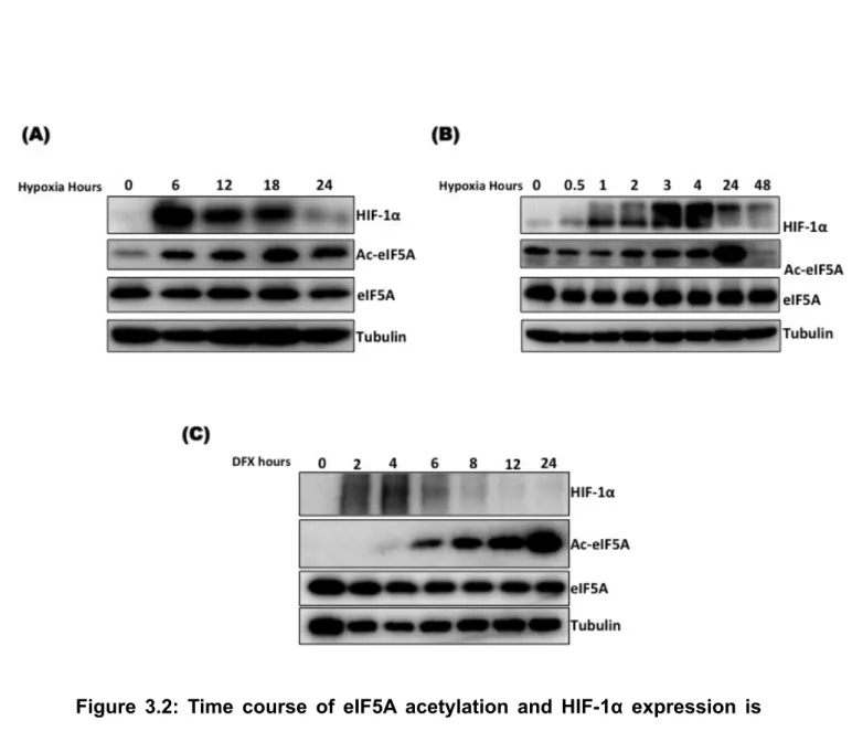
3.3.1 Time course of eIF5A acetylation and HIF-1α expression
is different
The seemingly common regulation of HIF-1α and eIF5A by hypoxia raised the question of whether the activities of HIF-1α prolyl hydroxylase (PHD) and eIF5A deoxyhypusine hydroxylase (DOHH) are inhibited at the same time point after induction of hypoxia? To address this question, I exposed A549 cells to hypoxia for different time periods as indicated. Quite interestingly the results indicated that expression of HIF-1α precedes the acetylation of eIF5A and HIF-1α expression is only transient which is significantly dropped as acetylated eIF5A (Defective eIF5A) is accumulated (Fig. 3.2). Induction of acetylation by hypoxia is presumed to be due to inhibition of eIF5A hypusination (DOHH). Transient induction of HIF-1α protein expression and its reduction with increased acetylation of eIF5A suggests that the presence of hypusinated eIF5A might be important for HIF-1α expression.
3.3.2 eIF5A is essential for HIF-1α expression in hypoxia
To test the involvement of eIF5A in the HIF-1α pathway, I used RNAi-mediated suppression of eIF5A protein expression. A549 cells were used for eIF5A knockdown. The results revealed suppression of eIF5A protein expression leads to suppression of HIF-1α expression as well. In order to further clarify the observation, I used several different types of siRNA oligos to target eIF5A and consistently all the potent siRNAs against eIF5A were able reduce the protein level HIF-1α as compared to scramble control siRNA-treated cells. Further I checked the effect of eIF5A targeting on HIF-1α expression in two other cell lines to check whether the effect is generalized or cell line specific. I used HeLa (cervical cancer) and PC3 (prostate cancer) cell lines. The results showed that eIF5A knockdown in these cells also negatively regulate HIF-1α protein expression in hypoxia. PC3 cells have detectable level of HIF-1α protein in normoxia [83] and eIF5A knockdown could reduce both hypoxic and normoxic HIF-1α protein level in PC3 cells (Fig. 3.3 C).
3.3.3 Functional eIF5A is important for HIF-1α expression in
hypoxia
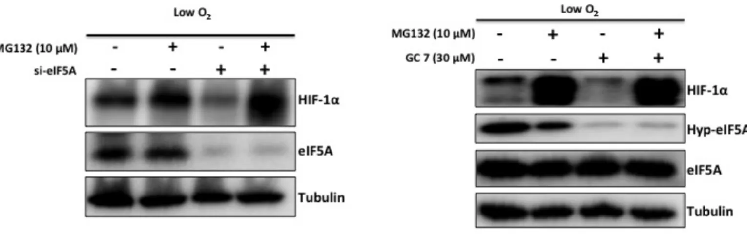
Hypusination is essential for the activity of eIF5A and only hypusinated eIF5A is the active form of this protein. Time course experiment (Fig. 3.2) showed that induction of HIF-1α in hypoxia is only transient with the increase in acetylation of eIF5A HIF-1α protein disappeared. Acetylated form of eIF5A is non-functional. Seemingly essential nature of eIF5A protein for HIF-1α protein induction in hypoxia was compelling and it was essential to find out which form of eIF5A plays a role in HIF-1α pathway. I used GC7 (N1-guanyl-1,7-diaminoheptane), an analogue of spermidine that potently inhibits of the first enzyme (DHS) of the hypsination pathway. GC7 inhibits hypusination thereby resulting in increasing acetylation level of eIF5A. I pre-treated A549 cells with GC7 (30 µM) for 12 hours in normoxia to inhibit hypusination and then exposed the cells to hypoxia for 12 hours. The result in Fig. 3.3 D shows that HIF-1α becomes undetectable in cells treated with GC7, suggesting that functional eIF5A (hypusinated) is important for HIF-1α accumulation in hypoxia.
3.3.4 GC7 treatment or eIF5A knockdown-mediated
suppression of HIF-1α is independent of proly hydroxylase
(PHD)
Prolyl hydroxylase (PHD) is responsible for hydroxylation of two proline residues (P402, P564) in oxygen-dependent degradation domain (ODDD) of HIF-1α. Prolyl hydroxylated HIF-1α is targeted for poloyubiquitylation by pVHL and ultimately degraded by the proteasome. PHD activity is dependent on oxygen, Fe2+ and 2-oxoglutarate [84]. HIF-1α protein level is elevated in hypoxia by inhibition of PHD therefore PHD inhibition in normoxia by inhibitors also leads to accumulation of HIF-1α (hypoxia-mimicking conditions). I used deferoxamine mesylate salt, DFX (Iron chelator) that can inhibit PHD and observed the effect of eIF5A knockdown or GC7 treatment in hypoxia mimicking conditions. The result showed that DFX was unable to raise HIF-1α protein level in cells treated with siRNA against eIF5A or GC7 compared to control treated cells (Fig. 3.3 E, F). This result suggests that eIF5A knockdown-mediated suppression of HIF-1α is independent of PHD activity.
3.3.5 Mechanism of HIF-1α suppression by GC7 treatment or
eIF5A knockdown
To find out the mechanism of eIF5A knockdown or GC7 mediated suppression of HIF-1α I used proteasome inhibition. eIF5A knockdown-mediated suppression of HIF-1α could be recovered by the proteasome inhibitor MG132 (Fig. 3.4 A). In my previous results I found that GC7 could suppress protein level of HIF-1α. Thus I also checked the effect of MG132 on GC7-mediated suppression of HIF-1α. Consistent with eIF5A knockdown result, GC7-mediated HIF-1α protein suppression could be recovered by MG132 (Fig. 3.4 B). Hypoxia is a hallmark of solid tumors and HIF-1α is a master regulator of adaptive response to hypoxic stress, thus contributes to angiogenesis, invasion, metastasis and metabolic reprograming of cancer cells. My study revealed that HIF-1α itself is dependent on active eIF5A in hypoxia. The role of eIF5A in cancer cell survival and proliferation is not yet fully understood and my study revealed a part of it. It remains to be determined how is HIF-1α degraded in the absence of functional eIF5A. hypusination of eIF5A is essential for its role in translation. The requirement of eIF5A hypusination for HIF-1α stability is pointing towards modulation of protein translation by eIF5A under hypoxia. The regulation of HIF-1α stability and eIF5A post-translational modifications seems quite similar. The activity of both, deoxyhypusine hydroxylase (DOHH) and prolyl hydroxylase (PHD) is dependent on the availability of oxygen and Fe2+. Like PHD, the activity of DOHH might also be blocked by hypoxia and as a result acetylation of eIF5A is induced (Fig. 2.2 B). I showed that expression of HIF-1α precedes acetylation of eIF5A (Fig. 3.2), and a negative correlation was observed between induction of eIF5A acetylation and HIF-1α protein expression. Based on these observations, eIF5A may be involved in translation of HIF-1α but it seems unlikely because the amino acid sequence of HIF-1α has no stretches of poly-proline residues, translation which requires eIF5A [32]. On the other hand MG132-mediated recovery of HIF-1α protein indicates a possible role of eIF5A in the stability of HIF-1α under hypoxia. In the absence of functional eIF5A, HIF-1α protein could not be observed in DFX treated cells (Fig. 3.3 E, F) suggesting that the role of eIF5A in HIF-1α stability is independent of PHD.
Summary
To sum up, upon hypoxia, HIF-1α protein level started to accumulate before eIF5A acetylation. Induction of eIF5A acetylation in the late phase of hypoxia had negative effect on HIF-1α protein level. Functional eIF5A was observed to be essential for HIF-1α expression in hypoxia. The activity of eIF5A in supporting HIF-1α was found to be independent of PHD activity. DFX, an inhibitor of PHD could not recover the level of HIF-1α protein in cells treated with siRNA against eIF5A or GC7. In the absence of hypusinated eIF5A, HIF-1α was degraded by the proteasome.
Figures Chapter 3:
Figure 3.1: Regulation of HIF-1α.
In the presence of oxygen (Normoxia), two proline residues (P402, P564) of HIF-1α are hydroxylated by prolyl hydroxylase (PHD). pVHL (E3 ubiquitin ligase) recognizes hydroxyl proline residues and induces polyubiquitylation and subsequent degradation of HIF-1α. In low oxygen (Hypoxia) the activity of PHD to hydroxylate HIF-1α is inhibited. HIF-1α escapes from polyubiquitylation and degradation. Stabilized HIF-1α translocates to the nucleus, dimerize with HIF-1β and binds to hypoxia response element (HRE) of HIF-1 target genes, and activates their transcription.
Figure 3.2: Time course of eIF5A acetylation and HIF-1α expression is
different. (A, B) A549 cells were grown in Hypoxia (1 % O2) for the indicated
periods of time. (C) A549 cells were treated with 200 µM DFX for the indicated periods of time.
Figure 3.3: eIF5A is essential for HIF-1 alpha expression in hypoxia. (A)
A549 cells were treated with siRNA against eIF5A for 72 hours, then allowed to grow in hypoxic chamber for 12 hours, and collected in lysis buffer, and equal amount of cell lysate was analyzed by SDS-PAGE. (B) Five different siRNA oligos against eIF5A were used to suppress eIF5A. (C) HeLa and PC3 cells were treated with scramble control or eIF5A siRNA for 72 hours and grown in hypoxia for 12 hours. (D) A549 cells were treated with vehicle control or 30 µM GC7 for 12 hours and then grown in normoxia or hypoxia followed by collection in cell lysis buffer and analysis by SDS-PAGE. (E) After A549 cells had been transfected with scramble control or eIF5A siRNA for 72 hours, cells were treated with 200µM DFX for four hours and collected in lysis buffer.
(F) A549 cells were treated with 30 µM GC7 for 12 hours and then treated
Figure 3.4: In the absence of functional eIF5A, HIF-1α protein is degraded by proteasome.
(A) A549 cells were transfected with scramble control siRNA or siRNA against
eIF5A. 72 hours post-transfection, cells were treated with vehicle control or 10 µM MG132 and grown in hypoxia for 12 hours. (B) A549 cells were treated with 30 µM GC7 for 12 hours. Then the cells were treated with vehicle control or 10 µM MG132 and grown in hypoxia for 12 hours.
Chapter 4:
eIF5A is required for HIF-1α-mediated
transcriptional activation in hypoxia
4.1 Introduction
Under hypoxic conditions HIF-1α is stabilized and translocates to the nucleus where it dimerizes with HIF-1β to constitute the HIF-1α transcription factor. HIF-1α binds to the hypoxia response element (HRE: 5’RCGTG3’) in promoters of HIF-1 target genes and activates HIF-1-mediated adaptive response to low oxygen stress [85]. The products of HIF-1 target genes are involved in glucose metabolism, cell survival, angiogenesis, and metastasis. In hypoxia, ATP generation is shifted from oxidative phosphorylation to glycolytic metabolism by HIF-1α-mediated transcriptional response. The less efficient glycolytic metabolism needs steady supply of glucose to meet energy demands. HIF-1α therefore enhances the expression of glucose transporters (GLUT1, 3) and PGK1 to ensure proper glucose supply [86]. On one hand HIF-1α enhances glycolysis and on the other hand it blocks tricarboxylic acid cycle (TCA cycle). During oxidative metabolism pyruvate is converted to acetyl-CoA by pyruvate dehydrogenase complex (PDH) but under hypoxic conditions, the activity of PDH is blocked by pyruvate dehydrogenase kinase 1 (PDK1) which is a product of HIF-1-target gene [85,87,88]. In tumor cells HIF-1α increases the production of NAD+ to be utilized in the following rounds of glycolysis and conversion of pyruvate to lactate is also enhanced by increased transcription of lactate dehydrogenase A (LDHA) [89].
My previous results showed that eIF5A knockdown or inhibition of its hypusination has a negative effect on protein level of HIF-1α. Furthermore, I studied the effect of eIF5A knockdown or inhibition of its hypusination on HIF-1 target gene expression using real-time quantitative PCR.
4.2 Materials and Methods:
4.2.1 Compounds, and cell culture:
GC7 was purchased from Biosearch Technologies, Inc. All cell lines were maintained in DMEM containing 10% heat inactivated Fetal Bovine Serum and antibiotics at 37°C, 5% CO2 in a humidified incubator.
4.2.2 Antibodies and immunoblotting:
A mouse monoclonal antibody against hypusinated eIF5A (Hyp-eIF5A) was raised as described previously[18]. Antibody against α-Tubulin (B-5-1-2) was purchased from Sigma. Monoclonal antibodies against eIF5A and HIF-1α were obtained from BD Bioscience. Immunoblotting was performed using whole cell lysates, prepared by collecting cells directly in 1x SDS PAGE sample loading buffer and heating at 95 °C for 5 min. Equal volumes of each whole cell lysate was resolved by SDS PAGE and transferred to a polyvinylidene difluoride membrane (Millipore) by electroblottting. Membranes were incubated with primary and secondary antibodies; immune complexes were detected using the Immobilon Western Chemiluminescent HRP substrate (Millipore); and luminescence was analyzed using a LAS-3000 image analyzer (GE Healthcare).
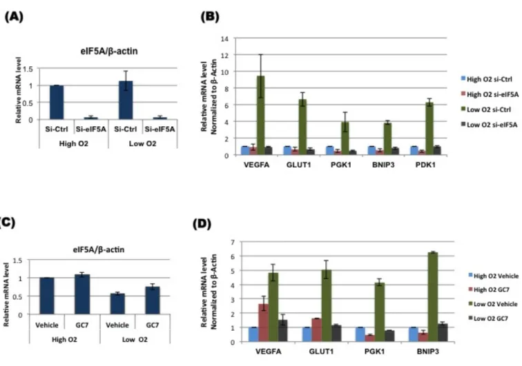
4.2.3 RNA interference, GC7 treatment and hypoxia:
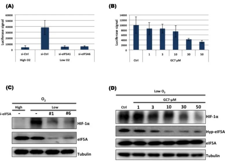
Small interfering RNAs (siRNAs) against eIF5A mRNA were obtained from NipponGene (siRNA #1), QIAGEN (siRNA # 2,3,4,5) and ambion® (Life technologies: siRNA # 6). All transfections were carried out according to manufacture’s instructions for 72 hours. A549 cells were transfected with control siRNA or siRNA targeting eIF5A for 72 hours. Cells were then grown in normoxia or hypoxia for 12 hours followed by RNA preparation. In case of GC7, A549 cells were treated with vehicle control or 30 µM GC7 for 12 hours. Cells were then allowed to grow in normoxia or hypoxia for 12 hours followed by RNA extraction. For luciferase assay, HT1080 cells stably expressing HRE-luciferase were collected in NETN buffer after treatments and luciferase activity was measured by Spectramax L (Molecular devices).
4.2.4 RNA extraction and qPCR
Total RNA was prepared with RNeasy Mini Kit (QIAGEN) and cDNA was prepared by reverse transcription with oligo dT primers using RNA PCR Kit (AMV) Ver.3.0 (Takara Bio Inc., Shiga, Japan). Expression of HIF-1 target genes was analyzed by qPCR SYBR Premix Ex Taq II (Takara BioInc.) and LightCycler 480 (Roche)
4.3 Results and discussion
HIF-1α is the master regulator of hypoxic response, which up-regulates the expression of its target genes. I checked the transcriptional activity of HIF-1α upon eIF5A knockdown or inhibition of hypusination by GC7. I found that eIF5A knockdown or GC7 treatment could suppress HRE-luciferase activity (Fig. 4.1 A, B), indicating that functional eIF5A is important for transcriptional activation by HIF-1α in hypoxia. It was also confirmed that suppression of luciferase activity by eIF5A KD or GC7 treatment in hypoxia is due to down-regulation of HIF-1α protein (Fig. 4.1 C, D). Transcript levels of representative HIF-1 target genes were analyzed in response to eIF5A knockdown or GC7 treatment, using quantitative real time PCR. The results indicated that functional eIF5A is required for HIF-1 alpha mediated transcription of HIF-1 target genes in hypoxia (Fig. 4.2 A-D). These results are consistent with the western blot results and reiterate that in the absence of eIF5A or in the presence of defective eIF5A, HIF-1α protein level drops resulting in down-regulation of HIF-1 target genes. Adaptive response to hypoxia is initiated by HIF-1α-mediated transcriptional induction of genes, which are necessary for cell growth and proliferation. Most solid tumors have regions of hypoxia. Tumors outgrow their vascular supply due to which severe gradient of nutrients is observed. To promote new blood vessel formation, HIF-1α up-regulates the expression of vascular endothelial growth factor (VEGF) to extend blood vessels and ensure supply of nutrients and oxygen. In hypoxic microenvironment cells cannot afford to continue oxidative energy generation so cellular metabolism is shifted to glycolysis. Shifting from TCA cycle to glycolysis for energy production requires sustained supply of glucose to obtain maximum energy by less efficient mechanism. HIF-1 target gene, GLUT1 expression is up-regulated to increase glucose transport to tumor cells for sustained glycolysis. Hypoxic tumor cells not only rely on increased glycolysis but also inhibit oxidative metabolism as well. Pyruvate dehydrogenase kinase 1 (PDK1) is up-regulated by HIF-1α to phosphorylate and block Pyruvate dehydrogenase (PDH), which is involved in conversion of pyruvate to acetyl-CoA to enter TCA cycle. Another HIF-1 target gene, lactate dehydrogenase A (LDHA) promotes conversion of pyruvate to lactate. Tumor cells utilize the
glycolytic pathway as a sole source of energy production thereby mitochondrial activity is completely suppressed. Not only mitochondrial activity but also the number of mitochondria is reduced. Autophagy (mitophagy) is induced by BNIP3, a HIF-1 target gene, in hypoxic tumor cells.
The role of HIF-1α is thus vital for tumor growth, angiogenesis, metastasis and survival. My results show that HIF-1α itself is dependent on functional eIF5A and in the absence of eIF5A, HIF-1α is unable to support growth and survival.
Summary:
eIF5A was found to be essential for HIF-1α-mediated transcriptional up- regulation of HIF-1 target genes. eIF5A knockdown led to down-regulation of the mentioned HIF-1 target gene expression in hypoxia. Cells treated with GC7 in hypoxia also showed a significant down-regulation of HIF-1 target gene expression. In hypoxia, functional eIF5A is essential for HIF-1 target gene expression.
Figures Chapter 4:
Figure 4.1: Functional eIF5A is essential for HIF-1α transcriptional activity. HT1080 cells stably expressing luciferase under the control of the
HRE promoter were transfected with scramble control or siRNA against eIF5A for 72 hours (A, C). For GC7 treatment, Cells were grown overnight followed by treatment with vehicle or indicated concentrations of GC7 for 12 hours (B, D). After treatments the cells were allowed to grow in normoxia (High O2) or
hypoxia (Low O2) for 12 hours. Luciferase activity was measured using a
Figures Chapter 4
Figure 4.2: Functional eIF5A is essential for HIF-1 target gene expression. A549 cells were transfected with scramble control or siRNA
against eIF5A for 72 hours. For GC7 treatment, Cells were grown overnight followed by treatment with vehicle or 30 µM GC7 for 12 hours. After treatments the cells were allowed to grow in normoxia (High O2) or hypoxia
(Low O2) for 12 hours. Expression of HIF-1 target genes was analyzed by real
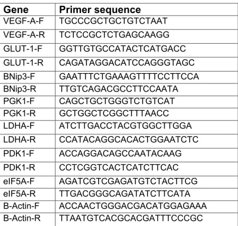
Table 4.1 List of primers
Gene
Primer sequence
VEGF-A-F TGCCCGCTGCTGTCTAAT VEGF-A-R TCTCCGCTCTGAGCAAGG GLUT-1-F GGTTGTGCCATACTCATGACC GLUT-1-R CAGATAGGACATCCAGGGTAGC BNip3-F GAATTTCTGAAAGTTTTCCTTCCA BNip3-R TTGTCAGACGCCTTCCAATA PGK1-F CAGCTGCTGGGTCTGTCAT PGK1-R GCTGGCTCGGCTTTAACC LDHA-F ATCTTGACCTACGTGGCTTGGA LDHA-R CCATACAGGCACACTGGAATCTC PDK1-F ACCAGGACAGCCAATACAAG PDK1-R CCTCGGTCACTCATCTTCAC eIF5A-F AGATCGTCGAGATGTCTACTTCG eIF5A-R TTGACGGGCAGATATCTTCATA B-Actin-F ACCAACTGGGACGACATGGAGAAA B-Actin-R TTAATGTCACGCACGATTTCCCGC
Table 4.1: Sequences of primers used for quantitative real-time PCR are
shown in the table. Sequences are shown 5’à3’. F: forward primer, R: reverse primer.
Chapter 5:
eIF5A is important for growth of tumor spheroid
5.1 Introduction
In the field of cancer drug discovery, it is very important to have model systems closely resembling the in vivo tumor microenvironment. Transplantable tumor model in rodents offered a great opportunity to study in vivo tumor biology and develop drug screening systems [90]. The cost and ethical issues associated with in vivo tumor models led scientists to think and develop new drug screening systems. The NCI60 program of the US National Cancer Institute was one such alternative. The NCI60 drug-screening platform was a high throughput screening system (HTS) system for cytotoxicity assays in vitro. Cell based assays combined with powerful computational methods can be used for mechanism of action and potential target identification of compounds. Such type of monolayer cell culture (2D culture) in drug screening has dominated but it has also been observed that cells in 2D culture do not represent truly the in vivo tumor microenvironment [91,92]. 3D culture was thus developed which shows in vivo-like growth and cellular interactions. 3D culture or tumor spheroid is the formation cell aggregate non-adherently. Spheroid resembles in vivo tumor in many traits, for example, growth pattern, nutrient gradient and interactions with ECM proteins [91,93,94]. The central region of spheroid is hypoxic like in vivo tumor and the activity of HIF-1α can be detected in that region. HIF-1α expression has been shown to be important for growth and proliferation of cells in a spheroid. Suppressing the expression of HIF-1α leads to reduction in size of spheroid [95]. In my study I found that eIF5A is essential for HIF-1α protein expression and transcriptional ability thus it was interesting to monitor the growth of spheroids in response to eIF5A knockdown or hypuisnation inhibition.
5.2 Materials and Methods:
5.2.1 Compounds, and cell culture:
GC7 was purchased from Biosearch Technologies, Inc.,. All cell lines were maintained in DMEM containing 10% heat inactivated Fetal Bovine Serum and antibiotics at 37°C, 5% CO2 in a humidified incubator.
5.2.2 Antibodies and immunoblotting:
A mouse monoclonal antibody against hypusinated eIF5A (Hyp-eIF5A) was raised as described previously[18]. Antibody against α-Tubulin (B-5-1-2) was purchased from Sigma. Monoclonal antibodies against eIF5A and HIF-1α were obtained from BD Bioscience. Immunoblotting was performed using whole cell lysates, prepared by collecting cells directly in 1x SDS PAGE sample loading buffer and heating at 95 °C for 5 min. Equal volumes of each whole cell lysate was resolved by SDS PAGE and transferred to a polyvinylidene difluoride membrane (Millipore) by electroblottting. Membranes were incubated with primary and secondary antibodies; immune complexes were detected using the Immobilon Western Chemiluminescent HRP substrate (Millipore); and luminescence was analyzed using a LAS-3000 image analyzer (GE Healthcare).
5.2.3 RNA interference and GC7 treatment:
Small interfering RNAs (siRNAs) against eIF5A mRNA were obtained from NipponGene (siRNA #1) and ambion® (Life technologies: siRNA # 6). All transfections were carried out according to manufacture’s instructions. A549 cells were transfected with control siRNA or siRNA targeting eIF5A. 24 hours post-transfection, cells (equal number) were transferred to U-bottom 96-well plate (PrimeSurface 96U from Sumitomo Bakelite Co., LTD) for spheroid formation. After 24 hours images were taken using celigo microscope (Day 0). Then images were recorded at two-day interval for two weeks. Spheroid size was measured using ImageJ software. In case GC7, A549 cells (equal number) were grown in U-bottom 96-well plate. After 24 hours images were recorded (Day 0) and treated with vehicle control or 30 µM GC7. At every 3rd day GC7 was added and every second day images were captured.
5.3 Results and discussion
5.3.1 HIF-1α knockdown leads to reduction in the size of
spheroid
Tumor spheroids are used as tumor models because of their structural and cellular arrangements. The central region of tumor spheroid becomes hypoxic as well as supply of nutrients to that area is limited (Fig. 5.1 A). The hypoxic region of spheroid is a site of HIF-1α expression, which is necessary for growth, and survival of cells in hypoxic stress. In order to reconfirm this observation, I used siRNA against HIF-1α (Fig. 5.1 B) for its effect on the growth of spheroid. A549 cells treated with scramble control or HIF-1α siRNAs and were allowed to form spheroid and the growth of spheroid was monitored for two weeks. It was observed that HIF-1α knockdown has a negative effect on spheroid growth (Fig. 5.1 C). Total area of spheroid was measured using ImageJ software and Fig. 5.1 D shows that HIF-1α knockdown had a significant (p<0.05) growth-limiting effect.
5.3.2 eIF5A knockdown has negative effect on spheroid
growth
Spheroid growth is dependent on HIF-1α to mediate adaptive response and survive in oxygen deficient environment. My previous results revealed that eIF5A is essential for HIF-1α function in hypoxia and eIF5A knockdown led to eventual proteasomal degradation of HIF-1α in hypoxia. To further verify my results, I used siRNA oligos-mediated suppression of eIF5A. A549 cells transfected either with scramble control siRNA or siRNAs (sieIF5A1, sieIF5A6) targeting eIF5A, were allowed to form spheroid. The growth of spheroids was monitored for two weeks. Consistent with my previous findings, it was observed that eIF5A knockdown had a significant (p<0.05) negative effects on spheroid growth (Fig. 5.2 A, B).
5.3.3 Functional eIF5A is essential for spheroid growth
I showed previously that HIF-1α expression in hypoxia is dependent on functional (hypusinated) eIF5A and inhibition of eIF5A hypusination resulted in downregulation of HIF-1α. I checked the effect GC7 (inhibitor of DHS) on growth of spheroid. The results (Fig. 5.3) indicated that like eIF5A knockdown,
blocking eIF5A hypusination leads to reduction of spheroid size. As I showed previously, HIF-1α is important for spheroid growth and siRNA-mediated knockdown of HIF-1α impairs spheroid growth, I checked the level of HIF-1α protein in spheroid. I collected the spheroids at day 10 and day 12 in cell lysis buffer and resolved the cell lysate on a polyacrylamide gel. Western blot results (Fig. 5.3 C) showed the expression of HIF-1α protein in spheroids treated with vehicle control or 3 µM GC7 but no expression in spheroids treated with 30 µM GC7. Consistent with previous results, it was observed that functional eIF5A is essential for growth of spheroid.
Summary:
It was found that HIF-1α is important for cell growth in 3D culture and HIF-1α knockdown had a negative effect on growth of spheroid. eIF5A being essential for HIF-1α expression, was found to be important for spheroid growth and siRNA-mediated knockdown of eIF5A significantly reduced spheroid growth. Hypusinated eIF5A was found to be important of spheroid growth and blocking eIF5A hypusination by GC7 resulted in reduction of spheroid size. In GC7 treated spheroids, the level of HIF-1α protein was dropped as compared to control spheroids.
Figures Chapter 5:
Figure 5.1: Spheroid growth is reduced in response to HIF-1α knockdown.
(A) A typical spheroid, which has central hypoxic (red) region and normoxic
region (green) at the periphery. (B) Western blot result indicates the efficiency of siRNA against HIF-1α. The 10 nM final concentration of siRNA was used.
(C) Spheroid size is suppressed by HIF-1α knockdown. (D) Line graph
represents the total area of spheroid (µM2). Error bars represent SEM, n=3. * p < 0.05, ** p < 0.01 as determined by students T-test.
Figures Chapter 5:
Figure 5.2: Spheroid growth is reduced in response to eIF5A knockdown.
(A) Spheroid size is suppressed by eIF5A knockdown. (B) Line graph
represents the total area of spheroid (µM2). Error bars represent SEM, n=3. *
Figures Chapter 5:
Figure 5.3: Spheroid growth is reduced in response to GC7 treatment. (A) Spheroid size is suppressed in response to GC7 treatment. (B) Line graph
represents the total area of spheroid (µM2). Error bars represent SEM, n=3.
* p < 0.01, ** p < 0.05 as determined by students T-test. (C) Western bot analysis of HIF-1α protein in spheroid.
Chapter 6:
Conclusions
In the current study I report a novel function of eIF5A in the HIF-1α pathway. eIF5A is an important cellular protein essential for cell growth and proliferation. eIF5A is the only known protein which undergoes hypusine modification, a post-translational modification essential for its activity. Previous studies revealed that reversible acetylation of eIF5A regulates subcellular localization of eIF5A and that acetylated eIF5A localizes to the nucleus, however the role of nuclear eIF5A is still unknown.
The current study was focused on the regulation of post-translational modifications of eIF5A. Based on the results, I concluded as follows.
A.
Acetylation of eIF5A is induced by glucose and hypoxia.B.
eIF5A is essential for HIF-1α induction in hypoxia.C.
Hypusine modification of eIF5A is essential for HIF-1α induction in hypoxia.D.
In the absence of functional eIF5A, HIF-1α protein is targeted for proteasomal degradation.References
1. Zanelli CF, Maragno AL, Gregio AP, Komili S, Pandolfi JR, et al. (2006) eIF5A binds to translational machinery components and affects translation in yeast. Biochem Biophys Res Commun 348: 1358-1366.
2. Aoki H, Dekany K, Adams SL, Ganoza MC (1997) The gene encoding the elongation factor P protein is essential for viability and is required for protein synthesis. Journal of Biological Chemistry 272: 32254-32259.
3. Park MH, Lee YB, Joe YA (1997) Hypusine is essential for eukaryotic cell proliferation. Biol Signals 6: 115-123.
4. Benne R, Hershey JW (1978) The mechanism of action of protein synthesis initiation factors from rabbit reticulocytes. J Biol Chem 253: 3078-3087.
5. Zanelli CF, Valentini SR (2007) Is there a role for eIF5A in translation? Amino Acids 33: 351-358.
6. Gregio AP, Cano VP, Avaca JS, Valentini SR, Zanelli CF (2009) eIF5A has a function in the elongation step of translation in yeast. Biochem Biophys Res Commun 380: 785-790.
7. Cracchiolo BM, Heller DS, Clement PM, Wolff EC, Park MH, et al. (2004) Eukaryotic initiation factor 5A-1 (eIF5A-1) as a diagnostic marker for aberrant proliferation in intraepithelial neoplasia of the vulva. Gynecol Oncol 94: 217-222.
8. Clement PM, Johansson HE, Wolff EC, Park MH (2006) Differential expression of eIF5A-1 and eIF5A-2 in human cancer cells. FEBS J 273: 1102-1114.
9. Jenkins ZA, Haag PG, Johansson HE (2001) Human eIF5A2 on chromosome 3q25-q27 is a phylogenetically conserved vertebrate variant of
eukaryotic translation initiation factor 5A with tissue-specific expression. Genomics 71: 101-109.
10. Park MH, Nishimura K, Zanelli CF, Valentini SR (2010) Functional significance of eIF5A and its hypusine modification in eukaryotes. Amino Acids 38: 491-500.
11. Caraglia M, Park MH, Wolff EC, Marra M, Abbruzzese A (2013) eIF5A isoforms and cancer: two brothers for two functions? Amino Acids 44: 103-109.
12. Park MH (2006) The post-translational synthesis of a polyamine-derived amino acid, hypusine, in the eukaryotic translation initiation factor 5A (eIF5A). J Biochem 139: 161-169.
13. Park MH, Wolff EC, Folk JE (1993) Hypusine: its post-translational formation in eukaryotic initiation factor 5A and its potential role in cellular regulation. Biofactors 4: 95-104.
14. Park MH, Wolff EC, Smit-McBride Z, Hershey JW, Folk JE (1991) Comparison of the activities of variant forms of eIF-4D. The requirement for hypusine or deoxyhypusine. J Biol Chem 266: 7988-7994.
15. Klier H, Csonga R, Joao HC, Eckerskorn C, Auer M, et al. (1995) Isolation and structural characterization of different isoforms of the hypusine-containing protein eIF-5A from HeLa cells. Biochemistry 34: 14693-14702.
16. Kim SC, Sprung R, Chen Y, Xu Y, Ball H, et al. (2006) Substrate and functional diversity of lysine acetylation revealed by a proteomics survey. Mol Cell 23: 607-618.
17. Lee SB, Park JH, Folk JE, Deck JA, Pegg AE, et al. (2011) Inactivation of eukaryotic initiation factor 5A (eIF5A) by specific acetylation of its hypusine