Acta Med. Nagasaki 39: 67-71
Eosinophil Cationic Protein (ECP) in Ulcerative Colitis
Kazufumi YAMASAKI, M. D. Kazuya MAKIYAMA, M. D.
Second Department of Internal Medicine, Nagasaki University School of Medicine, Nagasaki, Japan
One of the eosinophil granule proteins is eosinophil cationic protein (ECP) which is basic and cytotoxic. The association of ECP in ulcerative colitis (UC) was studied by immuno- histochemical method and RIA. ECP was localized in the lamina propria and the interstitium of the UC colon mucosa, showing a significant increase in distribution compared to the control. The ECP content, which was compared among the three groups according to the severity of UC classified by Matts' histopathological classification, disclosed an increas- ing tendency as the histological severity increased. The serum ECP level in active UC 17.68±12.25,µg/L (n = 21) was significantly higher than that in inactive UC. In view of these findings, it was speculated that ECP has a role, though partial, in the damage of the colonic mucosa in UC on the basis of its biological features.
Key words : Eosinophil cationic protein ; immunohisto- chemical study ; redioimmunoassay ; ulcera-
tive coltits
Introduction
The connection between ulcerative colitis (UC) and eosinophils has attracted attention so far. In UC, the extent of eosinophil infiltration in the colonic mucosa and the number of peripheral blood eosinophils have been under investigation in their relation to clinical stages, or as an factor in diagnosis. However, the precise role of the eosinophil has not been clarified as yet.
Recently, attention has been paid to the role of eosinophils, which infiltrate into tissues where an immedi- ate type hypersensitivity reaction is occurring. Such eosinophils had been believed to have an anti-inflam- matory effect, such as inactivation of chemical mediators.
It has been confirmed that several proteins such as eosinophil cationic protein (ECP)', major basic protein (MBP), eosinophil-derived neurotoxin (EDN), and eosino- phil peroxidase (EPO), are present in eosinophil specific granules. These proteins are considered to have a cytotoxic effect and eosinophils play a role as effector cells in the exacerbation of inflammation'). In 1985, Spry and his colleague proved the activated eosinophils with secreted ECP (EG2° staining positive) to exist in the UC colonic mucosa for the first time, pointing out its involvement in
the damage of the colon mucosa'). But the other paper that no increase in EG23) staining was seen in UC colonic mucosa was also reported'). And in the latest study, it was re- ported that the fecal ECP level was increased in UC6'.
Studies were conducted to elucidate the role of ECP in UC which is considered to be closely associated with immediate type hypersensitivity reactions. The distribu- tion of ECP in UC colonic mucosa was examined immunohistochemically and ECP concentrations in serum and colon mucosa were measured by ECP-RIA.
Materials and Methods
In this study the diagnosis of UC and Crohn's disease (CD) was based on typical clinical, endoscopic, histo- logical and rediological features of the diseases. No patient had been treated with steroids within one month prior to the study entry and/or other immunosuppressive therapy within six months without some UC patients in remission stage. The activity of UC was assessed by the method of Truelove and Witts'). The severity of CD was assessed using the I. O. I. B. D. index'). Patients with the score of less than 1 and CRP (-) were classified as inactive, and the rest as active.
1) Immunohistochemical study
The subjects were 42 patients with UC, 48 specimens (26 males and 16 females, mean (SD) age : 34. 1 (14.5) years), 9 patients with Crohn's disease, 9 specimens (5 males and 4 females, mean (SD) age : 30.4 (16.3) years), and 14 patients with control colitis, 14 specimens (5 with hemor- rhagic colitis, 4 with aphthoid colitis, 3 with infectious colitis, and 2 with ischemic colitis, 7 males and 7 females, mean (SD) age : 39.5 (15.9) years) as well as 12 normal control subjects, 12 specimens (8 males and 4 females, mean (SD) age : 57.8 (16.8) years).
In UC patients (9 total colitis, 27 left side colitis, and 6 proctitis), 6 patients were studied in both active stage and remission stage. And in remission stage, 10 patients were treated with steroids and/or azathioprine. In CD, patients were all in active stage (5 ileal, 3 ileocolonic, and 1 large intestinal disease).
Specimens were collected with biopsy forceps by colonofiberscopy. In patients with UC, 3 or 4 specimens from inflamed or noninflamed (visible vascular pattern, although irregular) regions were obtained by biopsy, as well as one or two specimens from regions with a worm- eaten appearance') in patients with CD. Similarly, 3 or 4 specimens from the inflamed areas in the control colitis patients and from the rectal mucosa of normal subjects were biopsied. The collected tissue was fixed in formalin and embedded in paraffin. After dewaxing paraffin sections, ECP was stained by an indircet immunoenzy- matic method. A mouse anti-human ECP antibody (EG2)3' (Sanbio Co. Heinsbergenstraat, Netherlands) was used as the primary antibody. Alkaline phosphatase- conjugated rabbit anti-mouse IgG antibody and alkaline phosphatase-conjugated goat anti-rabbit IgG antibody (Zymed Lab., San Francisco, USA) were used as the secondary and tertiary antibodies, respectively. Red colour was developed with Fast Red (Sigma Co., St. Louis, USA).
Sections were counterstained with hematoxylin and mounted in glycerol gelatin.
A biomicroscope (BHS-324, Olympus Co., Tokyo, Japan) and a colour image analyzer (SPICCA- 1 1, Nippon Avionics Co., Tokyo, Japan) were used for the quantitative analysis of ECP. In each specimen, the red reaction area per unit area (171,550 gm') in the regions of the most intensive reaction was measured.
2) ECP content of the colonic mucosa
The subjects were 9 patients with UC (7 left side colitis and 2 proctitis) (6 males and 3 females, mean (SD) age : 39.2 (20.6) years). All patients were in active stage. By colonofiberscopy, 3 or 4 specimens were collected with biopsy forceps from the inflamed and noninflamed (visible vascular pattern, although irregular) regions of the colon mucosa. After homogenization in physiological saline, the homogenate was centrifuged at 500G for 10 minutes at room temperature and the supernatant was measured with an ECP-RIA kit (Pharmacia diagnostics, Uppsala, Sweden).
The total protein content of the supernatnat was measured by Lowry's method to obtain the ratio of ECP to total protein.
3) Serum ECP levels
The subjects were 41 patients with UC (21 males and 20 females, mean (SD) age : 40.6 (16.5) years), 16 patients with CD (9 males and 7 females, mean (SD) age : 29.1 (11.8) years), 12 patients with bronchial asthma (8 males and 4 females, mean (SD) age : 52.2 (20.2) years), and 18 normal controls (10 males and 8 females, mean (SD) age : 35.6 (11.4) years). In UC patients (4 total colitis, 26 left side colitis, and 11 proctitis), 7 patients were studied in both active stage and remission stage. And in remission
stage 6 patients were treated with steroids and/or azathioprine. The CD patients were 9 with ileal, 5 with ileocolonic and 2 with large intestinal disease. Serum ECP concentrations were measured with an ECP-RIA kit.
Statistical Methods
For statistical analysis, Student's paired or unpaired t-test were used. One-way analysis of variance and Duncun's multiple range test were used in the study of [ Fig. 3 ] . P values less than 0.05 were considered as signifi- cant.
Results
1) Immunohistochemical analysis
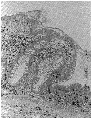
ECP was mainly localized in eosinophils in the lamina propria of UC colonic mucosa, and partially in the extracellular interstitium and also in the muscularis mucosa though small in number (Fig. 1). In control colitis patients, the findings were almost the same as in UC.
However, eosinophils were less common than in UC and the ECP content was lower accordingly. In the colonic mucosa
Fig. 1 The localisation of ECP in the colonic mucosa of UC.
Stained for activated eosinophils and secreted ECP/
EPX. Many activated eosinophils and discrete
deposits of ECP present within the mucosa. (x50)
Fig. 4 ECP concentration in the colonic mucosa of UC.
Comparison between active mucosa and noniflamed
mucosa in the same UC patients. (n = 9)
Fig. 2 ECP positive area in a unit (171,550 gm2) in various diseases and control subjects. Immunohistochernical
study.
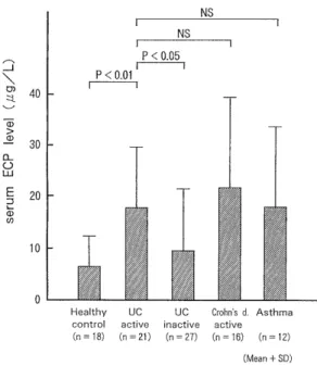
Fig. 5 Serum ECP levels in various diseases and control subjects.
Fig. 3 ECP positive area in a unit (171,550 ,u m2) by the 3 grades of histological severity according to Matts'
classification. Immunohistochemical study in UC.
of healthy controls, infiltration of eosinophils was hardly observed and the ECP content was nearly equal to zero.
The quantitative investigation of ECP content in the inflamed UC mucosa (n = 30) showed that the red reaction area per 171,550 ,um' was 1,448 ± 713 um2 (mean ± SD).
This was significantly higher than the level of 333 ±
3) Serum ECP concentration
The serum ECP concentration in active UC (17.68 ± 12.25 #g/L) (n = 21) was significantly higher than that in inactive UC (9.51 ± 11.71 #g/L) (n = 27) (p < 0.05) and in healthy controls (7.18 ± 5.10 ,ug/L) (n = 18) (p < 0.01).
However, no significant difference was observed when compared to active CD (21.37±17.92 ,ug/L) (n = 16), and bronchial asthma (17.93±15.91 ,ug/L) (n = 12) (Fig. 5).
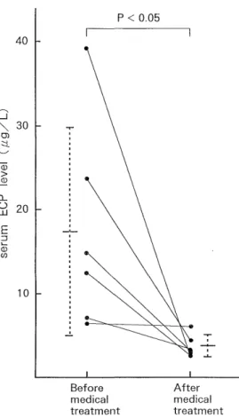
A significant decrease in the serum ECP concentration (n = 6) was observed after medical treatment (3.86 ± 1.24 ,ug/L) compared to that before medical treatment (1 7.42±12.52 ,ug/L) (p <0.05) (Fig. 6).
Fig. 6 Changes in serum ECP levels in 6 patients with active UC after medical treatment.
341,u m2 in inactive UC mucosa (n = 18), 316 ± 433 ,um' in the worm-eaten appearance region of CD mucosa (n = 9), 359±403 ,amt in control colitis mucosa (n = 14), and 120
±112 ,um2 in healthy controls (n = 12) (p<0.01) (Fig. 2).
The HE-stained specimens of the UC patients corre- sponding to the immunohistochemical stained sites were histologically divided into 3 groups [ Grade 1 (n = 4), Grade 2+3 (n = 36), and Grade 4+5 (n = 8) 1 according to Matts' histopathological classification, and the ECP content was compared among these groups. A significant increase in the distribution of ECP was observed between Grade (1) and both (2+3) and (4+5). The distribution of ECP showed a tendency to increase as the histological grade increased (Fig. 3).
2) ECP content of colonic mucosa
The ratio of the ECP content to the protein content in the inflamed colon mucosa (121 ± 128 X 10-s) was signif i- cantly higher than in the noninf lamed area (88.1 ± 58.9 X
10-6) (p < 0.01) (Fig. 4).
Discussion
The clinical symptoms of UC appear first as diarrhea with mucus and bloody stool. Edema, hyperemia, bleeding, and polyblennia are observed in the colonic mucosa. The onset and recurrence are paroxysmal. These are the phe- nomena that strongly suggest the association of immedi- ate type hypersensitivity caused by antigens in the intestinal tract.
Eosinophils, which infiltrate into the site of immediate type hypersensitivity, are considered not only to inactivate chemical mediators such as PAF, SRS-A, and histamine released from mast cells and/or basophils and thus inhibit inflammation, but also to have a role in tissue injury". In addition, eosinophil recruitment in the tissue is mediated by interleukin-5 (IL-5) from CD4+T-cells'o)
A series of researches on the eoinophils has detected ECP'', MBP, EDN and EPO, which are all eosinophil granule proteins. ECP originally isolated by Olsson et al.
in 1977 is a basic protein with a molecular weight of approximately 21,000 that is a constituent of the matrix of eosinophil granules'). The biological characteristics of this protein include destruction of Schistosomula, produc- tion of Gordon phenomenon, enhancement of XII factor- dependent pathways on coagulation, preactivation of plasminogen, and neutralization of heparin", 12) . And there is a report on the ECP release from human eosinophils
(eosinophil degranulation) induced by C3a and C5a'".
In the area of gastroenterology, the concentration of ECP in colorectal perfusate was measured"). As for UC, Spry reported in 1985 that ECP might be somehow associ- ated with the inflammation of UC after observing the distribution of activated eosinophils (ECP) in intestinal biopsy specimens'). In 1993, it was reported that the fecal ECP level was increased in UC and the ECP might be involved in the pathogenesis of UC6). But the opposite result that no increase in activated eosinophils staining was seen in lamina propria of UC was also reported').
We initially studied the distribution of ECP in the colon mucosa immunohistochemically. The antibody EG2 used in
this study reacts only with the secreted form of ECP (activated ECP, which is different in three-dimensional structure from storage form of ECP) and EPX, and does not react with inactivated ECP3). In the inflamed UC mucosa, eosinophils infiltrating into the mucosa were most positive for ECP and the interstitium and muscularis mucosa were also slightly positive (Fig. 1). As most of the eosinophils were EG2 positive, it was thought that they became the activated form for the most part. Further- more, the quantitative investigation showed a more significant increase of staining in inflamed UC mucosa than in other groups. Little staining was noted in the colonic mucosa of healthy subjects (Fig. 2). It was thought that the less distribution of ECP in control colitis than in active UC was due to the mild inflammation of control colitis compared with active UC. The HE-stained sections corresponding to the immunohistochemical stained sites were divided into three groups (Grade 1, Grade 2+3, and Grade4+5) by Matts' classification to compare the distribution of ECP. ECP staining mostly increased as inflammation became more severe (Fig. 3), suggesting a relationship between ECP and the severity of UC.
The ratio of ECP to total protein in the tissue was also significantly different (Fig. 4), being higher at the in- flamed sites. In addition, in the study measured with the ECP-RIA assay, both the activated and inactivated form of ECP are recognized")
The serum ECP level significantly increased in subjects with active phase UC when compared to those with inac- tive phase UC and healthy subjects (Fig. 5). The serum level of ECP was not parallel with the mucosal level studied immunohistochemically in Crohn's d., because the worm-eaten appearance') lesion was not main lesion of Crohn's d. but minimal lesion of the rectum. Furthermore, it was considered that the serum ECP concentration reflects the degree of inflammation, since the serum ECP level was decreased by medical treatment (Fig. 6). The serum ECP level has also been shown to increase in pa- tients with bronchial asthma (Fig. 5). These results suggest that an immediate type hypersensitivity reaction mediated by eosinophils also occurs in UC.
The present study on the distribution of ECP in the colonic mucosa and serum in patients with UC indicated that the pathophysiology of UC may be closely related to this protein. It is speculated that ECP has a role, though partial, in the damage to the colonic mucosa in UC on the
basis of its biological features.
Acknowledgements
The authors wish to thank Prof. Kohei Hara for his direction and review of this study, and also thank Prof.
Derek P Jewell for his correction of this article.
References
1) Olsson I, Venge I', Spitznagel JK, Lehrer RI. Arginine-rich cationic proteins of human eosinophil granules: comparison of human
eosinophils granules: comparison of the constituents of eosinophilic
and neutrophilic leukocytes. Lab. Invest., 36: 493-500, 1977.
2) Kay AB. Eosinophils as effector cells in immunity and hypersensitivity
disorders. Clin. Exp. Immunol., 62: 1-12, 1985.
3) Tai PC, Spry CJJ." , Peterson C, Venge P, Olsson I. Monoclonal antibodies distinguish between storage and secreted forms of eosinophil
cationic protein. Nature, 309: 182-4, 1984.
4) Spry CJF, Tai PC, Barkans J. 'T'issue localization of human eosinophil cationic proteins in allergic diseases. Int. Arch. Appl_ Immunol, 77:
252-4, 1.985.
5) Choy MY, Walker-Smith JA, Williams CB, MacDonald TT. Activated eosinophils in chronic inflammatory bowel disease. Lancet, 336: 126-7,
1990.
6) Berstad A, Borkje B, Riedel B, Elsayed S, Berstad A. Increased fecal eosinophil cationic protein in inflammatory bowel disease. I-Iepato
Gastroenterol., 40 : 276-8, 1993.
7) 't'ruelove SC, Witts LJ. Cortisone in ulcerative colitis. Final report on a therapeutic trial. B. VI. J., ii : 1041-8, 1955.
8) Myren J, Bouchier IAD, Watkinson G, Softley A, Clamp SE, deDombal FT. The O. M. G. E. multinational inflammatory bowel disease survey
1976-1982. A further report on 2657 cases. Scand. J. Gastroenterol., 95:
1-27, 1984.
9) Makiyama K, Bennett MK, Jewell DP. Endoscopic appearances of the rectal mucosa of patients with Crohn's disease visualised with a
magnifying colonoscope. Gut, 25: 337-40, 1984.
10) Nakajima H, Iwamoto 1, Tomoe S, Matsumura R, Tomioka II, Takatsu K, Yosida S. CD4+T-lymphocytes and interleukin-5 mediate
antigen-induced eosinophil infiltration into the mouse trachea. Am.
Rev. Respir. Dis., 146: 374-7, 1992.
11) Venge P, Dahl R, Fredens K, Hallgren R, Peterson C : Eosinophil cationic proteins (ECP) in health and disease. In: Yoshida T, Torisu
M, eds. Immunobiology of the eosinophil. 163-79. Elsevier Science
Publishing Co, New York 1983.
12) Fredens K, Dahl R, Vange P. In vitro studies of the interaction between heparin and eosinophil cationic protein. Allergy, 46: 27-9,
1991.
13) '[ akafuji S, Tadokoro K, Ito K, Dahinden CA. Degranulation from human eosinophils stimulated with C3a and C5a. Int. Arch. Allergy
Immunol., 104 (suppl 1) : 27-9, 1994.
14) Raab Y, Hallgren R, Knutson L, Krog M, Gerdin B. A technique for segmental rectal and colonic perfusion in humans. Am. J.
Gastroenterol., 87: 1453-9, 1992.
15) Peterson CGB, Jornvall H, Venge P. Purification and characterlization
of eosinophil cationic protein from normal human eosinophils. Eur. J.
Haematol., 40: 415-23, 1988.