原 著
多系統萎縮症における眼球運動障害
―50 例における経時的検討―
磯崎 英治
1)*飛澤 晋介
1)内藤 理恵
2)水谷 俊雄
3)松原 四郎
1) 要旨:多系統萎縮症における眼球運動障害の特徴を明らかにするため,50 例を対象に 9 種類の眼運動系パラ メーターの経時的変化を検討した.各パラメーターにおける異常出現率は,いずれも病型間(小脳失調先行型とパー キンソニズム先行型)で有意差をみとめなかった.また,経時的変化からパラメーターは 3 群―病初期から異常出 現率が高頻度にみとめられる群,経過とともに徐々に高率化する群,進行しても比較的低値にとどまる群―に大別 された.各群のそれぞれの代表として,頭位変換性眼振,視性抑制反応および温度眼振に注目し,これらの機能解 剖学的検討から,病変は小脳の背側虫部次いで片葉,そして前庭神経核から前庭皮質にもおよぶと考えた. (臨床神経 2012;52:218-226) Key words:多系統萎縮症,眼球運動障害,下眼瞼向き眼振,温度眼振,前庭皮質 はじめに 脊髄小脳変性症(SCD)では,眼振を始めさまざまな眼球 運 動 障 害 を き た す が,と く に 遺 伝 性 SCD に お い て は, Spinocerebellar ataxia(SCA)1 や SCA2 における Slowsac-cade1)∼3)あるいは SCA3 における前庭動眼反射(VOR)の低
下・消失4)などが特徴的な所見として知られている.なかで も,SCA2 患者における衝動性眼球運動の速度は,早期診断や 経過を追う上で surrogate marker になりうるとして注目さ れている2).一方,孤発性 SCD である多系統萎縮症(MSA)に ついては,欧米では小脳失調優位型(MSA-C)よりパーキン ソニズム優位型(MSA-P)の方が出現頻度が高く,したがっ てパーキンソン病との鑑別が問題となるが,この鑑別に眼運 動系パラメーターの所見が有用であるとされている5)∼7).本 邦においては MSA-P より MSA-C の方が出現頻度は高いが, この病型間の違いを眼球運動障害という観点から解析した報 告は少なく,経時的に検討した報告はさらに少ない.遺伝子型 が判明した SCA を除けば,本邦報告例の多くは,病型を問わ ずに SCD 全体を対象とした報告か,あるいは小脳型,脊髄小 脳型,脊髄型という大きなくくりによる分類にとどまった報 告が多く,MSA だけに注目した報告はまれである.こうした 背景から,本論文の目的は,9 種類の眼運動系パラメーターを もちいた 50 例の後方視的研究により,MSA における眼球運 動障害の自然史を明らかにすることである. 対象と方法 対象は,当科に入院し,一回以上神経耳科的検査がおこなわ れた 50 例の MSA 患者であり,12 例の剖検確定例をふくむ. 病型分類については,臨床上 MSA-C と MSA-P のいずれかに 分類することが必ずしも容易ではない例も存在することか ら,ここでは初発症状が小脳失調と思われる例を C 先行型 MSA,パーキンソニズムと思われる例を P 先行型 MSA とし て分類した.それにしたがって 50 例を分類すると,C 先行型 が 35 例(男 20 例,女 15 例,平均年齢 64±6.4 歳,平均罹病 期間 6.1±3.5 年),P 先行型は 15 例(男 5 例,女 10 例,平均 年齢 65±8.1 歳,平均罹病期間 5.7±3.8 年)である.検索した 眼運動系パラメーターは 9 種類であり,Table 1 にそれらの 検査項目および判定方法を示した.その内容は,フレンツェル 眼鏡下での頭位・頭位変換眼振検査(頭位の変化や座位・懸 垂頭位間での急激な体位変換時での誘発眼振),電気眼振図を もちいた注視および温度眼振検査(外耳道への冷水注入によ る誘発眼振), 視性抑制検査(Visual suppression test;VST, 明所固視下での反応),および衝動性・滑動性眼球運動検査で ある.延べ検査回数は 111 回であり,その内訳は 1 回のみが 14 例,2 回が 19 例,3 回が 11 例,4 回が 4 例,5 回が 2 例で ある.なお,VST は Takemori の方法8)にしたがい,その結果 は三段階―正常(40% 以上),低下(0∼40%),増強(0% 未 満)―で表した.各パラメーターは,Table 1 で示した 3 段階 * Corresponding author: 東京都立神経病院脳神経内科〔〒183―0042 府中市武蔵台 2―6―1〕 1) 東京都立神経病院脳神経内科 2) 同 神経耳科 3) 東京都立府中療育センター (受付日:2011 年 9 月 7 日)
Table 1 Eye movement parameters and their evaluations.
Eye movement
parameters Methods
Evaluations*
Normal Abnormal
type 1 type 2 type 3 Positional nystagmus Frentzel goggles no nystagmus antigeotropic direction-changing
nystagmus other types of nystagmus Positioning nystagmus Frentzel goggles no nystagmus downbeat nystagmus at
head-down position, more than 5 beats
upbeat nystagmus at head-down position, more than 5 beats Gaze nystagmus ENG no nystagmus gaze evoked nystagmus to either
horizontal or vertical direction
gaze evoked nystagmus to both horizontal and vertical directions Eye tracking test ENG normal borderline saccadic or dysmetric
Saccade test ENG normal borderline hypermetric or hypometric Caloric nystagmus test ENG normal (≧20%) depressive (1-20%) abolished (0%)
Visual suppression test ENG normal (≧40%) depressive (0-40%) increased (<0%)
*The evaluation of each parameter was divided into three types: normal (type 1) and two abnormal types (type 2 and type3).
ENG: electronystagmography.
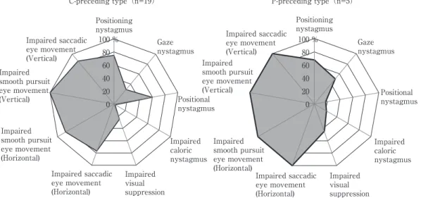
Fig. 1 Comparison of 9 eye movement parameters between C-preceding and P-preceding types
of MSA in the early stage with the duration less than 3 years. Gray area in each radar chart in-dicates the frequency of the abnormality in the 9 parameters. No significant differences were ob-served between the two disease types in any of the parameters.
0 20 40 60 80 100 %
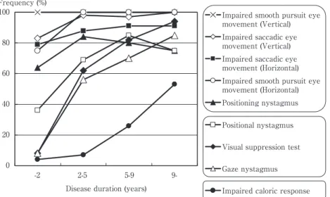
C-preceding type(n=19) P-preceding type(n=5) Positioning nystagmus Gaze nystagmus Positional nystagmus Impaired caloric nystagmus Impaired visual suppression Impaired saccadic eye movement (Horizontal) Impaired saccadic eye movement (Vertical) Impaired smooth pursuit eye movement (Vertical) Impaired smooth pursuit eye movement (Horizontal) Positioning nystagmus Gaze nystagmus Positional nystagmus Impaired caloric nystagmus Impaired visual suppression Impaired saccadic eye movement (Horizontal) Impaired saccadic eye movement (Vertical) Impaired smooth pursuit eye movement (Vertical) Impaired smooth pursuit eye movement (Horizontal) 0 20 40 60 80 100 % で評価された結果をもとに,異常出現率(延べ検査回数に対す る異常所見の出現回数の百分率)を算出した.この異常出現率 は,原疾患の進行にともなって高率化すると考えられるため, 病型間の比較をおこなうばあいには,発症早期(罹病期間が 3 年未満)と進行期(罹病期間が 8 年以上 11 年未満)に分け て検討し,また経時的変化を追跡するばあいには罹病期間を 4 つの期間(発症 2 年未満,2 年以上 6 年未満,6 年以上 10 年未満,10 年以上)に分けて検討した.統計学的有意差の判 定は,標本数の大きさにより,χ2検定あるいはフィッシャーの 直接確率計算法のいずれかをもちいて検定し,危険率 5% 未 満をもって有意差とした. 結 果 9 種類の眼運動系パラメーター毎に,発症早期(Fig. 1)お よび進行期(Fig. 2)における病型間の比較をおこなったとこ ろ,いずれの時期においても有意差はみとめられなかった (Table 2).そこで,病型を問わずに全症例を対象として各パ ラメーターの経時的推移を検討した結果,パラメーターは見 かけ上 3 群―異常出現率が病初期から高率にみとめられる 群,経過とともに徐々に高率化する群,進行しても比較的低率 にとどまる群―に大別することができた(Fig. 3).頭位変換眼 振検査は,衝動性および滑動性眼球運動障害に次いで病初期 から高率に異常を示し,その内容は大部分が懸垂頭位時の下 眼瞼向き眼振であり,上眼瞼向き眼振(3%)をふくめ他の眼 振を示した例はごく少数であった(Fig. 4A).これに対し,温 度眼振反応はよく保たれる傾向にあり(Fig. 1∼Fig. 3),発症 後 9 年以上の長期経過例でも異常出現率は 53% とほぼ半数 にとどまっており,機能廃絶にまで進行した例は 7% にすぎ なかった(Fig. 4B).一方,VST においては,正常反応を示す
Table 2 Frequency of the abnormalities of 9 eye movement parameters in the patients with MSA.
Patients from present study Anderson s studyPatients from 6)
Early stage
(Duration less than 3 years from onset)
Advanced stage (Duration between 8 and 11 years from onset)
C-preceding type P-preceding type p value* C-preceding type P-preceding type p value*
19 cases 5 cases 10 cases 4 cases 30 cases Positioning nystagmus 75% (15/20) 67% (4/6) n.s. 83% (10/12) 100% (2/2) n.s. 43% (13/30) Gaze nystagmus 29% (6/21) 50% (3/6) n.s. 67% (10/15) 60% (3/5) n.s. 40% (12/30) Positional nystagmus 60% (12/20) 17% (1/6) n.s. 92% (12/13) 75% (3/4) n.s.
Caloric nystagmus test 2% (1/44) 22% (2/9) n.s. 42% (10/24) 20% (2/10) n.s. 33% (10/30) Visual suppression test
Depressive type only 20% (9/44) 22% (2/9) n.s. 59% (10/17) 25% (2/8) n.s. Impaired VORS: 78% (23/30) Depressive or increased type 25% (11/44) 44% (4/9) n.s. 88% (15/17) 75% (6/8) n.s.
Impaired SEM (Horizontal) 77% (17/22) 100% (6/6) n.s. 83% (10/12) 100% (4/4) n.s. 87% (26/30) Impaired SPEM (Horizontal) 86% (19/22) 100% (6/6) n.s. 100% (12/12) 100% (4/4) ・ 90% (27/30) Impaired SPEM (Vertical) 100% (22/22) 100% (6/6) ・ 100% (13/13) 100% (4/4) ・ 87% (26/30) Impaired SEM (Vertical) 86% (19/22) 100% (6/6) n.s. 100% (8/8) 100% (4/4) ・ ・ Numerals in the parenthesis show the number of the times of abnormalities against the total number tested.
*statistical difference between C-preceding and P-preceding types of MSA, n.s.: not significant statistically.
SEM: saccadic eye movement, SPEM: smooth pursuit eye movement, VORS: vestibulo-ocular reflex suppression. ・: unable to analyze statitically.
Fig. 2 Comparison of 9 eye movement parameters between C-preceding and P-preceding types
of MSA in the advanced stage with the duration between 8 and 11 years from onset.
Both gray areas, indicating the frequency of the abnormality in the 9 parameters, increased with no significant differences between the two disease types in any of the parameters.
0 20 40 60 80 100 %
C-preceding type (n=10) P-preceding type (n=4) Positioning nystagmus Gaze nystagmus Positional nystagmus Impaired caloric nystagmus Impaired visual suppression Impaired saccadic eye movement (Horizontal) Impaired saccadic eye movement (Vertical) Impaired smooth pursuit eye movement (Vertical) Impaired smooth pursuit eye movement (Horizontal) Positioning nystagmus Gaze nystagmus Positional nystagmus Impaired caloric nystagmus Impaired visual suppression Impaired saccadic eye movement (Horizontal) Impaired saccadic eye movement (Vertical) Impaired smooth pursuit eye movement (Vertical) Impaired smooth pursuit eye movement (Horizontal) 0 20 40 60 80 100 % 割合は経過とともに急速に減少し,それに代わって増強型が 増え,9 年以上の例では 72% を占めた(Fig. 4C).Table 3 は,こうした VST の質的変化を,個々の症例ごとに経時的に 追跡した結果である.大部分の症例(28 例中 26 例)では進行 とともに正常型から低下型へ,低下型から増強型へ,あるいは 正常型から増強型へのいずれかをとるか,または不変であっ た.しかし,2 例(症例 C4 と C16)においては増強型から低 下型へ移行する時期(Table 3 における丸印)をみとめた. 考 察 本研究の結果をまとめると,1)9 種類の眼運動系パラメー ターの異常出現率は,いずれも病型間(C 先行型と P 先行型) において有意差をみとめない,2)頭位変換眼振検査時にみと められる懸垂頭位時の下眼瞼向き眼振は,病初期から高率に 出現し,一方温度眼振反応は,進行期にいたっても保たれる傾
Fig. 3 Chronological changes of the frequency of the abnormalities in the 9 parameters.
Parameters could be divided into three groups which are arranged in the right panel: those with high frequency from the early stage, those with gradual increase of the frequency, and those with less increase even in the advanced stage.
0 20 40 60 80
100 Impaired smooth pursuit eye movement (Vertical) Impaired saccadic eye movement (Vertical) Impaired saccadic eye movement (Horizontal) Impaired smooth pursuit eye movement (Horizontal) Positioning nystagmus Positional nystagmus Visual suppression test Gaze nystagmus Impaired caloric response Frequency (%)
Disease duration (years) -2 2-5 5-9 9-向を示す,3)異常出現率の経時的推移から,眼運動系パラメー ターは 3 群に大別されうる,の三点である.MSA では進行期 のみならず早期においても,眼運動系パラメーターは病型間 における有意差をみとめないという今回の結果は,P 先行型 であっても,すでに小脳脳幹の機能障害が病初期から潜在し ていることを示している.これは,欧米からの報告5)∼7)― MSA-P とパーキンソン病との鑑別に,眼運動系パラメーター が有用である―に矛盾しない結果である.今回の研究におい てとくに興味深い点は,眼運動系パラメーターは経時的に見 ると 3 つのグループに大別されうる点である.すなわち,異常 出現率が早期から高頻度にみとめられるパラメーター群,経 過とともに徐々に高率化するパラメーター群,および進行し ても比較的低値(ほぼ半数)にとどまるパラメーター群の三つ のグループである.そこで,それぞれのグループの代表として 頭位変換眼振検査,温度眼振検査および VST に注目し,これ らの検査でみとめられる眼振の特徴をより詳しく検討し,そ の結果から推測しうる責任病巣について検討した. 頭位変換眼振検査における懸垂頭位時の下眼瞼向き眼振 は,P および C 先行型に共通して発症早期から高頻度に出現 しており(C 先行型で 75%, P 先行型で 67%), この結果は, Anderson らの報告(43%)6)よりも高率であった(Table 2). 水野らによる報告でも9),同眼振の出現率は SCD 全体(40 例)では 80%,とくにオリーブ橋小脳萎縮症(16 例中 Menzel 型 1 例をふくむ)を対象とすると 88% と高率である.彼らの 報告は,1998 年の Gilman らによる MSA の分類10)が発表され る以前であり,分類上の問題はあるものの,オリーブ橋小脳萎 縮症すなわち MSA では,懸垂頭位時の下眼瞼向き眼振の出 現頻度が決して低くないことを示している.同眼振をみとめ た連続 50 例における原因(基礎疾患)を検討した報告でも11), 中枢性病変を有した 38 例の中で,MSA が 13 例ともっとも 多くを占めた.これに対し Yabe らは12),SCA6 では浮動感を ともなう懸垂頭位時の下眼瞼向き眼振を高頻度(84%)にみと めるが,MSA ではむしろまれ(16 例中 1 例,6%)としてお り,また,Lee らは MSA-C では 32%,MSA-P では 5∼16% と病型間での違いをみとめている7).報告者によるこうした出 現率の違いは,検査施行時の病期(罹病期間)の違いに加え, 本検査の異常値判定におけるカットオフ閾値の違いも一因と 思われる.すなわち,Lee らによる判定基準は,「誘発された眼 振における最初の 3 打の緩徐相速度の中央値が 5 度!秒以 上」と眼振図をもちいた方法7)で判定しているが,われわれは 「フレンツェル眼鏡下に,懸垂頭位時に 5 打以上の下眼瞼向き 眼振が,再現性をもって出現したばあい」を異常と判定してい る点でことなっている.両者における感度を客観的に比較す ることは難しいため,疾患対照として当院例において同一の 方法による判定基準で評価した SCA3 患者を検討した.その 結果,懸垂頭位時の下眼瞼向き眼振の出現率は 14 例中 3 例 (21%)であり,MSA にくらべ明らかに低値であった.以上 より,MSA における懸垂頭位時の下眼瞼向き眼振は,病初期 から高頻度に出現する眼振であると考えられる. これに対し,温度眼振検査は 9 種類のパラメーターの中で はもっとも障害をまぬがれるパラメーターであった.MSA における温度眼振についての報告は少なく,Anderson らの 報告での異常出現率は 33% であった6)(Table 2).罹病期間の 影響を避けるため,彼らの例における平均罹病期間(6.6 年)に 近い罹病期間(発症後 5 年以上 9 年未満)にある自験例と比較 したところ 26% であり,彼らとほぼ近似した値であった (Fig. 4B). 温度眼振は, 前庭動眼反射(VOR)の一つであり, 温度刺激ではなく頭部の回転刺激による VOR 検査をおこ なった報告では5),MSA では固視による VOR
Fig. 4 Chronological changes of the frequency of the abnormalities in the 3 parameters.
A: Downbeat nystagmus at hanging head position was the major abnormality in positioning nys-tagmus test.
B: Caloric response is apparently preserved as compared with positioning nystagmus test (A) and visual suppression test (C).
C: Frequency of the abnormality increased rapidly with the disease duration, associated with a qualitative change showing the predominancy of the increased type in visual suppression test.
normal normal normal depressed depressed increased increased increased depressed depressed
Visual suppression test 100% 75% 50% 25% 0 yrs Frequency C -2 2-5 5-9 9-normal normal normal depressed depressed depressed depressed normal abolished Caloric response 100% 75% 50% 25% 0 yrs Frequency B -2 2-5 5-9 9-normal normal normal upbeat others
Positioning nystagmus test 100% 75% 50% 25% 0 Disease duration Frequency yrs A -2 2-5 5-9 9-normal downbeat
downbeat downbeat downbeat
たれると述べられている.以上のことから,前庭神経核を介し て誘発される VOR は,MSA では進行期にいたるまで比較的 保たれると考えられ,早期から温度眼振が高度かつ高頻度に 障害される SCA313)とは対象的である. 一方,VST は経過とともに急速に異常を呈するようにな り,しかも低下型から増強型へと質的変化をともなっている ことが特徴的であった.例外的に,21 例中 2 例においてのみ, 増強型から低下型へと逆行した時期をみとめたが(Table 3), これらの例はいずれも検査施行時での罹病期間が長く(症例 C4 は 16∼17 年,症例 C16 は 8∼9 年),MSA でみとめられる 注意障害14)に基づく固視不良がその一因であるかもしれな い.MSA における上述した VST の質的変化は,すでに集団 的研究(mass study)においては指摘されていたが15)16),同一 症例での経時的追跡をおこなった本研究により,その事実を 確証することができた.なお,縄田ら17)が報告した 12 例のオ リーブ橋小脳萎縮症では,平均罹病期間が 2 年 3 カ月と比較 的短いにもかかわらず,増強型を示した割合が 58% と高率で あった.これに対し自験例では,早期(罹病期間が 3 年未満) に増強型を示したのは 15 例中 1 例のみであり(Table 3),ま た前述した Rascol らの報告5)でも,VST は MSA では低下型 を示すものの,増強型については記載されていない.VST の増強反応は,進行の速さの他,後述する障害部位の分布やそ の広さなどが関与しているのかもしれない. つぎに,上述した眼運動系パラメーターの障害パターンを もとに,MSA における責任病巣について検討した.動物実験 をふくめた多くの報告から,一つの眼運動系パラメーターの
Table 3 Chronological changes of the findings of visual suppression test in the 21 patients with MSA.
Patients* Disease duration (years)
-1 -2 -3 -4 -5 -6 -7 -8 -9 -10 -11 -12 -13 -14 -15 -16 -17 P1 1 1 P2 1 2 P3 2 3 P4 1 P5 2 3 P6 3 3 P7 1 2 C1 2 2 C2 3 3 3 C3 1 1 2 C4 2 3 3 C5 1 2 C6 2 3 C7 1 2 C8 1 1 2 2 C9 1 2 3 C10 1 3 C11 1 2 2 3 C12 2 2 2 2 C13 1 1 1 C14 1 2 C15 2 2 C16 3 3 C17 1 3 C18 1 1 2 2 2 C19 1 2 C20 2 2 3 C21 1 2 3
*P1 to P6 and C1 to C21 belong to parkinsonism- and cerebellar sign-preceding MSA, respectively.
Numerals show the evaluations of visual suppression test: 1=normal, 2=depressed, and 3=increased responses. Circle indicates the point of time when evaluation numerals decreased inversely from 3 to 2.
Table 4 Eye movement parameters and their corresponding lesions in the brainstem and the cerebellum.
Correspond-ing lesions Impaired saccadic eye movements Impaired smooth pursuit eye movements Positioning downbeat nystag-mus Depressive caloric response Deprressive type of visual suppres-sion test Increased type of visual suppres-sion test B r a i n s t e m level Superior collicu-lus, NRTP, PPRF, riMLF DLPN, NRTP, Vestibular nucleus, Y-Group Pontine parame-dian tract cells, Y-Group Vestibular nucleus, MLF, Interstitial nucleus of Cajal Superior collicu-lus, Y-Group PPRF Cerebellum level Dorsal vermis*, Fastigial nucleus, Hemisphere (VI・ VII) Dorsal vermis*, Fastigial nucleus, Flocculus, Paraflocculus, Hemisphere (VI・ VII) Flocculus, Paraflocculus, Nodulus/Uvula, Dorsal vermis Flocculus, Nodulus
*Dorsal vermis includes declive, forium vermis, and tuber vermis.
NRTP=Nucleus reticularis tegmenti pontis, PPRF=Paramedian pontine reticular formation, riMLF=rostral interstitial nucleus of medial longi-tudinal fasciculus, DLPN: dorsolateral pontine nucleus.
障害に対する責任病巣は一カ所のみとはかぎらず,また逆に 病巣は一カ所であっても複数のパラメーターに異常がおよぶ ことも報告されている.この一因には,病変の広がりや障害の 性質―興奮性!抑制性回路のいずれかあるいは両者―など各 パラメーターにおける機能解剖学的な裏づけがいまだ十分に は解明されていないことが考えられるが,パラメーター相互 間での影響を検討することは今後の重要な課題の一つと考え られる.本論文では,各パラメーターに対する小脳・脳幹レベ ルでの推定責任病巣をまとめ(Table 4),それをもとに MSA における病変部の進展について検討した.発症早期から,滑動 性および衝動性眼球運動障害と懸垂頭位時の下眼瞼向き眼振 の三者が高頻度にみとめられたが,前二者については脳幹お
よび小脳の様々な部位で出現しうる.これに対し,下眼瞼向き 眼振については,ある程度責任病巣は限定されており,とくに 片葉や小脳小節などがよく知られている18).しかし,片葉の障 害は,同時に VST の反応低下もきたしうることから18),この 時期での責任病巣としては考えにくく,また小節については MSA では萎縮をまぬがれる19)とされていることからやはり 否定的と考えた.一方,病理学的に検討した水谷らは20),MSA では山頂,山腹,虫部葉に変性が強いと報告しており,また Tsuchiya ら21)による報告でも,萎縮は山頂から始まり,経過 とともに前方(中心小葉)および下方(山腹)へと拡大する図 が描かれている.以上のことから,この時期における責任病巣 としては小脳の背側虫部(いわゆる oculomotor vermis に相 当する部位)が障害されるのではないかと考えた.MSA の早 期病変についてしらべてみると,発症 15 カ月の時点で剖検に 賦された MSA-C 例においては,橋核での神経細胞脱落が もっとも顕著であり,虫部前葉および下オリーブ核病変は軽 度にとどまり,虫部後葉(山腹,虫部葉,虫部隆起などのいわ ゆる背側虫部をふくむ領域)と小脳半球には異常をみとめな かったという22).また,拡散テンソル画像法により橋小脳路を 検討した報告例では23),発症後 1.5 年の時点ですでに同部に変 性所見がみとめられ,MSA の早期診断への可能性が指摘さ れている.これらの報告は,Eadie による仮説24)―オリーブ橋 小脳萎縮症における病変は,最初に橋小脳線維の軸索終末に おこり,その後 transsynaptic に小脳皮質を障害する―を支 持すると考えられる. 疾患の中期になると,VST における低下反応がめだって くるが,その責任病巣は片葉や小節18)のほかに上丘25)や y-group18)などが報告されている(Table 4).しかし,後二者は, それぞれ衝動性眼球運動や下眼瞼向き眼振なども生じうる部 位であることを考えると,この時期での VST 低下の責任病 巣は片葉に求めやすい.進行期にいたると,VST の異常はそ れまでの低下型から増強型に移行し,また温度眼振反応もほ ぼ半数例で低下∼消失してくる.一般に,温度眼振の障害は前 庭神経核を中心とした末梢ループにおける障害を反映する が,その最上位中枢は前庭皮質であり,最近の positron emis-sion tomography(PET)をもちいた研究から前庭皮質は,側 頭頭頂葉・島・被殻・帯状回前部など複数の領域に分散して いると考えられており26),また温度眼振検査での視性抑制時 には,視覚皮質のほかに側頭極や小脳虫部なども活性化され ることが報告されている26).空間認知に関する第 7 野も前庭 皮質の一つと考えられており27),サルにおけるこの部位の切 除実験により VOR が低下することが知られている28).これら の知見および MSA では進行期には大脳皮質にも萎縮がおよ ぶこと29)を考えると,VST の増強反応は従来考えられてきた 脳幹部病変(傍正中橋網様体)のほかに,前庭皮質病変も責任 病巣の一つとして考えられる. 以上の結果,パラメーターの経時的障害様式からみると, MSA における小脳病変は背側虫部から始まり,その後片葉 に進展し,さらに前庭神経核をふくめた脳幹から前庭皮質な どの大脳皮質へと拡大する可能性が考えられた. ※本論文に関連し,開示すべき COI 状態にある企業,組織,団体 はいずれも有りません. 文 献
1)Klostermann V, Zühlke C, Heide W, et al. Slow saccades and other movement disorders in spinocerebellar atro-phy type 1. J Neurol 1997;244:105-111.
2)Seifried C, Velázquez-Pérez L, Santos-Falcón N, et al. Sac-cade velocity as a surrogate disease marker in spinocere-bellar ataxia type 2. Ann NY Acad Sci 2005;1039:524-527. 3)Velázquez-Pérez L, Seifried C, Abele M, et al. Saccade
ve-locity is reduced in presymptomatic spinocerebellar ataxia type 2. Clin Neurophysiol 2009;120:632-635. 4)Gordon CR, Joffe V, Vainstein G, et al. Vestibulo-ocular
ar-reflexia in families with spinocerebellar ataxia type 3 (Machado-Joseph disease). J Neurol Neurosurg Psychia-try 2003;74:1403-1406.
5)Rascol O, Sabatini U, Fabre N, et al. Abnormal vestibu-loocular reflex cancellation in multiple system atrophy and progressive supranuclear palsy but not in Parkin-son s disease. Mov Disord 1995;10:163-170.
6)Anderson T, Luxon L, Quinn N, et al. Oculomotor func-tion in multiple system atrophy: Clinical and laboratory features in 30 patients. Mov Disord 2008;23:977-984. 7)Lee J-Y, Lee W-W, Kim JS, et al. Perverted head-shaking
and positional downbeat nystagmus in patients with mul-tiple system atrophy. Mov Disord 2009;24:1290-1295. 8)Takemori S. Visual suppression test. Adv
Oto-Rhinol-Laryngol 1983;29:102-110.
9)水野正浩. 脊髄小脳変性症における神経耳科学的所見. 耳 鼻臨床 1981;74:63-88.
10)Gilman S, Low PA, Quinn N, et al. Consensus statement on the diagnosis of multiple system atrophy. J Auton Nerv Sys 1998;74:189-192.
11)Bertholon P, Bronstein AM, Davies RA, et al. Positional down beating nystagmus in 50 patients: cerebellar disor-ders and possible anterior semicircular canalithiasis. J Neurol Neurosurg Psychiatry 2002;72:366-372.
12)Yabe I, Sasaki H, Takeichi N, et al. Positional vertigo and macroscopic downbeat positioning nystagmus in spi-nocerebellar ataxia type 6 (SCA6). J Nerurol 2003;250:440-443.
13)Yoshizawa T, Nakamagoe K, Ueno T, et al. Early vestibu-lar dysfunction in Machado-Joseph disease detected by caloric test. J Neurol Sci 2004;221:109-111.
14)Meco G, Gasparini M, Doricchi F. Attentional functions in multiple system atrophy and Parkinson s disease. J Neu-rol Neurosurg Psychiatry 1996;60:393-398.
15)水野正浩, 山根雅昭, 小山内龍一. 脊髄小脳変性症における visual suppression test に つ い て. 耳 鼻 臨 床
1988;81:165-171.
16)古川朋靖, 渡辺道隆, 正木義男ら. オリーブ橋小脳萎縮症の 神経耳科学的検討. 耳鼻臨床 1999;92:347-354.
17)縄田安孝, 古川朋靖, 渡辺道隆ら. 神経変性疾患における垂 直眼球運動障害について. 日耳鼻 1997;100:770-781. 18)Leigh RJ, Zee DS. Diagnosis of central disorders of ocular
motility. In: Leigh RJ, Zee DS, editors. Neurology of the eye movements. Fourth Edition. Oxford univ press; 2006. p. 598-718.
19)Wenning GK, Tison F, Elliott L, et al. Olivopontocerebel-lar pathology in multiple system atrophy. Mov Disord 1996;11:157-162.
20)水谷俊雄. 歯状核系変性症における眼球運動障害の神経病 理学的背景―とくに Slow Saccade について―. 神経進歩 1990;34:68-77.
21)Tsuchiya K, Watabiki S, Sano M, et al. Distribution of cerebellar lesions in multiple system atrophy : a topo-graphic neuropathological study of three autopsy cases in Japan. J Neurol Sci 1998;155:80-85.
22)Wakabayashi K, Mori F, Nishie M, et al. An autopsy case of early ( minimal change ) olivopontocerebellar atrophy (multiple system atrophy-cerebellar). Acta Neuropathol 2005;110:185-190.
23)藤盛寿一, 館脇康子, 清水 洋ら. 拡散テンソル画像法をも
ちいた多系統萎縮症における橋小脳路変性の検討. 臨床神 経 2011;51:271-274.
24)Eadie MJ. Olivo-ponto-cerebellar atrophy ( Dejerine-Thomas type). In: Vinken PJ, Bruyn GW, Klawans HL, editors. Handbook of clinical neurology. Vol 21. Amster-dam: Elsevier; 1975. p. 415-431.
25)Kato I, Kawasaki T, Aoyagi M, et al. Loss of visual sup-pression of caloric nystagmus in cats. Acta Otolaryngol 1979;87:499-505.
26)Bottini G, Sterzi R, Paulesu E, et al. Identification of the central vestibular projections in man: a positron emission tomography activation study. Exp Brain Res 1994;99:164-169.
27)Grüsser OJ, Pause M, Schreiter U. Localisation and re-sponses of neurons in the parieto-insular vestibular cor-tex of awake monkeys ( macaca fascicularis ) . J Physiol 1990;430:537-557.
28)Ventre J, Faugier-Grimaud S. Effects of posterior parietal lesions (area 7) on VOR in monkeys. Exp Brain Res 1986; 62:654-658.
29)Horimoto Y, Aiba I, Yasuda T, et al. Cerebral atrophy in multiple system atrophy by MRI. J Neurol Sci 2000;173: 109-112.
Abstract
Eye movement disturbance in multiple system atrophy: chronological study of 50 patients
Eiji Isozaki, M.D.1)
, Shinsuke Tobisawa, M.D.1)
, Rie Naito, M.D.2) , Toshio Mizutani, M.D.3)
and Shiro Matsubara, M.D.1) 1)
Departments of Neurology, Tokyo Metropolitan Neurological Hospital
2)Departments of Neuro-otology, Tokyo Metropolitan Neurological Hospital 3)
Tokyo Metropolitan Fuchu Medical Center for the Disabled
To clarify the features of the eye movement disturbance in the patients with multiple system atrophy (MSA), we retrospectively examined chronological changes of 9 oculomotor parameters as described below in 50 MSA pa-tients including 12 autopsied cases. Papa-tients with MSA were consisted of 35 papa-tients with cerebellar ataxia-preceding type and 15 patients with parkinsonism-ataxia-preceding type. Nine parameters include saccade test, eye tracking test, positioning!positional!gaze!caloric nystagmus tests, and visual suppression test. Each parameter was evaluated by three categories; normal and the two abnormal findings according to their characteristic fea-tures. In all of the 9 parameters, no significant differences were found between the cerebellar ataxia- and the parkinsonism-preceding types of MSA both in the early (disease duration less than 3 years) and in the advanced stages (duration between 8 to 11 years). From the chronological analysis, 9 oculomotor parameters could be di-vided into three groups: the first group with the higher frequency of the abnormality from the early stage, the second with gradual increase of the frequency, and the third with less increased frequency even in the advanced stage. We here focused on the three representatives corresponding with the above-described each group; position-ing nystagmus test mainly showposition-ing downbeat nystagmus as a first group, visual suppression test showposition-ing a quali-tative change from depressed into increased response as the second, and the caloric nystagmus test showing de-creased response as the third. Based on these chronological changes of the oculomotor parameters, we supposed that in MSA the dorsal vermis is involved at first, followed by the flocculus in the cerebellum, and then the degen-erative lesions might expand to the vestibular nucleus, and the cerebral cortex including the vestibular cortex.
(Clin Neurol 2012;52:218-226)
Key words: multiple system atrophy, oculomotor disturbance, downbeat nystagmus, caloric nystagmus test, vestibular cortex