長期間の持続性陽圧呼吸療法は閉塞性
無呼吸症候群患者の左心機能への影響を改善し,
2-dimensional speckled tracking echocardiographic imaging 法は 評価方法として有効である
昭和大学医学部内科学講座(循環器内科学部門)
福岡 裕人 茅野 博行* 松井 泰樹 太 田 礼 安達 太郎 小林 洋一
抄録:閉塞性睡眠時無呼吸症候群(Obstructive sleep apnea syndrome:OSAS)は心血管イ ベントを起こす独立する危険因子と言われている.OSAS に対し持続式陽圧呼吸療法(Contin- uous positive airway pressure:CPAP)は確立された治療法だが,左心機能に対する改善効 果は明らかではない.これまでの CPAP 治療の左心機能に対する報告はほとんどが従来の心 エコー図法を用いての評価であり,われわれは微細な変化を評価可能な 2D speckled tracking echocardiographic imaging 法 (2D-STE)により左心機能がどのように変化するか検討した.
対象は昭和大学病院で睡眠時ポリソムノグラフィを施行し無呼吸低呼吸指数 15 回 / 時間以上 の中等から重症の OSAS と診断された,壁運動異常を有さない(EF ≧ 55%)62 例(女性 46 例,
平均 61
±
13 歳).対象を CPAP 治療の使用状況により,Good-CPAP 群 (26 例,1 か月に 21 日以上かつ 1 日 4 時間以上使用),Fair-CPAP 群 (20 例,1 か月に 21 日未満もしくは 1 日 4 時 間未満使用) ,Non-CPAP 群 (16 例,CPAP 未使用)の 3 群に分けて左室収縮・拡張機能,動 脈硬化所見および身体所見の治療開始前と開始後平均 2.1±
0.5 年後の変化を比較検討した.治療開始後,治療開始前と比較し収縮期血圧は Good-CPAP 群で有意に改善した(vs Non- CPAP [p=0.0342],vs Fair-CPAP [p=0.0424]).CAVI(Cardio ankle vascular index)値 は 3 群間で変化は見られなかった.従来の心エコー図検査では左室拡張末期径,左室収縮末期 径,左室駆出率,左房圧での変化は 3 群間で差は見られなかったが,左房径は Good-CPAP 群 が Non-CPAP 群に比べ有意に縮小していた[p=0.005].2D-STE では Longitudinal strain で は Good-CPAP 群が Fair-CPAP 群・Non-CPAP 群に比べ有意に改善が見られた[p=0.0128,
p < 0.0001].Strain rate での解析でも収縮能[p=0.0274,p=0.0002]・拡張能[p=0.0288,
p < 0.0001]ともに同様の結果であった.Radial strain では 3 群間で有意差はなかったが,
strain rate では Good-CPAP 群が Non-CPAP 群に比べ収縮能[p=0.0087]・拡張能[p=0.022]
ともに改善していた.Circumferential strain は Good-CPAP 群が Non-CPAP 群に比べ有意に 改善していた[p=0.028].Strain rate でも Good-CPAP 群が Non-CPAP 群に比べ収縮能[p
=0.0039]・拡張能[p=0.0119]ともに有意に改善していた.OSAS 症例では CPAP 治療を 推奨時間使用することで血圧を下げることが可能である.また 2D-STE により従来の心エコー 図で評価できなかった左室収縮能,拡張能が改善することが正確に評価することができた.特 に長軸方向が顕著であり,CPAP を推奨時間行うことが重要だと言える.
キーワード:閉塞性無呼吸症候群,CPAP,左心機能,2-dimensional speckled tracking echo- cardiographic imaging 法
原 著
*
責任著者
緒 言
閉塞性睡眠時無呼吸症候群(Obstructive sleep apnea syndrome:OSAS)は糖尿病,高血圧やメ タボリック症候群の発症や増悪に関与していること が知られている1).また,OSAS では心血管イベン ト発症の独立した危険因子とも報告されている2). OSAS では夜間の間欠的な低酸素状態を引き起こす ことから,24 時間に渡る血圧の上昇および交感神 経活性化亢進状態が誘発され,左室収縮能・拡張能 障害が惹起され,最終的には心不全や心臓突然死を 発症すると報告されている3,4).持続式陽圧呼吸療 法(Continuous positive airway pressure:CPAP)
は OSAS に対する確立された治療法である5).しか し CPAP 治療の左心機能に対する改善効果が明ら かになっていない6,7).
これまでの CPAP 治療の左心機能に対する報告の ほとんどが心エコー図法を用いて評価したものであ るが,その評価項目は左室径(mm)や容積(ml)
および駆出率(ejection fraction;EF, %)を従来法
(M-mode 法および B-mode 法)で,局所収縮機能や 左房圧をカラードプラ法,組織ドプラ法で算出して いる8).近年,従来法に比較して角度依存性を受け ずに左室全体および局所心筋機能 (短縮,伸展率お よび率 / 時間) の微細な変化を評価可能な 2D speck- led tracking echocardiographic imaging 法 (2D-STE)
が臨床で広く利用されている9).そこでわれわれは OSAS 患者における CPAP 治療前後の左心機能の 変化について 2D-STE を用いて比較検討した.
研 究 方 法 1.対象
対象は 2011 年 10 月から 2014 年 11 月に昭和大学 病院で睡眠時ポリソムノグラフィー(Polysomno- graphy;PSG)検査を施行し,無呼吸低呼吸指数
(apnea-hypopnea index;AHI)15 回 / 時間以上の中 等から重症の OSAS と診断された,壁運動異常を 有さない(EF ≧ 55%)62 例(女性 16 例,平均 61
±
13 歳).対象を CPAP 治療の使用状況により,日本循環器学会の推奨する使用時間を満たしている Good-CPAP 群 (26 例,1 か月に 21 日以上かつ 1 日 4 時間以上使用),上記を満たしていない Fair-CPAP 群(20 例,1 か月に 21 日未満もしくは 1 日 4 時間未
満使用),Non-CPAP 群(16 例,CPAP 未使用)の 3 群に分けて左室収縮・拡張機能,動脈硬化所見お よび身体所見の変化を比較評価した.尚,Non-CPAP 群は CPAP 治療を拒否された 13 例と CPAP 治療を 開始したが装着不快感が強く脱落した 3 例である.
除外症例は残存する心筋虚血症例,心房細動症例,
中等度以上の心臓弁膜症例,Pacemaker 植込み症 例,腎不全症例(CKD G3a 以上),担癌症例であ る.本検討は後ろ向き縦断観察研究であり,ヘルシ ンキ宣言に遵守して行った.
2.従来心エコー図測定
左室拡張末期径(LVDd, mm), 左室収縮末期径
(LVDs, mm), 中隔壁厚(IVS, mm)と後壁厚(PWD, mm)およびその平均壁厚(mm), 左房径(LAD, mm) は 傍 胸 骨 長 軸 像 か ら 記 録 し た.Biplane Simpson 法で求めた左室駆出率(EF,%), パルスド プラ法(cm/s)で求めた左室流入速波形 (拡張早期 E 波 , 心房収縮 A 波), 組織ドプラ法(cm/s)で求め た中隔と側壁の僧帽弁輪移動速度(e ), 連続波ドプラ 法(m/s)で求めた収縮期右室右房圧較差(mmHg)
は心尖二腔および四腔像から記録した.左房圧
(mmHg)の指標として E/e (比) を計算した.
3.2D-STE 測定
左室心筋の収縮率・拡張率(strain, %)と収縮率 / 時間・拡張率 / 時間(strain rate,1/S)を 3 方向で 評価した.左室長軸方向(longitudinal strain/strain rate)は心尖四腔像から,壁厚方向(radial strain/
strain rate)と円周方向(circumferential strain/stra- in rate)は乳頭筋レベル左室短軸像から記録した.
三方向の心筋 Strain, Strain rate 値の計測は東芝社 製 Artida に内臓されている専用 software を用いて 自動算出した.
4.身体検査および動脈硬化所見測定
心エコー図検査直後に身長(m),体重(kg)を測 定し,Body mass index は体重 (kg)/ 身長(m)2で 算出した.同時に両上下肢の血圧,脈拍および動脈 硬化指標として血圧に依存しない血管固有の硬さ CAVI (cardio ankle vascular index) をフクダ電子 社製 VS-1500A を用いて測定した.
5.統計解析
全ての解析はJMP 10を用いて解析した.収縮期・
拡張期血圧,心拍数,CAVI 値,EF,E/e ,LAD,
longitudinal, radial, circumferential strain,
longitudinal systolic, diastolic strain rate は連続変 数であり全ての統計解析は Tukey-Kramer 法を用 いて ANOVA 検定を使い全ての群間において行っ た.統計学的有意差は p < 0.05 とした.
結 果
3 群間における CPAP 治療前の臨床背景を Table 1 に示す.AHI,身長,体重,BMI などの身体所見に 差は認めなかった.血圧,脈拍にも差は認めなかっ た.血液検査では BNP 値において Fair-CPAP 群は Non-CPAP 群に対して有意に低値であった.基礎 疾患(高血圧,脂質異常症,糖尿病)の有病率と嗜 好歴にも差は認めなかった.
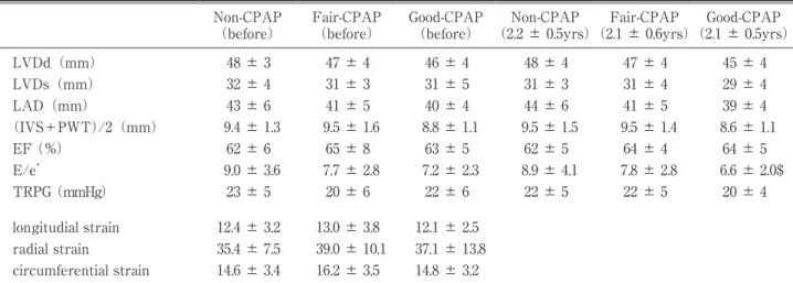
3 群間における CPAP 治療前と治療後(平均 2.1 年後)の心エコー図所見を Table 2 に示す.従来法 の心エコー図所見では CPAP 治療前は左室径,左
房径,平均壁厚,左室駆出率,左房圧(E/e ),右 房右室圧較差に差は認めなかった.CPAP 治療後で は左房径において Good-CPAP 群が Non-CPAP 群 に比して有意に低値であった.2D-STE においては longitudinal strain,radial strain,circumferential strain 3 方向全てにおいて有意差は認めなかった.
CPAP 治療前後における 3 群間の血圧と心拍数の 変化量を Fig. 1 に示す.収縮期血圧においては Good- CPAP 群では Fair-CPAP 群,Non-CPAP 群の両群に 比して有意に低下した.拡張期血圧は Good-CPAP 群では Non-CPAP 群に比して有意に低下した.脈拍 数においても同様に Good-CPAP 群では Non-CPAP 群に比して有意に低下した.
CPAP 治療前後の 3 群における CAVI 値の変化量 をFig. 2に示す.3群間で有意差は認められなかった.
CPAP 治療前後の 3 群間での従来の心エコー図法
Table 1 Baseline characteristics of this study population Non-CPAP
(n=16) Fair-CPAP
(n=20) Good-CPAP
(n=26)
AHI(n/h) 44.6 ± 26.2 52.9 ± 18.5 45.6 ± 24.8
age (yrs) 64 ± 12 58 ± 13 61 ± 14
male n(%) 12(75%) 15(75%) 19(73%)
height (cm) 166 ± 8 165 ± 8 165 ± 9
body weight (kg) 73 ± 13 80 ± 17 75 ± 21
BMI (kg/m
2) 26.5 ± 3.4 29.2 ± 5.8 27.7 ± 6.9
systolic blood pressure (mmHg) 137 ± 17 139 ± 16 142 ± 25 diastolic blood pressure (mmHg) 83 ± 12 88 ± 11 90 ± 19
heart rate(bpm) 66 ± 10 73 ± 10 70 ± 13
CAVI(ratio) 8.3 ± 0.8 7.6 ± 1.2 7.8 ± 1.5
BNP(pg/ml) 64 ± 75 17 ± 19$ 35 ± 53
Hb(g/dl) 14.5 ± 1.6 14.5 ± 1.8 14.0 ± 1.9
Alb(g/dl) 4.3 ± 0.3 4.4 ± 0.4 4.2 ± 0.3
24Ccr(ml) 92 ± 40 109 ± 45 99 ± 43
TG(mg/dl) 188 ± 170 161 ± 126 119 ± 46
HDL(mg/dl) 51 ± 9 49 ± 11 52 ± 15
LDL(mg/dl) 112 ± 32 105 ± 40 111 ± 28
HbA1c(%) 5.8 ± 0.7 5.8 ± 1.1 5.7 ± 0.7
hypertension n(%) 12(75%) 13(65%) 16(62%)
dyslipidemia n(%) 8(50%) 9(45%) 14(54%)
diabates n(%) 8(50%) 7(35%) 8(31%)
smoking n(%) 9(56%) 7(35%) 8(31%)
drinking n(%) 11(69%) 14(70%) 15(58%)
$
p < 0.05 vs Non-CPAP; AHI, Apnea hypopnea index; BMI, Body mass index
での計測値の変化量を Fig. 3 に示す.EF,E/e では 3 群間で有意差は認められなかった.LAD は Good- CPAP 群(‑1.1
±
1.5 mm)が Non-CPAP 群(0.8±
1.8 mm)に比べて有意に縮小していた(p < 0.01).
Fig. 4 に 3 方向の 2D-STE での変化量を示す.Lon- gitudinal strain では Good-CPAP 群(1.97
±
1.63%)Table 2 Acomparison of baseline and undergoing therapy echocardiography between 3 groups Non-CPAP
(before) Fair-CPAP
(before) Good-CPAP
(before) Non-CPAP
(2.2 ± 0.5yrs) Fair-CPAP
(2.1 ± 0.6yrs) Good-CPAP
(2.1 ± 0.5yrs)
LVDd(mm) 48 ± 3 47 ± 4 46 ± 4 48 ± 4 47 ± 4 45 ± 4
LVDs(mm) 32 ± 4 31 ± 3 31 ± 5 31 ± 3 31 ± 4 29 ± 4
LAD(mm) 43 ± 6 41 ± 5 40 ± 4 44 ± 6 41 ± 5 39 ± 4
(IVS+PWT)/2(mm) 9.4 ± 1.3 9.5 ± 1.6 8.8 ± 1.1 9.5 ± 1.5 9.5 ± 1.4 8.6 ± 1.1
EF (%) 62 ± 6 65 ± 8 63 ± 5 62 ± 5 64 ± 4 64 ± 5
E/e 9.0 ± 3.6 7.7 ± 2.8 7.2 ± 2.3 8.9 ± 4.1 7.8 ± 2.8 6.6 ± 2.0$
TRPG (mmHg) 23 ± 5 20 ± 6 22 ± 6 22 ± 5 22 ± 5 20 ± 4
longitudial strain 12.4 ± 3.2 13.0 ± 3.8 12.1 ± 2.5 radial strain 35.4 ± 7.5 39.0 ± 10.1 37.1 ± 13.8 circumferential strain 14.6 ± 3.4 16.2 ± 3.5 14.8 ± 3.2
There was no significant difference between 3 groups at baseline. p < 0.05 vs Non-CRAP ; LVDd, left ventricular diastolicdiameter; LVDs, left ventricular systolic diameter; LAD, left atrial diameter; IVS, interventricularseptum; PWD, posterior wall thickness ; EF, ejection fraction ; E, peak early diastolic left ventricular filling velocity ; e , peak early diastolic velocity of Mitral annulus(average oflateral and septum); TRPG; tricuspid regurgitation pressure gradient.
Fig. 1 Comparison of the change of BP and HR between beseline and after therapy
Left side is the change of systolic BP. Middle is that of diastolic BP. Right is that of HR. There was the
change at Non-CPAP(0.7 ± 17.2 mmHg), Fair-CPAP(‑0.7 ± 10.3 mmHg) and Good-CPAP(‑13.7 ±
22.0 mmHg) on systolic BP(p < 0.05, p < 0.05). There was the change at Non-CPAP(3.5 ±
13.3 mmHg) and Good-CPAP(‑7.8 ± 16.1 mmHg) on diastolic BP. There was the change at Non-
CPAP(3.0 ± 7.6 mmHg) and Good-CPAP(‑5.6 ± 7.4 mmHg) on HR.
$p < 0.05 vs Non-CPAP,
#p < 0.05
vs Fair-CPAP,
§p < 0.01 vs Non-CPAP; BP, blood pressure; HR, heart rate.
は Fair-CPAP 群(0.37
±
2.05 %),Non-CPAP 群(‑1.03
±
1.89%)に比べて有意に改善していた(p < 0.05,p < 0.0001,respectively).Radial strain にお いては 3 群間に有意差は認めなかった.Circumfer- ential strain は Good-CPAP 群(1.59±
2.73 %) は Non-CPAP 群(‑0.68±
3.04%)に比べ有意に改善 していた(p < 0.05).3 方向の心筋 Strain 値の変化の中で,最も CPAP
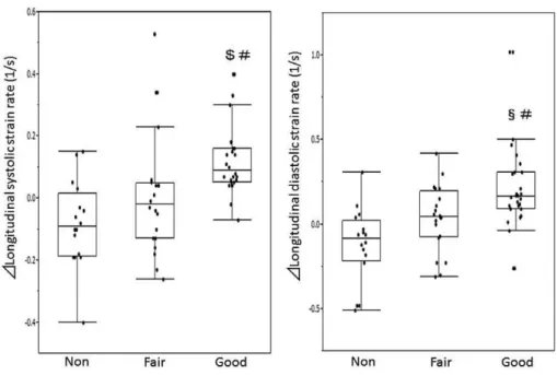
治療の効果を認めた Longitudinal 方向に対して収縮 能と拡張能を別々に Strain rate 法を用いて算出し,
Fig. 5 に示した.収縮能を表す Systolic strain rate では Good-CPAP 群(0.11
±
0.1)は Fair-CPAP 群(0±
0.19),Non-CPAP 群(‑0.08±
0.14)に比べて有 意に改善していた(p < 0.05,p < 0.0005,respec- tively). 拡 張 能 を 表 す Diastolic strain rate で は Good-CPAP 群(0.21±
0.03)は Fair-CPAP 群(0.03Fig. 2 Comparison of the change of CAVI between baseline and after therapy
There was no significant change between 3 groups.
Fig. 3 Comparison of the change of echocardiography parameter between beseline and after therapy
Left side is the change of EF. Middle is that of E/e . Right side is that of LAD. EF and
E/e is no significant difference between 3 groups. Good-CPAP has meked LAD smaller
than Non-CPAP.
$p < 0.01.
Fig. 4 Comparison of the change of 2D-STE parameter between baseline and after therapy
Left side is the change of longitudinal strain. Middle is that of radial strain. Right is that of circumferential strain.
There was the change at Non-CPAP(‑1.03 ± 1.89%), Fair-CPAP(0.37 ± 2.05%) and Good-CPAP(1.97 ± 1.63%)
on longitudinal strain. No significant change was detected on radial strain. There was the change at Non-CPAP
(‑0.68 ± 3.04%) and Good-CPAP(1.59 ± 2.73%) on circumferential strain(p < 0.05).
$p < 0.0001 vs Non-CPAP,
#
p < 0.05 vs Fair-CPAP,
§p < 0.05 vs Non-CPAP; 2D-STE, Two- dimensional speckle tracking echocardiography.
Fig. 5 Comparison of the change of longitudinal strain rate between baseline and after therapy
Left side is the change of longitudinal systolic strain rate. Right side is that of longitudinal diastolic strain rate.
There was the change at Non-CPAP(‑0.08 ± 0.14 1/s), Fair-CPAP(0 ± 0.19 1/s) and Good-CPAP(0.11 ± 0.1 1/s) on longitudinal systolic strain rate. There was the change at Non-CPAP(‑0.12 ± 0.22 1/s), Fair-CPAP(0.03
± 0.2 1/s) and Good-CPAP(0.21 ± 0.03 1/s) on longitudinal strain.
$p < 0.0005 vs Non-CPAP,
#p < 0.05 vs Fair-
CPAP,
§p < 0.0001 vs Non-CPAP.
±
0.2),Non-CPAP 群(‑0.12±
0.22)に比べて有 意に改善していた(p < 0.05,p < 0.0001,respec- tively).考 察
これまでの報告から OSAS は高血圧や糖尿病,メ タボリック症候群の発症や増悪に関連しており,心 血管イベントの危険因子と考えられている1,2).その 理由としては頻回な夜間低酸素状態や胸腔内圧の過 大な変動が交感神経活性の亢進や酸化ストレスを惹 起することで動脈硬化の進行に影響しているからと 報告されている10,12).OSAS は血管内皮機能の障害 を誘発することから高血圧を合併するとも報告され ている11).
今回の研究では Good-CPAP 群では治療前後で Non-CPAP 群に対して収縮期・拡張期血圧および心 拍数が有意に低下した.収縮期血圧においてはFair- CPAP 群に対しても有意に低下した.CPAP 治療は 時間,回数を十分に行うことで夜間低酸素状態,交 感神経活性亢進を有意に改善し血管緊張(tonus)が 低下することで血圧を低下させることができると推測 される.CAVI 値の変化は 3 群間で差は認めなかっ た.CAVI は血管内皮機能の早期障害を検出できる 鋭敏な検査法ではなく,進行した血管の硬化所見を 検出する方法であるため13),平均 2.1 年間という期 間では 3 群間に明らかな差は認めなかったと考えら れた.
CPAP 治療前後では 3 群とも LVDd,LVDs,EF の変化に差を認めなかった.本研究の対象は壁運動 異常がなく,心肥大も認めない心機能良好例が対象 であるため従来心エコー図法では微小な変化は検出 できなかったと推測される.しかし高血圧症例や肥 大心を有する症例では EF では検出できない潜在性
(非顕在性)収縮能低下を有すると報告されてい
る14‑16).正常収縮症例では血圧が低下して後負荷が
軽減すると収縮障害より拡張障害の改善が認められ ることが知られており17),本研究では左房径の有意 な縮小を認めた.これまでの報告で CPAP の 6 か 月間治療では EF や LAD に変化は見られないと報 告されているが18),本研究は 2.1 年間と比較的長期 間の評価であったために収縮能(EF)には変化を 認めなかったが,拡張能の指標である LAD は有意 に縮小したものと考えられた.
2D-STE は従来の心エコー図法では検出できない 微小な収縮能および拡張能障害を評価することが可 能である.本研究では Longitudinal strain におい て Good-CPAP 群 が Fair-CPAP 群,Non-CPAP 群 の両群に比べ有意に改善を認めた.Radial strain に は 3 群間に差は認めず,Circumferential strain は Good-CPAP 群は Non-CPAP 群に対してのみ有意差 を認めた.EF では検出できない心機能改善は Longi- tudinal 方向で最も顕著であった.これは心内膜心 筋の影響は Longitudinal 方向に最も反映されると報 告されていることに合致している19).Circumferen- tial 方向にも改善を認めているのは本検討が心筋障 害の小さい症例を対象としているからと考えられる.
そして Longitudinal 方向において,収縮能と拡張 能をより鋭敏に検出可能な strain rate 法を用いた 結果,Good-CPAP 群が Fair-CPAP 群,Non-CPAP 群の両群に有意な改善を認めた.これは十分な CPAP 治療は EF で検出できない微小な収縮および拡張機 能障害の改善を検出可能であり,非顕在性心機能障 害は長軸方向から改善すると考えられた.
結 語
閉塞性睡眠時無呼吸患者において長期的な CPAP 治療は潜在的心機能障害を改善する.その心機能評 価に2D-speckle tracking imaging法は有用であった.
利益相反
本研究に関し開示すべき利益相反はない.
文 献
1) Said S, Mukherjee D, Whayne TF. Interrela- tionships with metabolic syndrome, obesity
and cardiovascular risk. .
2016;14:415‑425.
2) Young T, Palta M, Dempsey J, . The occur- rence of sleep-disordered breathing among mid- dle-aged adults. . 1993;328:1230‑
1235.
3) Kim SH, Cho GY, Shin C, . Impact of ob- structive sleep apnea on left ventricular diastol- ic function. . 2008;101:1663‑1668.
4) Alchanatis M, Tourkohoriti G, Kosmas EN, . Evidence for left ventricular dysfunction in pa- tients with obstructive sleep apnoea syndrome.
. 2002;20:1239‑1245.
5) Weiss P, Kyger M. Positive airway pressure therapy for obstructive sleep apnea.
. 2016;49:1331‑1341.
6) Hammerstingl C, Schueler R, Wiesen M, . Impact of untreated obstructive sleep apnea on left and right ventricular myocardial function and effects of CPAP therapy. (Inter- net). 2013;8:e76352. (accessed 2013 Oct 11) https://
www.ncbi.nlm.nih.gov/pubmed/24146857 7) Craig S, Kylintireas I, Kohler M, . Effect of
CPAP on cardiac function in minimally symp- tomatic patients with OSA: results from a sub- set of the MOSAIC randomized trial.
. 2015;11:967‑973.
8) Butt M, Dwivedi G, Shantsila A, . Left ven- tricular systolic and diastolic function in ob- structive sleep apnea : impact of continuous positive airway pressure therapy.
. 2012;5:226‑233.
9) Galli E, Fournet M, Chanbanne C, . Prog- nostic value of left atrial reservoir function in patients with severe aortic stenosis: a 2D speck- le-tracking echocardiographic study.
. 2016;17:533‑541.
10) Kasai T, Bradley TD. Obstructive sleep apnea and heart failure: pathophysiologic and thera- peutic implications. . 2011;57:
119‑127.
11) Kasai T. Sleep apnea and heart failure.
. 2012;60:78‑85.
12) Davies CW, Crosby JH, Mullins RL, . Case- control study of 24 hour ambulatory blood pressure in patients with obstructive sleep ap- noea and normal matched control subjects.
. 2000;55:736‑740.
13) Namekata T, Suzuki K, Ishizuka N, . Es- tablishing baseline criteria of cardio-ankle vas- cular index as a new indicator of arteriosclero- sis : a cross-sectional study.
. 2011;11:51.
14) Kouzu H, Yuda S, Muranaka A, . Left ven- tricular hypertrophy causes different changes in longitudinal, radial, and circumferential me- chanics in patients with hypertension: a two- dimensional speckle tracking study.
. 2011;24:192‑199.
15) Smedsrud MK, Pettersen E, Gjesdal O, . Detection of left ventricular dysfunction by global longitudinal systolic strain in patients with chronic aortic regurgitation.
. 2011;24:1253‑1259.
16) Witkowski TG, Thomas JD, Debonnaire PJ, . Global longitudinal strain predicts left ven- tricular dysfunction after mitral valve repair.
. 2013;14:69‑76.
17) Parikh JD, Hollingsworth KG, Wallace D, . Normal age-related changes in left ventricular function: role of afterload and subendocardial dysfunction. . 2016;223:306‑312.
18) Craig S, Kylintireas I, Kohler M, . Effect of CPAP on cardiac function in minimally symp- tomatic patients with OSA: results from a sub- set of the MOSAIC randomized trial.
. 2015;11:967‑973.
19) Yamamoto T, Tanaka H, Kurimoto C, . Very early stage left ventricular endocardial dysfunction of patients with hypereosinophilic
syndrome. . 2016;32:
1357‑1361.
THE LONG-TERM CONTINUOUS POSITIVE AIRWAY PRESSURE IMPROVES LEFT VENTRICULAR FUNCTION ON OBSTRUCTIVE SLEEP APNEA SYNDROME
PATIENTS: 2-DIMENSIONAL SPECKLED TRACKING ECHOCARDIOGRAPHIC IMAGING IS EFFECTIVE FOR EVALUATION
Hiroto F
UKUOKA
, Hiroyuki KAYANO
, Taiju MATSUI
, Rei OOTA
, Tarou ADACHI
and Youichi KOBAYASHI
Department of Medicine, Division of Cardiology, Showa University School of Medicine
Abstract Continuous positive airway pressure (CPAP) is an established cure for OSAS, but it is controversial whether or not CPAP improves left ventricular function(LV). We examined how the left ventricular function changed on CPAP treatment by using 2D-speckle tracking imaging(2D-STE). The subjects were 62 patients (46 men, age: 61
±
13 yrs) diagnosed with moderate or severe OSAS (more than 15 times/hr of apnea hypopnea index) by polysomnography in Showa University Hospital without LV dysfunction(EF ≧ 55%). We divided the patients into 3 groups; Good-CPAP group (26 cases, CPAP> 21 days/mo; 4 hr/day), Fair-CPAP group (20 cases, using it 21 days/mo; 4 hr/day), Non-CPAP group
(16 cases; CPAP-free). We considered the change between baseline and follow-up(average of 2.1 years)
of LV systolic and diastolic function, arterial stiffness and vital signs. The systolic and diastolic blood pressure and heart rate were particularly improved in Good-CPAP. There was no significant difference in the change in CAVI level, LVDd, LVDs, EF, E/e among the three groups. Good-CPAP was significant- ly improved in Longitudinal strain in comparison with Fair-CPAP and Non-CPAP(p < 0.05, p < 0.0001, respectively). There was no significant difference among the three groups in Radial strain. Good-CPAP showed a significantly improved circumferential strain in comparison with Non-CPAP(p < 0.05). The Good-CPAP was significantly improved in longitudinal systolic strain rate in comparison with Fair-CPAP and Non-CPAP (p < 0.05, p < 0.0005, respectively). The Good-CPAP was significantly improved in com- parison with Fair-CPAP and Non-CPAP in longitudinal diastolic strain rate (p < 0.05, p < 0.0001, respec- tively). The long-term CPAP treatment improves a potential cardiac dysfunction for OSAS. The 2D- speckle tracking imaging was useful for cardiac function evaluation.
Key words: obstructive sleep apnea syndrome, continuous positive airway pressure, left ventricular
function, 2-dimensional speckle tracking imaging〔受付:1 月 5 日,受理:1 月 19 日,2017〕