Change of prefrontal cortex blood flow
during pain stimulation
Tomohiko Nishigami
1, Tatsunori Ikemoto
2, and Takahiro Ushida
31Rehabilitation Center, Kochi Medical School Hospital 2Department of Orthopedic Surgery, Kochi Medical School
3Multidisciplinary Pain Center, Aichi Medical University
[Received 2 February 2010, Accepted 9 March 2010] Abstract
The prefrontal cortex (PFC) is widely associated with cognitive and emotional functions, including attention, decision making, goal–directed behavior, and working memory. In a previ-ous study, neural activity and morphological alterations in PFC were detected in patients with chronic pain. In addition, these patients had impaired performance on emotional decision– making tests and increased incidence of depressive and anxiety disorders. However, little is known about the time course of PFC blood flow during pain stimulation. Therefore, we exam-ined the time course of PFC blood flow and the relationship between PFC activation and the subjective pain score during thermal stimuli.
Fifteen healthy right–handed subjects participated in this study. Thermal stimuli were delivered to right dorsal forearm for 30 sec by a 5 × 5-mm peltier device (Unique Medical, Japan; UDH-300). Pain intensity was assessed using a visual analog scale (VAS) after the stimuli were delivered, and PFC blood flow data were obtained using near–infrared spectroscopy. The oxyhemoglobin (oxyHb) waveform was divided into four segments: the “baseline” segment for 10 s before the task period, the “ first” segment for 10 s from the task period, the “ middle” segment for 10 s from the end of the first segment, and the “last” segment of 10 s from the end of the middle segment. OxyHb data were averaged across the four segments and analyzed using Tukey's post hoc test. We calculated Spearman's correlation coefficient between the pain VAS score and oxyHb in each segment (first, middle, and last) during thermal stimuli. In addition, high–sensitivity (5 subjects) and low–sensitivity (5 subjects) groups were divided using the VAS score, and the differences in average oxyHb in each segment (first, middle, and last) between the high– and low–sensitivity groups were assessed using the Mann–Whitney U test. Statistically significant differences were expressed as p values less than 0.05.
Furthermore, oxyHb of the last segment in the left dorsolateral PFC significantly decreased compared with those of the baseline and first segments. OxyHb of the first segment in the left side PFC decreased significantly in the high-sensitivity groups (mean VAS rating = 72.2 ± 5.6) compared with the low–sensitivity groups (mean VAS rating = 25.8 ± 8).
Decrease in PFC blood flow during pain stimulation may induce atrophy and change in neural activation in PFCs of patients with chronic pain.
はじめに
前頭前野は記憶26),意思決定5),情動4)な どの高次脳機能に関与しているが,慢性疼痛患 者においては前頭前野が萎縮することや局所脳 神経機能が低下するといった神経活動に変調が 引き起こされていることが明らかになってい る2,11,12,13)。同部位の機能低下が慢性疼痛患者 における意思決定能力の低下などを引き起こし ている可能性が指摘されており1),慢性疼痛患 者において治療がより難渋する要因にもなって いる。また,持続的な痛みが前頭前野の形態学 的変化や意思決定能力の低下を引き起こすこと はモデル動物においても確認されている20,23)。 臨床においては,慢性期のみならず,外傷後や 外科的手術後のような急性期においても,痛み とともに易怒性,意欲の低下などが生じること を経験することがあり,痛みによって前頭前野 を含めた脳神経機能に即時的な変調が生じてい る可能性が考えられる。 痛み刺激に対する前頭前野の反応について は,先行研究において,Apkarian
はメタ分析か ら,健常者に対して侵害刺激を与えると前頭前 野の活動は増加すると報告しているが3),一方 で,健常者24),慢性的な外科的手術後の歯痛患 者8),関節リウマチ患者16)において痛み刺激 によって前頭前野の活動が減少することも報告 されており,痛み刺激と前頭前野の脳血流量の 時間的関係は未だ明らかでない部分も多い。痛 み刺激に対する前頭前野の即時的な反応が明ら かではないために,慢性疼痛患者における前頭 前野の萎縮や神経活動の変化がどのような経過 で生じているか不明である。また,急性期にお ける情動の変化に前頭前野がどのように関わっ ているか不明である。これまでの痛み刺激に対 する脳活動を検討した研究では,陽電子放射断 層 撮 影 装 置 (Positron Emission Tomography:
PET
),機能的核磁器共鳴画像法(functional
magnetic resonance imaging: fMRI
)が主に用いられているが,これらの特徴として空間分解能 は高いが時間分解能は低いことが挙げられる。
一方で,近赤外分光イメージング装置(
near
infrared spectroscopy: NIRS
)は 空間分解能はfMRI
より劣り大脳皮質レベルの測定しか行え ないが,時間分解能はfMRI
より高く,脳活動 の時間経過を測定することに適していることが 報告されている14,27)。そこで,本研究では痛 み刺激に対する前頭前野の脳血流変化の時間経 過と経験した痛みの強さとの関係をNIRS
を用 いて検討した。 Key words : Pain ; Prefrontal cortex ; NIRS ; Chronic painPAIN RESEARCH 25 (2010) 127–134
痛み刺激による前頭前野の即時的な脳血流量変化
西上 智彦1/池本 竜則2/牛田 享宏3 1高知大学医学部附属病院 リハビリテーション部 2高知大学医学部 整形外科教室 3愛知医科大学医学部 学際的痛みセンター方 法
対象は利き手が右側の健常成人15 名(男性 9 名,女性6 名)で,平均年齢は 27.
3±
3.
0 歳で あった。対象者には本研究の趣意を十分に説明 し,文書にて同意を得た。なお,本研究は理学 療法科学研究倫理委員会の承認をうけて行った (承認番号H
21-
1)。痛み刺激は温・冷型痛覚計 (ユニークメディカル社製,UDH-
300)を用い て,49℃の熱刺激をペルチエ素子製のプローブ (5×
5mm
)にて右前腕に行った。測定終了後に痛みの程度を
visual analog scale
(VAS
)にて評価した。なお,
VAS
の範囲は全く痛みがない 状態を0,最悪の痛みを 100 とした。実験にはKoyama
ら18)のデザインを参考にして,測定 中に1 回の熱刺激のみを提示するsingle-epoch
design
を採用し,10 秒間のベースライン後に 30 秒間の熱刺激を行った。脳血流酸素動態はNIRS
(fNIRS
,島津製作所製,OMM-
3000)にて測定した。測定部位は前頭前野とし,脳波計測 国際10
-
20 法を参考に,最も下端のプローブをFp
1-Fp
2 に沿って固定し,計 45 チャネルで計 測した。各プローブの位置の解剖学的な位置の 推定はOkamoto
22),Suda
ら30)の方法を参考 に,前頭前野背外側部(右側:ch
28,
29,
34,
35,
41,
42。左側:ch
30,
31,
37,
38,
43,
44),前頭前 野腹外側部(右側:ch
27,
33,
40。左側:ch
32,
39,
45)の位置を推定し,解析の対象とした(Fig.1)。 測定開始前は安静とし,酸素動態が安定した後 に測定を開始した。 解析対象は測定開始からの10 秒間(ベースラ イン),刺激開始からの10 秒間(初期),刺激開 始10 秒後からの 10 秒間(中期),刺激開始 20 秒後からの10 秒間(後期)の酸素化ヘモグロビ ン(oxyHb
)のそれぞれの平均値とした。oxyHb
の変化はfMRI
におけるBOLD
信号の変化と 1 2 3 4 5 6 7 8 9 10 11 12 13 14 15 16 17 18 19 20 21 22 23 24 25 26 27 28 29 30 31 32 33 34 35 36 37 38 39 40 41 42 43 44 45 Fpz Fp2 Fp1Dorsolateral prefrontal cortex
Dorsomedial prefrontal cortex
F4 Fz F3
Light sources
Detectors
相関性が高いこと29)や,
oxyHb
は脳の神経活 動と相関性が高いこと15)が報告されているた め,解析の対象とした。 統計処理は,まず,痛み刺激に対する脳血流 量の時間経過を検討するため,多重比較検定と してTukey
法を用いて,ベースライン,初期, 中期,後期間のoxyHb
の有意差をそれぞれ求 めた。次に,痛み刺激に対する主観的痛みと脳 血流量の関係を検討するため,初期,中期,後 期のoxyHb
とVAS
の相関関係をSpearman
検定を用いてそれぞれ求めた。さらに,主観的痛 みが強い人と弱い人での脳血流量の違いを検討
Fpz
Fp2
Fp1
Fig.2 Waveforms of oxyHb in prefrontal cortex during pain stimulation. Table 1 Time courses of prefrontal blood flow during pain stimulation (mean ± SD)
Baseline First Middle Last
Dorsolateral prefrontal cortex Right ch28 -0.0020±0.0026 0.0017±0.0068 0.0018±0.0094 -0.0043±0.0094 ch29 -0.0011±0.0042 0.0009±0.0076 -0.0011±0.0107 -0.0041±0.0112 ch34 -0.0021±0.0043 0.0016±0.0077 0.0011±0.0112 -0.0052±0.0112 ch35 -0.0014±0.0047 0.0028±0.0055 0.0027±0.0073 -0.0010±0.0097 ch41 -0.0029±0.0067 -0.0011±0.0077 -0.0017±0.0151 -0.0058±0.0146 ch42 -0.0027±0.0048 -0.0007±0.0077 -0.0038±0.0103 -0.0087±0.0125 Left ch30 -0.0015±0.0051 -0.0009±0.0066 -0.0033±0.0075 -0.0054±0.0074 ch31 -0.0019±0.0085 0.0003±0.0108 -0.0017±0.0113 -0.0060±0.0140 ch37 -0.0036±0.0050 -0.0008±0.0085 -0.0035±0.0106 -0.0074±0.0105 ch38 0-0.0011±0.0095 0.0026±0.0121 0.0006±0.0166 -0.0054±0.0163 ch43 -0.0021±0.0049 -0.0008±0.0071 -0.0074±0.0098 -0.0131±0.0089 ch44 0-0.0007±0.0052 0.0047±0.0110 0.0029±0.014 -0.0036±0.0115 Dorsomedial prefrontal cortex Right ch27 -0.0011±0.0033 -0.0001±0.0088 -0.0009±0.0128 -0.0053±0.0123 ch33 -0.00003±0.0042 0.0006±0.115 -0.0007±0.0134 -0.0034±0.0122 ch40 -0.0008±0.0031 0.0005±0.0115 -0.0002±0.0116 -0.0038±0.0092 ch32 -0.0008±0.0073 0.0003±0.113 -0.0027±0.0125 -0.0086±0.0127 Left ch39 -0.00005±0.0034 0.0031±0.0116 0.0022±0.0141 -0.0021±0.0132 ch45 -0.00002±0.0066 0.0053±0.0183 0.0031±0.0177 -0.0029±0.0144
*
OxyHb of the last segment in ch43 (left dorsolateral prefrontal cortex) significantly decreased compared with the baseline and first segment. *P<0.05
するため,初期,中期,後期における右側全体 (
ch
27,
28,
29,
33,
34,
35,
40,
41,
42 のoxyHb
を 平均)と左側全体(ch
30,
31,
32,
37,
38,
39,
43,
44,
45 のoxyHb
を平均)の値をMann-Whitney's
U
検定にて,痛みが少ない下位5 名(痛みが少 ない群,VAS:
25.
8±
8.
3mm
)と痛みが強い上 位5 名(痛みが強い群,VAS:
72.
2±
5.
6mm
)で 比較した。統計解析はSPSS ver.
11.
5J
を用い, 有意水準5%
未満で判定した。結 果
VAS
は平均51.
6±
20.
6(14〜83)であった。多 重比較検定にて,左側の前頭前野背外側部 (ch
43)のoxyHb
がベースライン,初期より後 期において減少していた(Table 1,Fig.2)。ま た,初期,中期,後期のoxyHb
とVAS
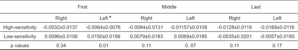
の相関 関係は認めなかった。痛みが強い群は痛みが少 ない群より初期における左側の前頭前野のoxyHb
が有意に減少していた(Table 2)。考 察
本研究では痛み刺激に対する前頭前野の脳血 流の時間経過をNIRS
を用いて検討した。結 果,左側の前頭前野背外側部のoxyHb
がベー スライン,初期より後期において減少してい た。左側の前頭前野背外側部のみに有意な減少 を認めた要因としては,C
線維のみ刺激したと きと比較してA
δ線維のみを刺激したときには, 刺激対側の前帯状回の活動は増加することが報 告されており25),本研究で用いた熱刺激は末 梢神経においてC
線維だけでなくA
δ線維も活 性化するため7,32),A
δ線維の影響によって左 側の前帯状回の神経活動が増加し,脳血流を必 要としたため,前帯状回の隣接部位である前頭 前野から脳血流が流入し,相対的に左側の前頭 前野背外側部の脳血流量が減少したのかもしれ ない。左側の前頭前野背外側部の脳血流量が減 少した他の要因として,痛み刺激が加わると扁 桃体が前頭前野の神経活動を抑制するため21), 前頭前野に血流が送られる必要がなくなった可 能性が考えられる。 また,うつ病患者においてうつ症状と左側の 前頭前野の脳血流量は負に相関していることが 認められており6),慢性疼痛患者で認められる 抑うつ状態9)は,本研究で認められた左側の前 頭前野背外側部の脳血流量の低下が長期間にわ たって続いた結果かもしれない。本研究では右 側前腕に痛み刺激を与えているが,左側や体幹 に痛み刺激を与えたときに同様に左側の前頭前 野の脳血流量が減少するかは不明であり,今後 の検証が必要である。 次に,VAS
と初期,中期,後期のoxyHb
とTable 2 Comparison of oxyHb in prefrontal cortex between high- and low-sensitivity groups
First Middle Last
Right Left Right Left Right Left High-sensitivity -0.0032±0.0137 -0.0064±0.0076 -0.0084±0.0131 -0.01157±0.0108 -0.0128±0.0119 -0.0169±0.0116 Low-sensitivity 0.0096±0.0106 0.0150±0.0156 0.0079±0.0183 0.0069±0.0185 -0.0035±0.0201 -0.0057±0.0185
p values 0.34 0.01 0.11 0. 07 0.11 0.17
*
OxyHb of the first segment in the left side prefrontal cortex decreased significantly in the high-sensitivity groups compared with the low-sensitivity groups. *P<0.05
の相関関係は認めなかった。痛み刺激として電 気刺激を加えると,左右の前頭前野背外側部と
pain catastrophizing scale
は負に相関することが報告されている28)。本研究で用いた
VAS
は痛 みの強度の指標であるが,pain catastrophizing
scale
は痛みの破局的思考の指標であり31),こ の指標の違いが結果に影響したのかもしれな い。VAS
の結果から痛みが強い群と少ない群 を分けると,初期における左側の前頭前野にお いてoxyHb
は痛みが強い群が少ない群より減 少していた。これまでに,Placebo
33)や痛みを 抑制しようと念ずると10),痛みが減少し,そ のときには前頭前野背外側部が活性化すること が明らかになっており,前頭前野が下行性疼痛 抑制系の賦活に関与することが示唆されてい る。痛みが強い群はこのような前頭前野の下行 性疼痛抑制系の機能が低い可能性があり,主観 的痛みの個人差の一要因として前頭前野の活動 の関与が示唆される。 先行研究において,侵害刺激に対して前頭前 野の活動は増加すると報告されていることが多 いが,実験デザインとして侵害刺激を10 秒間 程度繰り返すmultiple-epoch design
を用いてい ことが多い17,19)。本研究では,局所のシグナ ル変化を検出することにより感度が高いsingle-epoch design
18)を採用し,持続的な痛み刺激(30 秒間)を1 回のみ行った。ch
43 をみてみると, 痛み刺激開始直後では前頭前野の脳血流量は増 加する傾向であるが,その後,経時的に減少し ていくことが認められた。慢性疼痛症例では本 研究で用いたような持続的な痛みを感じること も多く,痛み刺激に対する左側の前頭前野背外 側部の脳血流量の減少はより臨床に即した結果 かもしれない。 本研究結果から得られた仮説として,持続的 な痛み刺激に対しては前頭前野の血流量が減少 し,一次的に前頭前野の機能が減弱し,情動な どの変化を惹起し,また,末梢からの痛み刺激 が続くと二次的な前頭前野の萎縮,神経活動の 変化などが生じて,慢性痛患者特有の社会的認 知能力の低下,意欲の低下などを引き起こして いることが考えられる。今後,これらの仮説を 検証していき,急性期における痛みが慢性痛に つながるメカニズムをさらに明らかにしていく 必要がある。結 語
痛み刺激に対する前頭前野の即時的な脳血流 変化を検討した。結果,左側の前頭前野背外側 部のoxyHb
が経時的に減少した。また,痛み が強い群は痛みが少ない群より初期における左 側の前頭前野のoxyHb
が有意に減少していた。 このような痛み刺激に対する前頭前野の脳血流 量の減少の継続が慢性疼痛患者の前頭前野の萎 縮や神経活動の変化をもたらしているかもしれ ない。 文 献1) Apkarian, A.V., Sosa, Y., et al., Chronic pain patients are impaired on an emotional decision-making task, Pain, 108 (2004) 129-136.
2) Apkarian, A.V., Sosa, Y., et al., Chronic back pain is associated with decreased prefrontal and thalamic gray matter density, J. Neurosci., 24 (2004) 10410-10415.
3) Apkarian, A.V., Bushnell, M.C., et al., Human brain mechanisms of pain perception and regulation in health and disease, Eur. J. Pain, 9 (2005) 463-484. 4) Beauregard, M., Lévesque J., et al., Neural cor
-relates of conscious self-regulation of emotion, J. Neurosci., 21 (2001) RC165.
5) Bechara, A., Damasio, H., et al., Different contri-butions of the human amygdala and ventromedial prefrontal cortex to decision-making, J. Neurosci., 19 (1999) 5473-5481.
6) Bench, C.J., Friston, K.J., et al., Regional cerebral blood flow in depression measured by positron emission tomography: the relationship with clinical dimensions, Psychol. Med., 23 (1993) 579-590. 7) Campbell, J.N., Meyer, R.A., Sensitization of un
-myelinated nociceptive afferents in monkey varies with skin type, J. Neurophysiol., 49 (1983) 98-110. 8) Derbyshire, S.W., Jones, A.K., et al., Cerebral responses to pain in patients suffering acute post-dental extraction pain measured by positron emis-sion tomography (PET), Eur. J. Pain, 3 (1999) 103-113.
9) Dersh, J., Polatin, P.B., et al., Chronic pain and psychopathology: research findings and theoretical considerations, Psychosom. Med., 64 (2002) 773-786.
10)Freund, W., Klug, R., et al., Perception and suppression of thermally induced pain: a fMRI study, Somatosens. Mot. Res., 26 (2009) 1-10. 11)Geha, P.Y., Baliki, M.N., et al., The brain in
chronic CRPS pain: abnormal gray-white matter interactions in emotional and autonomic regions, Neuron, 60 (2008) 570-581.
12)Grachev, I.D., Fredrickson, B.E., et al., Abnormal brain chemistry in chronic back pain: an in vivo proton magnetic resonance spectroscopy study, Pain, 89 (2000) 7-18.
13)Grachev, I.D., Thomas, P.S., et al., Decreased levels of N-acetylaspartate in dorsolateral prefrontal cortex in a case of intractable severe sympathetical-ly mediated chronic pain (complex regional pain syndrome, type I), Brain Cogn., 49 (2002) 102-113. 14)Hanaoka, N., Aoyama, Y., et al., Deactivation and activation of left frontal lobe during and after low-frequency repetitive transcranial magnetic stimula-tion over right prefrontal cortex: a near-infrared spectroscopy study, Neurosci. Lett., 414 (2007) 99-104.
15)Hoshi, Y., Kobayashi, N., et al., Interpretation of near-infrared spectroscopy signals: a study with a newly developed perfused rat brain model, Appl. Physiol., 90 (2001) 1657-1662.
16)Jones, A.K., Derbyshire, S.W., Reduced cortical responses to noxious heat in patients with rheuma-toid arthritis, Ann. Rheum. Dis., 56 (1997) 601-607. 17)Kong, J., White, N.S., et al., Using fMRI to disso-ciate sensory encoding from cognitive evaluation of heat pain intensity, Hum. Brain Mapp., 27 (2006) 715-721.
18)Koyama, T., McHaffie, J.G., et al., The single-epoch fMRI design: validation of a simplified paradig m for the collection of subjective ratings, Neuroimage, 21 (2004) 99-111.
19)Kurata, J., Thulborn, K.R., et al., Early decay of pain-related cerebral activation in functional magnet-ic resonance imaging: comparison with visual and motor tasks, Anesthesiology, 96 (2002) 35-44. 20)Metz, A.E., Yau, H.J., et al., Morphological and
functional reorganization of rat medial prefrontal cortex in neuropathic pain, Proc. Natl. Acad. Sci. USA, 106 (2009) 2423-2428.
21)Neugebauer, V., Galhardo, V., et al., Forebrain pain mechanisms, Brain Res. Rev., 60 (2009) 226-242. 22)Okamoto, M., Dan, H., et al., Three-dimensional
probabilistic anatomical cranio-cerebral correlation via the international 10-20 system oriented for trans cranial functional brain mapping, Neuroimage, 21 (2004) 99-111.
23)Pais-Vieira, M., Mendes-Pinto, M.M., et al., Cognitive impairment of prefrontal-dependent deci-sion-making in rats after the onset of chronic pain, Neuroscience, 161 (2009) 671-679.
24)Porro, C.A., Cettolo, V., et al., Temporal and intensity coding of pain in human cortex, J. Neurophysiol., 80 (1998) 3312-3320.
25)Qiu, Y., Noguchi, Y., et al., Brain processing of the signals ascending through unmyelinated C fibers in humans: an event-related functional magnetic resonance imaging study, Cereb. Cortex, 16 (2006) 1289-1295.
26)Rypma, B., Prabhakaran, V., et al., Load-dependent roles of frontal brain regions in the maintenance of working memory, Neuroimage, 9 (1999) 216-226. 27)Sato, T., Ito, M., et al., Time courses of brain
acti-vation and their implications for function: a multi-channel near-infrared spectroscopy study during finger tapping, Neurosci. Res., 58 (2007) 297-304. 28)Seminowicz, D.A., Davis, K.D., Cortical responses to pain in healthy individuals depends on pain cata-strophizing, Pain, 120 (2006) 297-306. .
29)Strangman, G., Culver, J.P., et al., A quantitative comparison of simultaneous BOLD fMRI and NIRS recordings during functional brain activation, Neuroimage, 17 (2002) 719-731.
30)Suda, M., Fukuda, M., et al., Subjective feeling of psychological fatigue is related to decreased re -activity in ventrolateral prefrontal cortex, Brain Res., 1252 (2009) 152-160.
31)Sullivan, M.J., Bishop, S.R., et al., The Pain Catastrophizing Scale: Development and Validation, Psychol. Asses., 7 (1995) 524-532.
32)Treede, R.D., Meyer, R.A., et al., Myelinated me -cha nically insensitive afferents from monkey hairy skin: heat-response properties, J. Neurophysiol., 80 (1998) 1082-1093.
33)Wager, T.D., Rilling, J.K., et al., Placebo-induced changes in FMRI in the anticipation and experience of pain, Science, 303 (2004) 1162-1167.
Address for correspondence: Tomohiko Nishigami
Department of Physical Therapy, Faculty of Nursing and Rehabilitation, Konan Women's University
6-2-23 Morikitamachi, Higashinada-ku, Kobe 658-0001, Japan E-mail: [email protected]