Fukushima Medical University
福島県立医科大学 学術機関リポジトリ
This document is downloaded at: 2021-11-08T00:40:28Z
Title Usefulness of new EUS criteria for diagnosis of chronic pancreatitis, especially early chronic pancreatitis( 本文 )
Author(s) 佐藤, 愛
Citation
Issue Date 2014-03-25
URL http://ir.fmu.ac.jp/dspace/handle/123456789/613
Rights This is the pre-peer reviewed version. Published version is
"Endosc Ultrasound. 2018 Mar-Apr;7(2):110-118. doi:
10.4103/2303-9027.209870", used under CC BY-NC-SA.
DOI
Text Version ETD
Usefulness of new EUS criteria for diagnosis of chronic pancreatitis, especially early chronic pancreatitis
Ai Sato
Department of Gastroenterology and Rheumatology, Fukushima Medical University School of Medicine
Abstract
Background and Aim: Endoscopic ultrasonography (EUS) is a well-established and less-invasive modality for chronic pancreatitis (CP) diagnosis. Recently, the Rosemont classification was developed for EUS diagnosis of chronic pancreatitis. Although the Rosemont classification was developed to improve the early diagnosis for CP, it is more complicated than the traditional scoring system. Aims of the study were to compare our new convenient EUS criteria and Rosemont classification in early CP having mild changes, and to evaluate whether the new one can work or not.
Methods: This study examined 177 consecutive patients who had undergone both EUS and endoscopic retrograde pancreatography (ERP) within 2 months, excluding patients with either pancreatic tumor or lower biliary ductal cancer. Clinical symptoms included patients with and without abdominal or back pain. The obtained EUS images were classified retrospectively according to the Rosemont criteria and our new modified criteria based on the traditional criteria. The latter one was added a criterion, fine-reticular pattern (F-RP) as a normal pancreatic parenchyma, to the modified traditional criteria. The ERPs were evaluated using the Cambridge classification.
Results: Results show that 132 patients had normal or equivocal findings on ERP; 113 patients (85.6%) had F-RP on EUS. In contrast, F-RP was found in only 6 (13.3%) of 45 CP cases on ERP (p<0.0001). A diagnostic accuracy of new criteria was superior to the traditional criteria related to the diagnosis of normal pancreas (84.1% vs. 62.9%). In the no-ERP grade, no significant difference was found between results obtained using the new criteria and using Rosemont classification.
Conclusion: The new criteria proposed for addition to the F-RP findings will prevent overdiagnosis without decreasing sensitivity. We believe that they will be useful for the diagnosis of CP, especially in early stages.
Introduction
Chronic pancreatitis (CP) is an irreversible and progressive inflammatory process featuring pathological modifications of fibrosis, inflammatory infiltration, and destruction of exocrine and endocrine tissue, resulting in characteristic morphological changes in the parenchyma and pancreatic ducts. These modifications vary in intensity and distribution (diffuse or patchy). This unrelenting course engenders a high rate of morbidity and mortality over a 20–25-year period. For that reason, current efforts emphasize the establishment of an early diagnosis to commence intervention that can positively affect or halt the natural progression of the diseasei.
For defining and grading the severity of CP, the Cambridge classificationii of endoscopic retrograde pancreatography (ERP) is an accepted and internationally recognized system. However, the usefulness of ERP is limited by the fact that it visualizes only the ductal systemiii,iv. Consequently, it is difficult to diagnose early stages of CP with mild changes using ERP alone because changes of CP occur from the parenchymav.
Endoscopic ultrasonography (EUS) is well suited for assessment of the pancreas because of its high resolution and the proximity of the transducer to the pancreas. In contrast to ERP, EUS can detect abnormalities that are suggestive of CP in the pancreatic parenchyma and ductal system, which are not visible on any other imaging modality. In fact, EUS can become an imaging method that is able to detect both early and late pancreatic abnormal changes with low risk of complications. To date, several investigators have reported the use of EUS for the diagnosis of CP. For the diagnosis of CP, EUS is now regarded as the most sensitive imaging modality because of its ability to place the transducer in proximity to the pancreasvi,vii,viii,ix,x. Therefore, EUS is anticipated for use as a modality for early and accurate diagnosis of CP. Now we can use two criteria for diagnosing CP: traditional criteria and Rosemont classificationxi. The traditional EUS criteria for CP are recognized as follows: hyperechoic foci, hyperechoic strands, parenchymal lobularity, irregular pancreatic duct margins, hyperechoic pancreatic duct margins, visible pancreatic side branches, pancreatic duct dilation, shadowing calcifications, and cysts. Opinions vary among researchers, but the presence of CP was diagnosed when EUS revealed at least 2–3 of the features described above. In addition, the EUS can evaluate the severity of CP with high sensitivity and specificity depending on the number of criteria present. The disease severity is classified as mild (2 or 3–4 features), moderate (5–6 features), or severe (more than 7 features) based on ERP findings as a gold standard. The Rosemont classification system was an attempt to standardize the EUS features and thresholds for the diagnosis of chronic pancreatitis and to define them more explicitly, with grouping of criteria into major and minor importance categories. Regarding the traditional EUS diagnostic criteria described above, the disease severity classification is accomplished merely from the number of CP findings. However, the value of each EUS finding differs according
to the level of CP severity. The EUS findings observed on each practical CP stage are not considered in traditional criteria, but Rosemont classification assigns each finding a grade (major or minor) related to the value of findings.
The Rosemont classification therefore presents consensus-based criteria for an EUS diagnostic system for CP that examines the existing body of evidence and the experience of experts. Although Rosemont classification was developed to decrease interobserver variation and to improve the early diagnosis for CP, it is more complicated than the traditional scoring systemxii.
Therefore, we proposed new simple EUS criteria for CP to improve the diagnostic value for early CP with mild changes. Our new criteria add the normal pancreatic parenchymal feature to the traditional criteria: as a criterion, normal parenchyma is considered to be intermingled in the early stage CP because changes of CP will occur irregularly. This study was conducted to compare our new convenient EUS criteria with a normal pancreatic parenchyma pattern and Rosemont classification in early CP having mild changes, and to evaluate whether the new one can work or not.
Patients and Methods 1. Patients
During December 2000 – December 2011, 177 consecutive patients (106 men and 71 women; mean age 61.2 years, range 20–84) who had undergone both EUS and ERP within 2 months in Fukushima Medical University Hospital for suspicious of some pancreatic, bile duct or gall bladder abnormal lesions on CT/ US or by symptoms were enrolled. Patients were excluded if they had a pancreatic tumor or lower biliary ductal cancer. Clinical symptoms included patients with and without abdominal or back pain. In all cases, the endoscopists who prospectively performed EUS and ERP were aware of the patient’s history and other findings, including CT, and these findings were retrospectively analyzed.
2. Methods
1) Endoscopic ultrasonography (EUS)
EUS was performed before or after ERCPs using a radial or curved linear arrayed scope (UM 2000, UCT240-AL5, UCP240-AL5, UCT260 or UE260 system; Olympus Medical Systems Corp., Tokyo, Japan) with 7.5–12 MHz frequencies. All patients were placed in the left lateral position. Sedation was accomplished using either intravenous diazepam (5 mg) or midazolam (5 mg). Using radial arrayed EUS, the head of the pancreas was examined via the duodenum, and body to tail was scanned via the stomach.
Using curved linear arrayed EUS, the head of the pancreas was scanned via the duodenum and stomach, the body to tail of the pancreas was observed via the stomach, according to previous report.
All EUS images obtained during the procedure were stored on a computer as
electronic images. We retrospectively confirmed and analyzed these images through discussion by 2 experienced endosonographers (A.I., A.S.). In cases with at least 1 alteration, the EUS was classified positive.
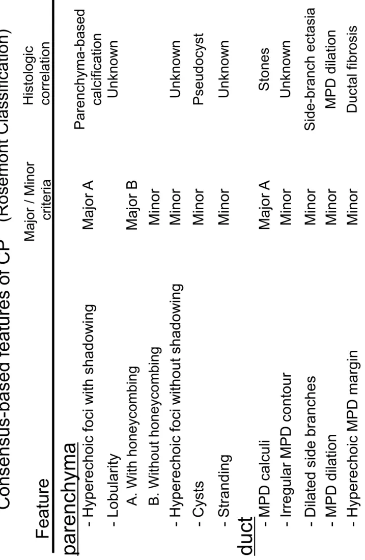
The obtained EUS images were classified according to the Rosemont criteria (Tables 1, 2) and our new modified criteria based on traditional criteria (Table 3). A criterion was added to the latter one, a fine-reticular pattern (F-RP) as a normal pancreatic parenchyma (Fig. 1), to the modified traditional criteria. In this new proposed criteria, CP consisted of the EUS images according to traditional criteria as follows (Table 4): hyperechoic foci (Fig. 2-A), hyperechoic strands (Fig. 2-B), lobularity (Fig.
2-C), cysts as parenchymal features, and hyperechoic ductal margin, dilated main pancreatic duct, duct irregularity, dilated side-branches, stones in the duct as ductal features. In this new proposed criteria, we defined lobularity as findings with honeycombing. The lobularity without honeycombing excluded the findings of lobularity. Each item was counted as 1 point. If the F-RP was identified in the pancreatic parenchyma, then 1 point was deducted from the amount of the point. We defined the diagnostic criteria as follows (Table 5): suggestive/consistent CP in more than 4 points, indeterminate CP in 2–3 points, unlikely in 1 point, and normal in -1 or 0 points.
2) Endoscopic retrograde pancreatography (ERP)
ERP was performed using oblique duodenoscopes (JF240, TJF240, TJF260V:
Olympus Medical Systems Corp., Tokyo, Japan) and a triple lumen ERP catheter (Tandem XL; Boston Scientific Japan, Tokyo, Japan). After cannulation into the pancreatic duct, contrast medium (60% meglumine sodium amidotrizoate) was injected slowly until side branches were contrasted. All patients were placed in the abdominal position, with sedation accomplished using either intravenous diazepam (5 mg) or midazolam (5 mg). Pentazocine (15 mg) was administered as needed.
The results for each patient were analyzed and evaluated in agreement by 2 investigators (A.I. and A.S.). Cambridge classification was used to assess the severity of CP as follows: normal, no abnormal features; equivocal, fewer than three abnormal side branches; mild, more than three abnormal side branches with normal main duct;
moderate, abnormal main duct and branches; severe, same as moderate disease except with one or more additional abnormalities (large cavity, ductal stone/filling defect, duct obstruction/stricture, gross irregularity).
3) Approach to investigation
Firstly, we evaluated F-RP whether it could be adapted as the normal pattern or not.
Frequency of F-RP appearance was analyzed by each ERP grade. Then, it was investigated based on not only ERP, but also EUS. Since EUS is well-known to the most sensitive modality for assessment of the pancreas, EUS was added to this evaluation.
F-RP would be observed more frequently in cases without EUS abnormal findings than
in cases with those ones. Frequency of hyperechoic foci and/or hyperechoic strands was also examined as abnormal findings because they are findings of pancreatic parenchyma, which were not observed by other modalities.
We subsequently compared the accuracy of new EUS criteria to traditional criteria.
Since EUS findings are sometimes controversial because of its possibility of overdiagnosis especially in early stages, normal ERP was needed to take as a normal pancreas. True boundary between normal and early chronic pancreatitis would exist between ERP normal and EUS normal or EUS subtle abnormal. Finally, we made a comparison of the accuracy between new EUS criteria and Rosemont classification.
4) Statistical analysis
Statistical analysis was performed with the Fisher’s exact test. A p value of less than 0.05 was regarded as significant.
Results
1. Meanings of fine-reticular pattern on EUS
Patients were classified by ERP as normal (normal or equivocal) or having CP (mild, moderate, severe) based on Cambridge classification. Normal or equivocal findings on ERP were obtained for 132 patients; 113 patients (85.6%) had F-RP on EUS.
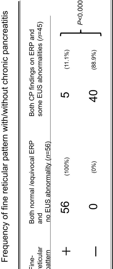
In contrast, F-RP was found in only 6 cases (13.3%) of 45 CP cases on ERP (p<0.0001) (Table 6). Additionally, we evaluated whether F-RP, as a normal finding on EUS, was observed or not in patient with or without CP (Table 7). In this assessment, the apparent CP was defined as having both ERP findings more than mild on Cambridge classification and parenchymal hyperechoic abnormalities on EUS (n=56). Normal was defined as both normal/equivocal ERP findings and no EUS abnormalities (n=45). F-RP was identified in all patients without CP. In contrast, only five patients (11.1%) among CP patients were identified among findings of F-RP. Significant difference was found between them (p<0.0001). These findings suggest that F-RP on EUS was a specific finding in normal pancreatic parenchyma.
2. Meanings of parenchymal hyperechoic abnormalities
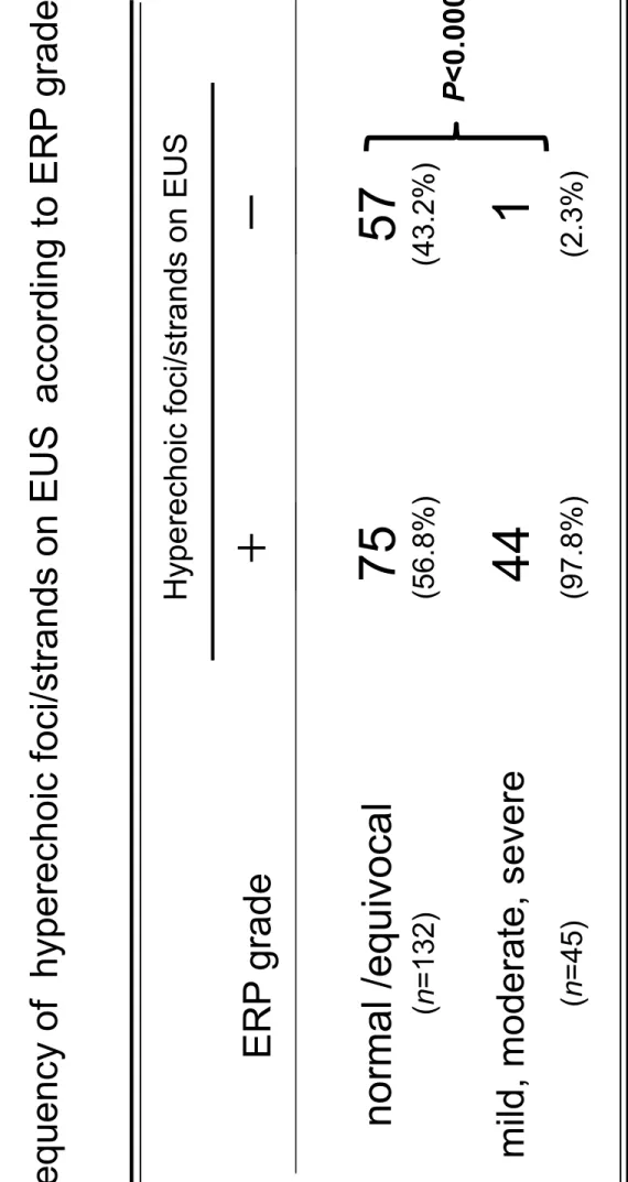
Next, we investigated the frequency of having hyperechoic abnormalities (hyperechoic foci and/or Stranding, excluding lobularity) on EUS (Table 8). The reason why lobularity was excluded from this inspection was that lobularity was the major finding among Rosemont criteria (hyperechoic foci and strands were categorized in minor items on Rosemont criteria). Hyperechoic foci and strands were observed in 75 of 132 cases (56.8%) in patients with normal/equivocal pancreatic duct on ERP. However, they were observed in 44 (97.8%) among the patients (n=45) having CP on ERP.
Significant difference was found between them. These results indicated that the findings of hyperechoic foci/strands are equivocal as the item for diagnosis of CP, although these
EUS findings are identified frequently in patients with obvious CP.
3. Diagnostic ability for normal pancreas in new proposed criteria
Based on these results, F-RP is done to reduce the overdiagnosis of CP, especially early/mild CP. We added the item of F-RP to the traditional criteria and made a proposition of the new EUS criteria for the diagnosis of CP. Subsequently, we investigated whether our new criteria have a high diagnostic capability of normal pancreas in comparison with traditional criteria. In cases where fewer than two points were defined as normal, a diagnostic accuracy of new criteria was superior to the traditional criteria related to the diagnosis of normal pancreas (84.1% vs. 62.9%) (Table 9). Results indicated that the addition of F-RP as a normal pattern was useful to avoid overdiagnosis.
4. Diagnostic capability for CP in the new proposed criteria in comparison with Rosemont classification
Finally, we compared our new criteria with those of Rosemont classification (Table 10). In no ERP grade was significant difference found between the new criteria and Rosemont classification. Consequently, it is considered that our proposed new criteria will in no way be inferior to Rosemont classification.
Discussion
CP has been defined as an inflammatory disease of the pancreas characterized by irreversible morphological (parenchymal and ductal) changes causing loss of both endocrine and exocrine functions. However, some cases of CP in early stages might be reversible or avoid worsening by treatment. It is crucial to diagnose chronic pancreatitis in an early stage to prevent many complications including main pancreatic duct stenosis, diabetes mellitus and pancreatic cancer. The diagnosis of advanced CP is not difficult using various imaging modalities such as CT, MRI, and ERP. However, the definition of early CP is wrapped in obscurity because of its slight changes and the lack of a gold standard. Consequently, its diagnosis remains a challengexiii. Agreement among various diagnostic methods is poor.
In fact, EUS is a well-established modality for the diagnosis of pancreatic disease.
The higher imaging resolution provided by EUS allows detection of subtle pancreatic abnormalities such as hyperechoic foci and strands items of EUS criteria for CP, which are undetectable using other modalities. Previous reports suggest that EUS can be a sensitive modality for diagnosing CP when compared to ERCP findingsiv,viii,ix,xiv. In this study, 19 cases with normal/equivocal ERP and without F-RP actually suggested that there were some abnormalities, which could not be detected by ERP but could only be EUS (table 6). Accordingly, it is anticipated for use with EUS for the diagnosis of early CP. However, aging is regarded as a factor of parenchymal/ductal changes on EUS
similar to CP, in fact, many patients have pancreatic parenchymal and ductal changes that are identifiable using EUS in individuals with no history or symptom of pancreaticobiliary diseasesxv. In addition, Rajan et al.xvi reported that isolated EUS abnormalities occur frequently in adults older than 60 years old without pancreatic disease. Moreover, the presence of more than three EUS abnormalities, ductal or intra-parenchymal stones, ductal narrowing or ductal dilation is more likely to represent disease than age-related changes. In this study, pancreatic parenchymal changes, hyperechoic foci and strands, were frequently identified in normal pancreas on ERP.
Referring to the Rosemont classification, these findings were categorized in minor items.
Consequently, as described above, mild parenchymal EUS features can have poor specificity, resulting in the ‘overdiagnosis’ of CP.
Because EUS criteria for CP were first described by Jones et al. and further refined by Wiersema et al.iv,xvii several criteria were proposed. Traditional EUS criteria assessed the total number of criteria and required the threshold number of criteria to diagnose CP. These systems are extremely convenient, but it is considered that each criterion such as ‘”pancreatic stone” and less specific findings such as “hyperechoic foci/strands”, are evaluated as the same value. In other words, the EUS findings observed on each practical CP stage are not considered in traditional criteria. However, in the Rosemont classification, which is an attempt to standardize the EUS features and thresholds for the diagnosis of CP and define them more explicitly, each finding is assigned a grade (major or minor) in consideration of the value of findings. Although it is apparently more reasonable, it is much more complicated.
Because the minimal changes that are not visible on any other imaging modality are difficult to interpret, the potential exists for ‘overdiagnosis’ of CPxviii. Even though EUS can detect the abnormal features on an early CP precisely, the distinction between
‘normal’ and ‘early CP’ continues to pose a challenge. Therefore, we inferred that including the normal EUS findings in the EUS criteria for CP might remove uncertainty of diagnosis of early CP. Although F-RP is a well known finding as normal pancreatic parenchyma, no apparent evidence exists in relation to F-RP. In this study, F-RP was visible in many cases of normal ERP and was significantly less in cases of mild to severe ERP, which indicated that F-RP can be regarded as a normal EUS feature.
Based on this result, we added F-RP as a criterion, i.e., - 1 point was provided when there was an area with F-RP. Consequently, cases with fewer than 2 points were interpreted as normal pancreas, and 2 or more points were regarded as CP including early stages. Using our new criteria, the diagnostic accuracy of normal pancreas was improved in comparison with results obtained using traditional criteria. The new criteria will be useful for avoiding overdiagnosis without decreasing the sensitivity. In addition, the whole diagnostic value for CP in our new simple criteria was quite similar to that obtained using the more-complicated Rosemont classification.
In the course of CP, some patients develop structural changes before functional
abnormalities, or vice versaxix. The borderline between the normal pancreas and the early/mild changes of CP is ambiguous in the first place, which might imply the importance of detecting not only abnormal features but also normal patterns of the pancreas to distinguish “early CP” from a normal pancreas. Simple and easy criteria added to the normal EUS features of the pancreatic parenchyma can be helpful to diagnose early CP.
There is one more issue, however, the correlation of EUS findings of mild/early pancreatitis with histologic diagnosis remains an unanswered questionxx. Pancreatic biopsy on a routine basis is thought to be risky and impractical; therefore, it cannot be justified, especially in asymptomatic patients. Moreover, CP is often focal in
distribution, and a random biopsy specimen may be falsely negative. In addition, a feature-to-feature correlation of EUS and histopathology is not known. There are several studies which analyzed these issue. Although Dewitt et al.xxi performed EUS-guided Truecut biopsy (EUS-TCB) of the pancreatic body in 16 patients with non-focal CP, agreement between EUS and ERCP with EUS-TCB were poor and fair, respectively. They concluded that EUS-TCB is not currently recommended for
evaluation of these patients because of potential complications and limited diagnostic yield. Varadarajulu et al.xxii evaluated the correlation EUS criteria for noncalcific CP with histology from surgical specimens. In the study, there was a significant correlation between the number of EUS criteria and severity of noncalcific CP on histology in 42 patients. The most of patients underwent resection for pancreatic cancer, making it difficult to extrapolate the results to the typical patients with abnormal painxxiii and how this fact affects the ‘‘findings’’ at EUS xx. This limitation also referred to this study.
In conclusion, EUS provides a safe and non-invasive method of obtaining detailed structural information related to the pancreatic parenchyma and ducts. The new criteria added the F-RP finding will avoid overdiagnosis without decreasing diagnostic
sensitivity. We believe that it will be useful for the diagnosis of CP, especially in early stages.
Figure legends Figure 1
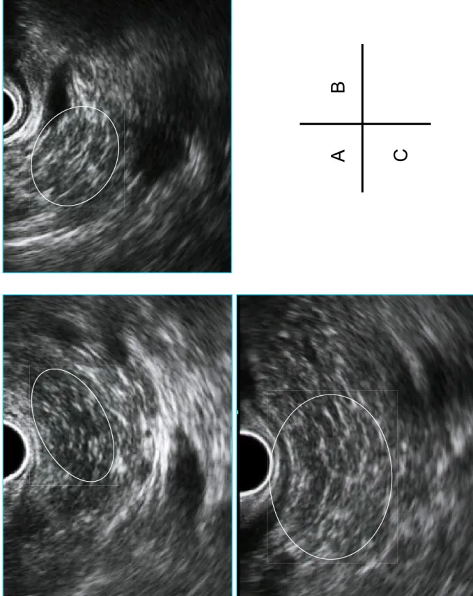
Normal pancreatic parenchyma on EUS. A homogeneous and fine-reticular pattern (F-RP) is visible in both images in parenchyma without dilated ducts (circle).
Figure 2
A: Hyperechoic foci in the pancreatic parenchyma. Many hyperechoic small dots are identified (circle)
B: Hyperechoic strands. Many linear hyperechoes are visible (circle).
C: Lobularity. Pancreatic parenchyma is lobulated by linear hyperechoes (circle).
References
i Raimondo M. What is the role of EUS in screening for chronic pancreatitis? Nat Clin Pract Gastroenterol Hepatol. 2007; 4: 530–531.
ii Axon AT, Classen M, Cotton PB, Cremer M, Freeny PC, Lees WR. Pancreatography in chronic pancreatitis: international definitions. Gut. 1984; 25: 1107–1112.
iii Catalano MF, Geenen JE. Diagnosis of chronic pancreatitis by endoscopic ultrasonography.
Endoscopy. 1998; 30 Suppl 1: A111–A115.
iv Wiersema MJ, Hawes RH, Lehman GA, et al. Prospective evaluation of endoscopic ultrasonography and endoscopic retrograde cholangiopancreatography in patients with chronic abdominal pain of suspected pancreatic origin. Endoscopy. 1993; 25: 555–564.
v Sahai AV, Mishra G, Penman ID, Williams D, Wallence MB, Hadzijahic N, Peason A, Vanvelse A, Hoffman BJ, Hawes RH. EUS detect evidence of pancreatic disease in patients with persistent or nonspecific dyspepsia. Gastrointest Endosc. 2000; 52: 153–9.
vi Dancygier H. Endoscopic ultrasonography in chronic pancreatitis. Gastrointest Endosc Clin N Am. 1995; 5: 795–804.
vii Bhutani MS. Endoscopic ultrasound in pancreatic diseases. Indications, limitations, and the future. Gastroenterol Clin North Am. 1999; 28: 747–770.
viii Sahai AV, Zimmerman M, Aabakken L, et al. Prospective assessment of the ability of endoscopic ultrasound to diagnose, exclude, or establish the severity of chronic pancreatitis found by endoscopic retrograde cholangiopancreatography. Gastrointest Endosc. 1998; 48:
18–25.
ix Catalano MF, Lahoti S, Geenen JE, et al. Prospective evaluation of endoscopic ultrasonography, endoscopic retrograde pancreatography, and secretin test in the diagnosis of chronic pancreatitis. Gastrointest Endosc. 1998; 48: 11–17.
x Irisawa A, Katakura K, Ohira H, Sato A, Bhutani MS, Hernandez LV, Koizumi M.
Usefulness of endoscopic ultrasound to diagnose the severity of chronic pancreatitis. Journal of Gastroenterol. 2007; 42: 90–94.
xi Catalano MF, Sahai A, Levy M, Romagnuolo J, Wiersema M, Brugge W, Freeman M, Yamao K, Canto M, Hernandez LV. EUS-based criteria for the diagnosis of chronic pancreatitis: The Rosemont classification. Gastrointest Endosc. 2009; 69: 1251–1261.
xii Kalmin B, Hoffman B, Hawes R, Romagnuolo J. Conventional versus Rosemont endoscopic ultrasound criteria for chronic pancreatitis: Comparing interobserver reliability and intertest agreement. Can J Gastroenterol. 2011; 25: 261-4
xiii Kalhl S, Glasbrenner B, Leodolter A, Pross M, Schulz HU, Malfertheiner P. EUS in the diagnosis of early chronic pancreatitis: a prospective follow-up study. Gastrointest Endosc.
2002; 55: 507–11.
xiv Bhutani MS. Endoscopic ultrasonography: changes of chronic pancreatitis in asymptomatic and symptomatic alcoholic patients. J Ultrasound Med. 1999; 18: 455–462.
xv Natterman C, Goldschmidt AJW, Dancygier H. Endosonography in chronic pancreatitis: A
comparison between endoscopic retrograde pancreatography and endoscopic ultrasonography.
Endoscopy 1993; 25: 565.
xvi Rajan E, Clain JE, Levy MJ, Norton ID, Wang KK, Wiersema MJ, Vazquez-Sequeiros E, Nelson BJ, Jondal ML, Kendall RK, Harmsen WS, Zinsmeister AR. Age-related changes in the pancreas identified by EUS: a prospective evaluation. Gastrointest Endosc. 2005; 61:
401–406.
xvii Jones SN, Lees WR, Frost RA. Diagnosis and grading of chronic pancreatitis by morphological criteria derived by ultrasound and pancreatography. Clinical Radiology. 1988;
134: 453–459.
xviii Raimondo M, Wallence MB. Diagonosis of early chronic pancreatitis by Endoscopic
Ultrasound. Are we there yet? JOP 2004; 5: 1–7.
xix Albasir S, Bronner MP, Parsi MA., Walsh RM, Stevens T. Endoscopic Ultrasound, Secretin Endoscopic Pancreatic Function Test, and Histology: Correlation in Chronic Pancreatitis. Am J Gastroenterol. 2010; 105: 2498–2503.
xx Kaul V, Catalano MF. EUS and chronic pancreatitis: seeing is believing? Gastrointest Endosc. 2007; 63: 510–512.
xxi DeWitt J, McGreevy L, LeBlanc J, McHenry L, Cummins O, Sherman S. EUS-guided Truecut biopsy of suspected non focal chronic pancreatitis. Gastrointest Endosc. 2005; 62:
76–84.
xxii Varadarajulu A, Eltoum I, Tamhane A, Eloubeidi MA. Histopathologic correlates of noncalcific chronic pancreatitis by EUS: a prospective tissue characterization study.
Gastrointest Endosc. 2007; 66: 501–509.
xxiii Stevens T, Parsi MA. Endoscopic ltrasound fot the diagnosis of chronic pancreatitis. World
J Gastrpenterol. 2010; 16: 2841-2850.