− 28 −
加齢に伴う皮膚脂質の変化に対するリピドーム解析
Human skin is exposed to some environmental stress. Because phospholipid (PL) and cholesterol esters (CE) are major component of cellular membrane, it is expected that the lipid change is a cause of the morphological change of the human skin. To analyze the morphological change and the lipid change of the skin according to aging would be important.
In this study, to investigate the correlation of histology change and lipid change of skin, we analyzed lipid class, fatty acid composition using HaCaT cells.
The major component of lipid extracted from HaCaT cells was PL. The relative amount of CE was also high. The total relative amount of saturated fatty acid and unsaturated fatty acid was the almost same, but it of poly unsaturated fatty acid was low. Elaidic acid and/or oleic acid (18:1 ; carbon numbers : degree of unsaturation) would be the key component of skin lipid. The major molecular species of phosphatidyl choline (PC) were 32:1PC, 34:1PC, 34:2PC and 36:2PC. These molecules contained more than one unsaturated fatty acid acyl chain. We will examine the structural analysis for triacylglycerol (TG) molecular species and PC molecular species and these oxidation products of skin tissue.
Lipidomic analysis of skin lipid changing by aging
Mayuko Morita
Molecular Gastroenterology and Hepatology, Kyoto Prefectural University of Medicine
1 緒 言
皮膚は異物との接触や、紫外線、乾燥など、常にストレ スを受けている組織である。皮膚脂質に関して脂質過酸化 物による皮膚炎の発症、コレステロール酸化物の発がん性 などが報告されている1)。脂質過酸化物は加齢による量的 変動を示し2)、リン脂質やコレステロールは生体膜の構成 成分であることから、脂質変化が皮膚の形態変化の一因で あることが予想される。皮膚では加齢に伴って表皮、真皮、
脂肪層が薄くなり、弾力を損失するなど形態が変化する。
皮膚脂質を網羅的に解析し、脂質変動の全体像のプロファ イリングから加齢に伴う皮膚の形態変化および脂質変動を 解析することが重要であると考えられる。
本研究では質量分析法を用いて皮膚のリピドーム解析を 行い、皮膚が受けるストレスに起因する皮膚の組織学的な 変化と脂質変動の相関関係を明らかにすることを目的とし、
皮膚細胞を用いてリピドーム解析へ向けた予備検討を行っ た。
2 実 験 2-1 脂質抽出
クロロホルム‐メタノール混合溶液を用い、細胞から脂 質を抽出した。
2-2 脂質クラス分析
脂質クラス分析はTLC-FID法に従った3, 4)。活性化させ たクロマロッド(Chromarods SⅢ,三菱化学メディエン ス)に脂質サンプルを1mLスポットし、溶媒中で展開した。
脂質種の検出はイアトロスキャン(Iatroscan MK- 6 三菱 化学メディエンス)を用いた。各脂質クラス同定は、標準 物質との保持時間の比較により行った。
【TLC条件】
・展開溶媒
ヘキサン:ジエチルエーテル:酢酸=80:20:1(v/v/v)
【検出器条件】
・水素流量 160mL/min、空気流量1500mL/min
・スキャンスピード 30sec/scan
2-3 脂肪酸組成分析
脂肪酸組成分析はGC-MSを用いて行った。抽出した脂 質を三フッ化ホウ素・メタノール法5)に従ってメチルエス テル化し、ガスクロマトグラフィー・質量分析計(GC- 17A-HP5000(Shimadzu))に供した。脂肪酸メチルエス テルの同定は標準試薬(Supelco 37 Component FAME Mix, SUPELCO)の保持時間との比較およびライブラリサ ーチによって行った。
【GC-MS条件】
・カラムDB-23(Agilent Technologies)
・カラム温度 170℃ 1分間→4℃ /minで230℃まで昇温 →230℃で5分間保持
・キャリアガス ヘリウム(流速 1mL/min)
・スプリット比 40:1
・気化室温度 270℃
・インターフェイス部温度 290℃
京都府立医科大学 消化器内科
守 田 麻 由 子
− 29 −
加齢に伴う皮膚脂質の変化に対するリピドーム解析
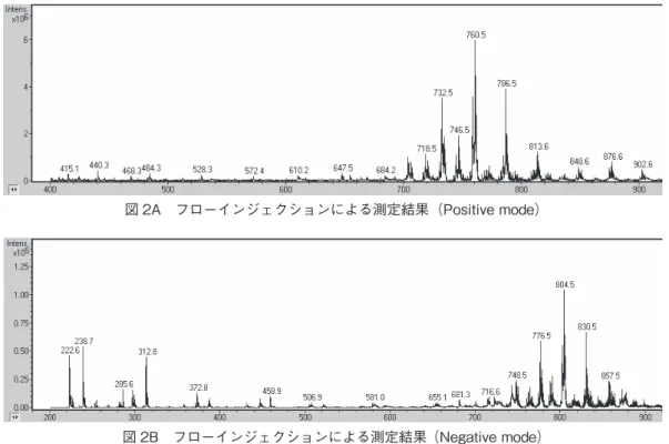
脂質の割合が高かったが、リン脂質の中でも細胞膜の構成 成分であるPCが主要成分であった。Positive modeで検出 された主なPC分子種、TG分子種を図2A、Table 3 に示 した。飽和脂肪酸だけで構成されたPC、TG分子種の Intensityは弱く、多くが分子内に1個以上の不飽和脂肪酸 を含むPC、TG分子種であった。Negative modeで検出さ れた主なPC分子種をTable 4 に示した。遊離脂肪酸(〜
m/z400)はほとんど検出されず、検出された主な脂質は PCであった(図2B、Table 4)。
4 考 察
HaCaT細胞を用いて皮膚脂質の解析を行った。細胞抽 出脂質の主な脂質成分はリン脂質であり、質量分析計によ る分析により、リン脂質の中でも特にPCを多く含んでい 2-4 質量分析
質量分析法を用いて、脂質カラムの代わりにユニオンを 接続した、フローインジェクションを行った。
【測定条件】
・イオン化法 エレクトロスプレーイオン化法
・質量分析計 esquire HCT plus(BRUKER DALTONICS)
・溶媒 メタノール:水=90:10(pH=7.0,1% ギ酸)
・流速 20mL/min
・測定モード Positive mode、Negative mode 3 結 果
3-1 脂質クラス分析
HaCaT細胞から抽出した脂質の脂質クラスをTable1に 示した。HaCaT細胞抽出脂質ではリン脂質の割合が高く、
次いでステロール類、TGの割合が高かった。
3-2 脂肪酸組成分析
HaCaT細胞抽出脂質の脂肪酸組成をTable2に示した。
主な脂肪酸は16 : 0(ステアリン酸)、18 : 0(パルミチン酸)、
18 : 1(エライジン酸、オレイン酸)であった。エライジン 酸の割合はオレイン酸と同等であった。HaCaT細胞抽出 脂質は、飽和脂肪酸と一価不飽和脂肪酸の割合がほぼ同じ であり、高度不飽和脂肪酸の割合は低い(図1)。
3-3 脂質の網羅的解析
HaCaT細胞抽出脂質では、Table 1に示したようにリン
Table 1 HaCaT 細胞抽出脂質の脂質クラス ① ② ③ Ave.
CE 11.20 12.15 16.19 13.17±2.65 TG 8.52 5.73 10.34 8.20±2.32 FFA 6.40 3.83 6.02 5.42±1.39 Ch1+DG 6.21 5.85 7.49 6.52±0.86 PL 67.67 72.44 57.94 66.1±7.38
Table 2 HaCaT 細胞抽出脂質の脂肪酸組成 ① ② ③ Ave.
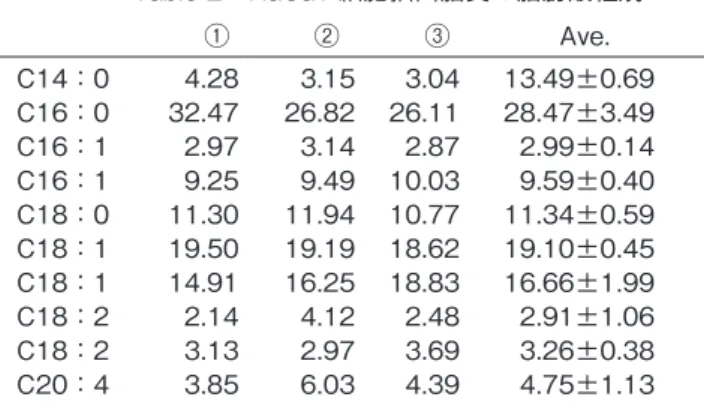
C14:0 4.28 3.15 3.04 13.49±0.69 C16:0 32.47 26.82 26.11 28.47±3.49 C16:1 2.97 3.14 2.87 2.99±0.14 C16:1 9.25 9.49 10.03 9.59±0.40 C18:0 11.30 11.94 10.77 11.34±0.59 C18:1 19.50 19.19 18.62 19.10±0.45 C18:1 14.91 16.25 18.83 16.66±1.99 C18:2 2.14 4.12 2.48 2.91±1.06 C18:2 3.13 2.97 3.69 3.26±0.38 C20:4 3.85 6.03 4.39 4.75±1.13
Table 3 HaCAT 細胞抽出脂質の主な PC、TG 分子種(Positive mode)
PC[M+H]+ m/z Intensity 30:1PC 706.5 951998 32:1PC 732.5 4201270 32:2PC 730.5 939616 34:0PC 762.5 1093622 34:1PC 760.5 7121840 34:2PC 758.5 4265662 34:3PC 756.5 517890 36:1PC 788.5 1346654 36:2PC 786.5 4647872 36:4PC 782.5 529874 38:2PC 814.5 967667 38:3PC 812.5 545545 38:4PC 810.5 541770
TG[M+NH4] m/z Intensity 46:0TG 796.5 313125 48:0TG 824.6 316770 48:1TG 822.6 385241 48:2TG 820.5 327803 50:1TG 850.6 524523 50:2TG 848.6 665469 52:1TG 878.6 283877 52:2TG 876.6 939291 52:3TG 874.6 656301 54:2TG 904.6 287452
Table 4 HaCAT 細胞抽出脂質の主な PC 分子種(Negative mode)
PC[M+HOO]+ m/z Intensity 30:1PC 750.5 18760 32:0PC 778.5 33925 32:1PC 776.5 58922 32:2PC 774.5 13071 34:0PC 806.4 15502 34:1PC 804.5 103839 34:2PC 802.5 54970 36:1PC 832.4 17334 36:2PC 830.5 66767 38:2PC 858.5 19594 図 1 HaCaT 抽出脂質の脂肪酸組成
43.29%
48.35%
飽和脂肪酸
一価不飽和脂肪酸 多価不飽和脂肪酸
10.93%
− 30 −
コスメトロジー研究報告 Vol.20, 2012ることが示された。脂肪酸組成の特徴として、飽和脂肪酸 と一価不飽和脂肪酸の割合がほぼ同じであること、エライ ジン酸の割合が高いことが挙げられる。フローインジェク ションによる網羅的測定において、ポジティブモード、ネ ガティブモードともにPCが顕著に検出され、それらは分 子内に1個以上の不飽和脂肪酸をアシル基にもつPC分子 種であった。
細胞膜の主要構成成分であるPCの組成変化は、加齢に 伴う皮膚組織の形態変化の指標になると考えられる。特に 一価不飽和脂肪酸を含むPC分子種やTG分子種、これら に由来する化合物や酸化生成物が加齢に伴う皮膚脂質の指 標になると考えられる。
(参考文献)
1) Black HS, DR Douglas: Formation of carcinogen of natural orgin in the etiology of ultraviolet light-induced carcinogenesis, Cancer Res, 33, 2094-2096, 1973.
2) 金子孝夫,田原正一,松尾光秀: 老化に伴う皮膚脂質の
過酸化物量の加齢変化
3) N.C.Shantha: Thin-layer chromatography- flame ionization detection Iatroscan system. J.
Chromatography, 624, 21-35, 1992.
4) Laurent Striby, Raymond Lafont, Madeleine Goutx: Improvement in the Iatroscan thin-layer chromatographic-flame ionization detection analysis of marine lipids. Separation and quantitation of monoacylglycerols and diacylglycerols in standards and natural samples. J. Chromatogr. A, 849, 371-380, 1999.
5) Shirai N, Suzuki H, Tokairin S, Ehara H, Wada S:
Dietary and seasonal effects on the dorsal meat lipid composition of Japanese (Silurus asotus) and Thai catfish (Clarias macrocephalus and hybrid Clarias macrocephalus and Clarias galipinus). Comp Biochem Physiol A Mol Integr Physiol, 32, 609-619, 2002.
図 2B フローインジェクションによる測定結果(Negative mode)
図 2A フローインジェクションによる測定結果(Positive mode)