哺 乳卵 研誌
(J.Marnm.Ova Res.) Vol.7 No.2 61-651990
Age-Related Changes
in the Amount of Lipids in Mouse Oocytes
Akira NARITA*, Sueo NIIMURA and Kazuo ISHIDA
* Graduate School of Science and Technology,
Niigata University, Niigata-shi 950-21,
and Faculty of Agriculture, Niigata University,
Niigata-shi 950-21
Abstract: Using mice of the ICR strain, sudanophilic lipids,
lipoids and neutral fats were histochemically detected in the
oocytes immediately after ovulation, and the amounts were compared
among 30-day-old, 60- to 90-day-old and 180- to 210-day-old ones.
Sudan IV -positive lipids and Ciaccio-positive lipoids were
demon-strated in all the oocytes as droplets of different sizes, showing
no significant difference among different age groups. Concerning
nile blue sulfate-positive neutral fats observed as droplets of
varied sizes, none was detected in the oocytes of 30-day-old mice,
but came to appear in 1.0 % of the oocytes from 60- to 90-day-old
mice, and extended to 6.3 % of those from 180- to 210-day-old
mice, even showing a significant difference between 180- to 210-
day-old mice and 30-day-old or 60- to 90-day-old mice. These
results show that neutral fats in mouse oocytes first come to
exist when the animals become mature, and the percentages of
neutral fat-containing oocytes tend to be higher with the advance
of animals' ages; while the amounts of sudanophilic lipids and
lipoids do not change with aging.
Introduction
It is a well-known fact that mammalian oocytes and early
embryos usually have either one, two or all of such inclusions;
proteinous structures1,2), glycogen granules3-8) and lipid
droplets9-12). As for mouse oocytes and early embryos, it is
ascertained that they possess all the inclusions described above.
It is also said, in general, that these inclusions gradually get
accumulated while the eggs are growing as oocytes, and then are
utilized as energy sources in the course of embryonic development
1,4,6,7,10) .
As for age-related changes of such inclusions in mammalian
oocytes and early embryos, Narita et al. 13,14) observed proteinous
structures and glycogen granules in mouse oocytes, and Parkening
and Soderwall8) examined glycogen granules in hamster blastocysts.
Generalizing the above mentioned investigations, an increase of
proteinous structures and a decrease of glycogen granules are
observed in mouse ooeytes13-14), while no fluctuation was seen in
the amount of glycogen granules in hamster blastocysts8). But,
no one has studied age-related changes in the amount of lipids
哺乳卵研誌
(J.Mamm.
Ova Res.)
第7巻 第2号 1990年10月This histochemical investigation dealt with age-related
changes in the amounts of sudanophilic lipids (lipids in general),
lipoids and neutral fats in the mouse oocytes immediately after
ovulation.
Materials and Methods
Seventy two female mice of the ICR strain were used in this study out of three age groups of 30-day-old, 60- to 90-day-old and 180- to 210-day-old animals. They were fed in cages in a room at 24•Ž and lit 14 hours a day, 4 a.m. through 6 p.m. They were superovulated with 5 i.u. PMSG (Serotropin, Teikoku Hormone Manufacturing Co. Ltd., Tokyo, Japan), and 48 hours later with 5 i.u. hCG (Gonatropin, Teikoku Hormone Manufacturing Co. Ltd.). In order to detect general lipids, as well as neutral fats, the oocytes were collected at 13 hours after hCG injection by tearing oviducts in a 0.1 M phosphate buffer solution (pH 7.4), and then fixed in a 10 % formalin phosphate buffer solution (pH 7.4); while to detect lipoids, the oviducts were taken at 13 hours after hCG injection, and then were fixed in a solution composed of 8 ml 5 % potassium bichromate solution, 2 ml formalin and 5 ml acetic acid. Naked oocytes, suspected to be abnormal, were excluded from the object of study.
For the demonstration of general lipids, the oocytes were stained with sudan IV15); for neutral fats, with nile blue sulfate 15); and for lipoids
, by the Ciaccio method15), in which oocyte-containing oviducts were embedded in paraffin, sectioned at the thickness of 6 ƒÊm, and then stained with sudan IV .
A statistical analysis was carried out on the percentages of the oocytes in each age group, using the t test after the angle transformation.
Results
The presence of general lipids was
observed as reddish-orange droplets of
different sizes in the cytoplasm, when
the oocytes were stained with sudan IV
(Fig. 1). The presence of lipoids was
shown as similar-looking droplets when
treated by the Ciaccio method (Fig. 2).
The presence of neutral fats was shown
as varied-sized pink droplets, when
stained with nile blue sulfate (Fig.
3). The number of oocytes containing
either small or large amounts of such
inclusions are given in Table 1.
As shown in Table 1, a small or a
large amount of sudanophilic lipids and
lipoids were contained in all the
oo-1
Fig. 1. A large amount of
droplets of general lipids
is seen in an oocyte
ob-tained from an 180-day-old
mouse. Sudan PV stain.
•~ 200.
哺 乳卵 研 誌 (J.Mamm.
Ova Res.)
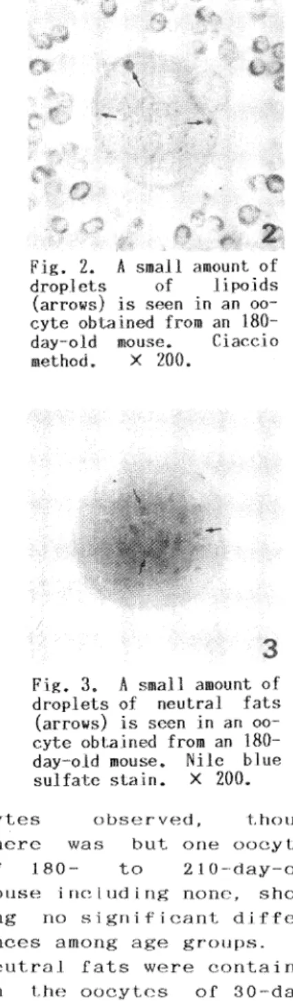
第7巻 第2号 1990年10月Fig. 2. A small amount of
droplets of lipoids
(arrows) is seen in an oo-
cyte obtained from an 180-
day-old mouse. Ciaccio
method. •~200.
3
Fig. 3. A small amount of
droplets of neutral fats
(arrows) is seen in an
oo-cyte obtained from an 180-
day-old mouse. Nile blue
sulfate stain. •~200.
cytes observed, though
there was but one oocytes
of 180- to 210-day-old
mouse including none,
show-ing no significant
differ-ences among age groups. No
neutral fats were contained
in the oocytes of
30-day-old mice, while those were
found in 1.0 % of the
oo-cytes of 60- to 90-day-old
mice, and in 6.3 % of the
oocytes of 180- to
210-day-old mice, showing a
signif-icant difference between
哺乳卵研誌
(J.
Mamm.
Ova Res. )
第7巻 第2号 1990年10月Discussion
In mouse oocytes, sudanophilic lipids first appeared in those
in secondary follicles9,10), and the lipids always existed in the
embryos up to the blastocyst stage10,11). According to Niimura
and Ishida10) who examined histochemical characteristics of
sudanophilic lipids in early mouse embryos, the lipids were
composed of lipoids and neutral fats, though the latter was not
contained in 1-cell eggs10).
In the present investigation, sudanophilic lipids in the
oocytes of 30-day-old mice were made only of lipoids, while only a
few oocytes of 60- to 90-day-old and 180- to 210-day-old mice came
to contain neutral fats though slightly. The percentage of the
oocytes containing a little neutral fats became higher with the
advance of animals' ages.
Kaler and Haensly16) histochemically studied age-related
changes in the amount of fettrot-positive neutral fats in urinary
tubule cells of the kidney of 60- to 2700-day-old cats, and
reported of an increase in the amount of neutral fats with aging,
which results agreeing to the present one obtained of mouse
oocytes.
References
1) Enders, A.C. (1971). The fine structure of the blastocyst. In
The Biology of The Blastocyst, Edited by Blandau, R.J.,
Chicago and London, Univ. of Chicago Press, P. 73.
2) Sasaki, H., Niimura, S. and Ishida, K. (1979). Lamellar or
fibrous inclusions in mammalian oocytes. Jpn. J. Zootech.
Sc., 50, 88-94.
3) Ishida, K. (1952) Histochemical studies of rat ova with
special reference to glycogen of them after ovulation. Tohoku
J. Agr. Res., 3, 39-49.
4) Ishida, K. (1954). Cytochemical studies of tubal ova. Tohoku
J. Agr. Res., 5, 1-11.
5) Ishida, K. (1962). Differential counts of glycogen-free and
glycogen-laden ova of the sow and rabbit with special
refer-ence to the tubal ova. Jpn. J. Fertil. Steril., 7, 251-256.
6) Thomson, J.L. and Brinster, R.L. (1966). Glycogen content of
preimplantation mouse embryos. Anat. Rec., 155, 97-102.
7) McReynolds, H.D. and Hadek, R. (1972). Periodic acid
Schiff-positive material in hamster preimplantation embryos. J.
Reprod. Fert., 30, 173-175.
8) Parkening, T.A. and Soderwall, A.L. (1974). Histological
localization of glycogen in preimplantation and implantation
stages of young and senescent golden hamsters. J. Reprod.
Fert. , 41, 285-295.
9) Ishida, K. (1960). Histochemical studies of lipids in the
ovaries of domestic animals. Arch. Hist. Jap., 19, 547-556.
哺乳卵研誌
(J.
Mamm.
Ova Res.)
第7巻 第2号 1990年10月of lipid droplets in mammalian eggs during the early
develop-ment. Jpn. J. Anim. Reprod., 26, 46-49.
11) Ozdzenski, W. and Czolowska, R. (1980). Lipid inclusions in
oocytes and preimplantational embryos of different strains of
mice. Folia. Morphol., 39, 497-502.
12) Hishinuma, M., Nakata, H., Urano, K., Takahashi, Y. and
Kanagawa, H. (1985). Histochemical observations of lipid
droplets in mouse embryos. Jpn. J. Vet. Res., 33, 51-57.
13) Narita, A., Niimura, S. and Ishida, K. (1990). Ultrastructural
changes of oocytes with the advancement of age of mice. Jpn.
J. Anim. Reprod., 36, 83-87.
14) Narita, A., Niimura, S. and Ishida, K. (1990). Age-related
changes in the percentage of glycogen-laden oocytes in mice.
Jpn. J. Fertil. Steril., 35, 444-449.
15) Mallory, F.B. (1961). Pathological Technique, New York, Hafner
Publishing Co., P.117.
16) Kaler, L.W. and Haensly, W.E. (1977). Kidney in the aging cat:
neutral lipids and adenyl cyclase histochemistry. Am. J. Vet.
Res., 38, 897-901.