長崎大学教育学部自然科学研究報告第20号45‑56 (1969)
ジャコウネズミにおけるジャコウ腺の
電子顕微鏡的観察
森田真一,陣野信孝
Electron Microscopic Observation on The Seba‑
ceous Gland in The Scent Organ of Suncus
Murinus Temminckii Fitzinger.
Shin‑Ichi MORITA and Nobutaka JINNO
Biological Laboratory, Faculty of Education, Nagasaki University, Nagasaki.
Abstract
The sabaceous gland in the scent organ of Suncus murinus temminckii Fitzinger was observed by electron microscope.
The sebaceous cells are generally distinguishable three characteristic steps owing to the centripetal continum of the sebaceous transformation, (1) the undifferentiated peripheral cell (UC), (2) the partially differentiated cell with some transitional types (PDC), and (3) the fully differentiated mature cell (FDC).
The peripheral cell (UC) contains no lipid droplets but small vesicles (smooth‑surfaced end oplasmic reticulum‑SER) and large amount of free ribo‑
somes are scattered through the cytoplasm, as well as the round or ovate mitochondria. With the sebaceous transformation of the cell the lipid droplets increase in size and number but the small vesicle is most abundant in PDC, although the ribosome decreases in amount and electron density of cell matrix decreases as the cell grows.
Rough‑surfaced end oplasmic reticulum (RER) is very scanty. It is short rod‑like and not lamellar. Most of the mitochondria contains one to several round and high dense intramitochondrial granules. Golgi apparatus is cistern‑
like or vesicular.
1.昭和45年度日本動物学会九州支部大会発表
2.本種の学名については,前報まではSuncus murinus riukiuanus Kurodaとしたが,新日本動物図 悲(下) (北隆館,昭和40年初版) P668の黒田長礼博士にしたがって上記の学名を揮用した。
46 森田 真一・陣野 信孝
緒 言
皮脂腺の電顕的研究は,マウス(Rogers,1957)一,ヒト(Kitamura and Kurosumi,1959
;Charles,1960;Hibbs,1962;Henrikson,1965;Zelickson,1967)等でなされているが 脂質の分泌機構に関してはまだ不明な点が多く, また脂質の起源についてもcytoplasm
(Charles,1960),mitochondria(Rogers,19571Kurosumi et a1.,1960;Kurosumi,
1961),Golgi apparatus(Hibbs,19621Ellis and Henriksol1,1965),Glycogen(Ilen−
rikson,1965),滑面小胞体(黒住,1967)など統一された見解に達していない。
ジャコウネズミのジャコウ腺は,極めて肥大した皮脂腺が多数集まってできた特殊な腺であ る(Fig.1)。このジャコウ腺は,特有の臭気をだし分泌腺として興味のある腺である。この ジャコウ腺の細胞分化と脂質滴にっいて電顕的に観察した。ここでは正常な個体の電顕像につ いての観察結果を報告する。
材料と方法
長崎市および近郊で捕獲したジャコウネズミ(S%%o郷7朋7伽% 6脚2∫n6履Fitzinger)
の成体雌5匹,雄10匹を用いた。捕獲後数週間飼育したもの,または捕獲当日のものをピスし すぐに材料を切りとり,0.1Mリン酸緩衝液でpH7.5にした5%グルタルアルデヒド溶液で,
40Cにて2時間固定した。リン酸緩衝液で5〜6回,60分間良く洗瀧した後,0.1Mリン酸緩 衝液でpH7.3にした1.4%のオスミック酸溶液で2時問半再固定した。 その後リン酸緩衝液で 簡単に洗い,アセトンにて脱水した。包埋には.Epon812を用いた。超薄片作製には,日本電 子J UM−5A型を用いた。染色は酢酸ウラニル,硝酸鉛二重染色を施し,カーボン蒸着後,
日本電子T S−6型電子顕微鏡で観察した。
観察結果
ジャコウ腺を構成する皮脂腺は,腺体部と頸部とに区別される。腺体部と頸部付近の腺細胞 の構造は,電顕的には大きな相異は認られず,また雌雄についても著しい相異は認られない。
ここでは腺体部の中央部における観察結果について報告する。腺細胞は周辺部から中心部また は腺腔にかけて,未分化の細胞(Undifferentiated peripheral cell l UC),一部分または 比較的分化した細胞 (Partially differentiated cell l PDC),十分に分化した細胞(Fully differentiated mature cell;FDC)の三つに分けられる。
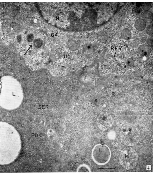
1.未分化細胞(Undifferentiated peripheral ce11;UC) (Fig.2,5,4,5,6)
この細胞は,腺の周辺部にあり,基底膜に接しており,まれにPDCの間に入り込んだもの もある。細胞は小さく,半円形,三角形など多様で腺腔の方にのびている。核は楕円形または 円形を呈しており,細胞の占める割合は他の何れの細胞に比べてもかなり大きい。細胞質の基 質の電子密度はPDC,FDCに比べて低い(Fig.4,5,6)。
ミトコンドリアは,細胞全体に均等に1分布しているが,多数集まっているところもある
(Fig.2)。大きさは,0.4〜1μで多くは円形または楕円形を呈し方向性はない。クリスタ は少なくて発達は良くない。 多くのミトコンドリア内には, 電子密度の高い明瞭なintra−
mitochondri田g田nule(IMG〉が1〜数個含まれている9このIMGは孕ほぼ円形でその
ジャコウネズミにおけるジャコウ腺の電子顕微鏡的観察 47 直径は0.05〜0.5μであり,大きいもの程電子密度が高く明瞭である(Fig.5,6)。また1 個のミトコンドリアに含まれるIMGの数は,PDCの場合よりも少ないようである(Fig.5,
6)。細胞質には全体にリボゾームが散在するが,それが集まって大きさ0.01・μ位のcluster となるものもある(Fi喜.2)。
小胞体S ERは,大きさが0.1〜0.3μで,ほとんどのものがほぼ円形を呈しsmall vesicle となっている(Fig.2)。R ERはその数も少なく短かい棒状(0.4〜1.5μ)のものが,わず かに存在するにすぎない(Fig.4,5)。
ゴルジ体Golgi apParatus(GA)は核周辺に存在するがあまり発達していない。lamella 状のものとvesicle状のものとが集合した形態を示す(Fig,5,4)
2.一部分または比較的分化した細胞(Partially differentiated cell l PDC)(Fig.4,
576)
この細胞は,基底膜に接するものと,UCに接するものとがあって電子密度がUCに比べて 高レ・(Fig・4,5,6)。細胞も肥大して細胞質の量が増加している・形成初期の脂質滴を含 む細胞から,脂肪滴が増加肥大して細胞質のかなりの部分を占めるに至ったものまでいろいろ な段階の細胞があり,腺体の大部分を占める。細胞は中心に向うもの程肥大して大きくなって いる。細胞内の脂質滴は,ほとんどのものが均質であり,中心に近い細胞のもの程大きく,電 子密度は低くなる傾向にある。 また中には脂質滴がぬけて空胞状になったものもある(Fig.
5)。
細胞質内に散在するミトコンドリアは,基本的な形態に変化は認められないが,UCのそれ Pに比べてやや大きく,直径0、4〜1.5μで円形または長楕円形など多様で不定形なものもある。
ミトコンドリアの数または分布の仕方は,UCのそれと著しい変化は認めがたい。IMGは4
〜5個のものが普通で,12〜13個の多数のI MGを含むものもあって,UCのミトコンドリア に比べてその数がやや増加しているようである(Fig.5,6)。しかもこのPDCのI MGは
1μにも達する大きなものもあってミトコンドリアをうめつくしているようなものも見られる
(Fig.6) 。
リボゾームは,UCに比べてややその数が少なくなっているが,個々の大きさ,分布の仕方 はUCのそれと変らないようである。
小胞体S ERはsmall vesicleをなしUCのそれに比べて数が多くなり,このような細胞で は脂質滴が出現し,その数も増加してくる(Fig.4,5,6)。更に分化の進んだ細胞では,
small vesicleが著しく増加し細胞質をうめつくす程で,脂質滴も更に増加し大きくなってい る(Fig.4,5,6)。
R ERは少なくて1amella状のものはなく,短棒状で0。5〜1μの大きさのものが少数認め られるにすぎず,UCと比べて著しい変化は見られない。
ゴルジ体はUCにおけると同じように発達は悪く核周辺に認められる。lamella状のものと vesicle状のものとが集合した形態を呈するのもUCのそれと同じである。
3.十分に分化した細胞(Fully differentiated mature cel1;FDC) (Fig.7)
皮脂形成過程で,細胞が破壊直前の細胞で,若い腺体の中心部はこの細胞でみたされてい る。皮脂分泌中で腺控を具えた腺では,破壊しつつある細胞に接しており,やがて破れて皮脂 の一部として分泌されるもので皮脂形成過程で最も肥大した細胞である,
48 森田 真一・陣野 信孝
脂質滴は更に大きくなり不定形となり,また癒合しあって細胞質の大部分を占めに至る。脂 質滴の電子密度はやや低くなり,脂質滴がぬけて空胞状になっているものもある。
細胞質は,大きな脂質滴の間隙をうめる索状物となってはなはだ疎な網目状構造となってい る。細胞膜も区別できにくくなる。
核は濃縮し電子密度は多少高くなり,著しく大きくなった脂質滴によって圧迫されて不定形
となる。
シトコンドリアは,膜,クリスタが不鮮明になり,その特徴的な構造が認めにくくなってく る。しかしIMGは膜が全く消失したものでも残っているものが多い。
リボゾームは,その存在がすこぶる不鮮明となり,cluster状のものは認められず,わずか に散在しているにすぎない。
小胞体はSER(smallvesicle)のみとなり,その数も少なくなり形も不鮮明になってくる。
またゴルジ体は,構造が不鮮明になり全く認められない場合もある。
やがてこの細胞は,全分泌過程に入って腺腔に面する細胞膜が破れて,脂質が流出し,いわ ゆる皮脂を形成し腺腔を満たすに至る。
考 察
ジャコウ腺の細胞における皮脂形成過程に見られる細胞の変化は,全般的にはヒト,マウス などのそれと著しい相異はない。
細胞質の基質の電子密度は,細胞の分化が進むにつれて高くなる。これはリボ核蛋白の減少 によるものと考えられ(Hibbs,19621Montagna,1956), また組織化学的にも周辺部の細 胞程basophilicである。
Hibbs(1962)はヒトの皮脂腺で,脂質滴の電子密度は,分化の進んだ細胞のもの程高くな ると報告しているが,ジャコウ腺では逆に分化の進んだ細胞のもの程低くなる傾向にある。こ れは脂質滴の成分そのものの変化によると考えられ,また脱水時の人工産物とも考えられる。
Rogers(1957)がマウスの皮脂腺で,Kurosumi(1961)がヒトの皮脂腺で, ミトコンド リアは脂肪形成に直接に関係していると報告している。またRogers(1957)はI MGは脂質 形成の過程に関係していると報告している。ジャコウ腺ではI MGはかなり多く存在し,電子 密度が高く明瞭で特徴的で,中にはすこぶる大きいI MGが溶出しているようなものもみられ
るが,脂質形成にいかに関係しているか明らかでない。
Palay(1958)はラットの特殊な脂質分泌腺meibomian glandにはゴルジ体は見られない と報告しているが,ジャコウ腺には発達は悪いが各段階の細胞に見られる。またHibbs(19 62),Ellis and Henrikson(1965)はヒトの皮脂腺について,脂質滴はゴルジ体に集められ,
貯留すると報告している。ジャコウ腺では,ゴルジ体の近くにそれに由来すると考えられる電 子密度の高いvesicle状のものがしばしば認められるが(Fig.5,4),それが脂質滴に発達 するのかどうか不明である。
またHenrikson(1965)はグリコーゲンが脂肪形成に関係していると報告しているが, ジ ャコウ腺ではグリコーゲン特有の形態をもったものは認められない。また組織化学的にもグリ コーゲンは検出されない。
黒住(1967)は,脂質は滑面小胞体の内に貯留され脂質滴が形成されると報告している。ジ ャコウ腺では細胞の分化が進むにつれてsmall vesicleが増加し,それと平行して脂質滴が出
ジャコウネズミにおけるジャコウ腺の電子顕微鏡的観察 49
現しはじめ,更に増加,肥大してくる。またPDCにおいてsmall vesicle内にやや電子密 度の高い小穎粒が認められることがあるが,これが脂質滴に発達するのかどうか不明である。
以上のことからジャコウ腺の脂質形成には,smooth membrane system(SER,Golgi apparatus)が関係していると考えられるが,尚詳細な観察結果をまちたい。
要 約
1)ジャコウ腺は多数の皮脂腺が集合してできた特殊な腺である。
2)ジャコウ腺の腺細胞は,電顕的には周辺部から中心部にかけて未分化の細胞(UC),一 部分または比較的分化した細胞(PDC),十分に分化した細胞(FDC)の三つに分け
られる。
5)ジャコウ腺細胞における細脂質の基質の電子密度は,分化が進むにつれて高くなる。
4)細胞の分化に伴いsmall vesicleが増加し,それと平行して脂質滴が出現してくる。
本稿を草するにあたり,電顕資料作製ならびに写真撮影について御指導賜わった長崎大学教 養部柴田昇助教授,長崎大学医学部末松正技師に感謝の意を表します。
文 献
1.Charles,A.1960Electron microscpic observation of the human sebaceous gland.Z 動泥s .Dθ耀観.,35,51.
2. Ellis,R.A.,and Henrikson,R.C.1965Advans in Biology of Skin.IV.The sebaceous glnds(W.Montagna,R。A。EIlis and A。F.Silber。Eds.),New York,PergamonPress,94.
5.Fawcett,M.D.1966An Atlas of Fine structure.THE CELL.W.B.Saunders Company,Philapelphia,and London
Henrikson,R.C.1965Glycogen and transformation。刀η∂8s!.jDθ7卿α!..44,455.
Hibbs,R,1.1962Electron microscpy of human axillary sebaceous glands。∫1卿θs∫.
pθ燗α .,38592
Kitamura,T.,and Kurosumi,K.1959P700.6!hハ400励8甲ακ吻如η!o〃 84.Soo.1.
Kurosumi,K.,kitamura,T.,and Kano,K.1960Electron microscpy of the human
sebaceous gland.ノ170h.ノノσs!.力ρ,,20,255.
8. Kurosumi,K l961Electron microscopic observatiion analysis of the secretion mechanism.乃2 .R8銑C罪oJ.,II,1.
9.黒住一昌,1967,細胞の微細構造と機能(12)一腺細胞の微細構造からみた分泌機能.遺伝,21(10).
10。Montagna,W.1956The Structure and Function of Skin.Academic Press,New Yovk.
11. Palay S.L.1958Frontiers of Cytology.New Haven,Yale University Press.505.
12. Rogers,G。E.1957Electron microscopic observation on the structure of sebaceous
gland・E諾ρ」.C6ZJ Rθ3・,13, 51ワ.
15. Zelickson,A。S.1967Ultrastructure of normal and abnormal skin。Lea and Febiger,
Philadelphia.
l ;FEI :‑. 4'‑' "‑"=' 50
. ・**.* *+ '..*
!/'* ; ・ j
' ' i..***; * * ; ^ ・・ ;:
*** . * * *.**=+!+*.. * i**:
' ;is: .!;/;S if'.;" ..!"f:S"" ・...."" ;/;;; ;・=・・..
* +' . * ・ i : *"**:*・・・S. * '
*+**・.
Fig. l
. i
* * *i*** '.**.:*=>:
;* * "*・*.
Longitudinal section of the scent organ consisting enlarged sebaceous glands.
*・*.
+ i i
' :!
=*+++.<+#
>': .!
'!.S * S
of many remarkably
51 :! T . ; :' }C 5( ;> 1 7 CD' =* ; f :A l } ; '.
:" : i'"s$; ; :'S:: : 't i '{
' ' iSs" ' ' t
.#' ' '#S #i
# e ; ̲ " i i * i* ". . ; ' #
' is :
: i4
"' "' ' 1{ ‑ ' # I ;' 'F F t" S ' ' . ;i* ** '$ ;. : '
, ; :* # ^ s S ' ; : '
" # f ; :'
i; :F, * * t
," i '
i"' F *
i ' ̲ ' ‑ :t'
i :' "' . d' i " :̲# ' : i ;'; ' i ; i ; ;;;::$Ji :" . #'s
...< i '. . i'
' ' : '
's' '> " . . ' *:+
j' ?S
* : * ' :s
++ fy **
< ' i : 'W;sS
^ ; $ j':. ": "' l . ‑ ' '::"̲':iel ' ' " ' l' " ' 's i #'#
' : . ' ii:' ss 1
' e ! ' i i!
'" ^ ;
j', s
:'s * '
j' p :jrs 'lLS:;S" ;:;;s5{"i' ;^ ;; ::; c '. 2Fig' 2 Undifferentiated cell'
Round or ovate mitochondria are observed through the cytoplasm' many of them contain intramitochondrial granule' Arrow shows cluster of ribosome'
Abbreviation used N, nucleus.
M, Mitochondria.
PDL; , Partially differentiated cell.
CT, Connective tissue
IMG. Intramitochondrial granule.
LU, Central lumen of gland.
GA(GC), Golgi apparatus L, Lipid droplet UC, Undifferentiated cell
R, Free ribosome
RER. Rough‑surfaced end oplasmic reticulum IF. Infolding.
SER, Smooth‑surfaced end oplasmic reticulum
+* *FEi 1 f ‑ l]*. / 52
;'{‑
T
Fig. 5 Undifferentiated cell.
High dense vesicular granule seemed to be originated from Golgi apparatus is clearly observed. (arrow)
55 :J 7 ・ ; :' }C j(y ; ; ::! I ) 'R {f : i ; A l !. i+ '.
Fig. 4 Undifferentiated cell and partially differentiated cell. Lipids increase in size and number with the increase of small vesicle (SER). High dense vesicular granule seemed to be orlginated from Golgi apparatus is observed (arrow)
EEI :=‑.P p / 54
' s " ' ' * x ji i :i'i ‑ :' !#'; ;‑ ' ' ' ' . i ' = ‑ i ‑; ' ' ;' ; ' :/' '. = ' ' = ^ >"t ' ' : ^ ̲' .' '. ' ;=
' ; i' <‑ ‑ '‑ ?
' i ' " ; " ' 'Ts' {i *F' # '; s*' ' ' ̲ :' ' '^ <‑ <: '
' i ; '' ' 7‑$#' ‑ SSR ‑ ‑ . ' ' '"' '' ; * ; ; < ' ' ' ' ‑" ^ ' s#' ' .:
^ * ‑ +*
e ! " '" i ='x i '" " <' $‑ '* *'* * '
' ' ' * f :
':s ‑' ' :' ' ' $ " *
' :' s 'y i"+ s * * ̲ ' ** '‑* ' " 's; '>*'‑' ' ‑* * *s s; ' '* 4 't ‑f‑' " '‑ '* ' ' :$" "#: **' ' ' '*"' ' ' ; ' # ' ^' = ' ' s' * < "‑ '. . e += *' ' ‑' " " *$i ‑=
' ' i ' ‑ " ' ‑ ‑" '" i :: '>"... . . , ?': ' ' ' ' +' ' s' * ' ' ' '= i' *' ; ' :"'
{ *';; i 's'*' :' * i =** ': * ' *
} F"‑ ' "' + " * * " # *#i s ' + *' ' ; :i { ' ' ^t
++".+ * * ,
f' = "i ' ' ' "' ' # : ' :̲ :"t
^ ¥・i, .' : ' ::< ': >‑ '"s = "' '‑' ' ;'=" ' '
* * * ‑ s : i :
' : # w s'" : '*' 'i= *: i i < * ='* ; " s
̲ ̲Si"! "' ' s‑ S.i^: .;; ; ::::;;;̲! ::i;:1:: {::,$.} ' ^ i " : s}'*' " *
= <: ' t'^+ i # ; ' ' ' ti . ., : s" '$ ; s‑ *s
< ' ' = #' : ; 't it ' :" '
'" ' ' = ' "' se ‑ ‑ ; ' ' *F ' ' e: ' ' t= ' =*"
i
" {{ i;s s " ":: . # :/ . t " ' ‑ " ' ' ' . .' { i ; {..,.ti S
" ^# ' = ‑ ' L ̲ s
;̲ $ 's '$ '"" ' i *
‑ 'j'* +** ;: j ^ s :' ; ;;: ;+*"< '
̲' i . ' ̲' ':' '>' ̲ s4'
' ;'
. ;' i" l'
: :
'‑
i'; " i "' i"
' ' st'! *"f' : ̲ '
* ' '* = $ ' ' s ' ' ' "(:' : #"'
,. . . ' '
^
's '
: : ̲1'> ' ' ' $' ' s F" ' '" ' ' ""t ;si ' "* ;' *'^ s * ' 'ts' '* *s? "
' *s' * #i :; *+" 's'* :* ' + :‑' i* c * * ; i"*
'" : * *" i * F
f" ' ?'1i' : * ' ' t' #t {'1 ' i' e! : :',. ss '!" '1$' ;‑ { : : ; i ' ; s
; , r :; < 1$:' '
' ' <'t L'#: ; :: ' ' ' # =i ' 't { :
‑S: ' ;i;S. .. 5
' i 'i >'
Fig' 5 Undifferentiated cell and Partially differentiated cell' Large amount of small vesicles (SER) scatter through the cytoplasm'
55 ) :r 7 ' ; :' IC i(y ) > ] 7 )***t !if : '; : . . "/j+'
$ ' # :' ;
;'i '^{ il{:?{ ?:;;" ;' :;:'}:i i!; i:S: ;: :‑;' ;' "i $
i'iE ;<' *' i" H ' * '*'. :i "
**' :' * ‑
"
'* ' ' ' ;" ; t
' *; *'
.
# * . *
* * * ・
x '#j
!
Fig. 6 Undifferentiated cell and partially differentiated cell. Prominent small vesicles and large intramitochondrial granule. Mitochondria are swollen and limiting membrane is distinct.
‑ :
F EI 1 i:‑. ? /'‑'‑'‑・‑
. 56
"" {‑s'""" '"' <. ̲i"=i ;
i .#;; sj'. ; ' ' ' .
̲;:'#i ;
;:1!i:::,;': ::;(t ;( i);:;4 ";' s;';;1 :; '
* *' *' *"' *' i'=:' :* t; '
;:'# '
": (;:; := 's '; " :t' #' ' ' c
^; *'
;:;ti;if;S ;:;:i ; ' : ' '‑: s ' ' $" ;! ' ;"; '" '# " #"ih'fj ;//') '/ i '; : ' :;:' ;st : :' i
' 's " :1 ;::S ;ssii :j is; ̲ :
' ' ' ' '{ s' s: sc'* ;! i : ' *
i'"
4; : i
#: ""' '
j
' ':" '; f s* "
* * F ;s ;'f . : *, ;1 "s' # ii :; '
'
;. i' :' s ' s { !' : i;' ;' ' #s ' ?
.$
'" ;" '
) :f s{"'""i =
' i'
* :*'f
' i' !' s ""(' * ss: '
s/"' ; '. =' s ;" /!; il : + fi' " t'" "'...: :;(;1::;;:;; ;:;S::lTj i;'
"#:'
"' '
' ' " "'!
t S:r:;i; ;t, " i :"" '*: ' '{'i : .' {.!{ F' s# i ;: ' ''
* * ' ::.:'*""""'
'/*'s!: s s! i" : ' '}#s '"
"' " ; { !/ : ;'!' {='< ' ' ';
; si ;" ^ } :; f '{/'#: . . ' i = ' :: :{ ;; ;'$1 *'
'; ' ""= "'!' :$
;;;
; ' '
. * <"i * '* :;. { *"" *‑ *' . .* * *
"'* . =*: * ; .i : j**' ;*i*
' '* .'.
" "・ '"S. i',f. . . .
Flg. 7 Remarkably enlarged liPid droplets in fully differentiated cell. Some of them are in fusion to each other. Small vesicles (SER) are observed only around the lipid droplets.