Naturally occurring and artificially synthesized polyquinanes composing of fused five-membered car-bocyclic framework have attracted considerable atten-tion from the viewpoints of challenging targets for ste-reocontrolled syntheses and their biological activities.1, 2) In the course of our continuous investiga-tions on stereoselective syntheses of heterocyclic an-gular polyquinane analogues from active urazoles(N-phenyl-1, 2-dihydrocinnoline 1, 2-dicarboximide derivatives) 1a and 1b,3-12) we isolated heterodi-quinane-based tricycle 3 from the photochemical
reac-tion of urazole 1b with diethyl 1, 3-acetonedicarboxyl-ate 2 in acetonitrile (Scheme 1). Since the 1H and 13C-NMR analyses did not permit an identification of the framework and the stereochemistry of the com-pound, the crystal structure was determined by X-ray analysis.
Experimental
General procedure. 1H-NMR(90 MHz)and
13C-NMR (22.5 MHz) spectra were recorded on a
Hit-Crystal Structure of Heterodiquinane Containing a Triazine,
an Indoline, and a Pyrrolidine Skeletons Prepared by Photoreaction
of 7-Methoxy-3-[1-(methoxyimino)ethyl]-N-phenyl-1,
2-dihydro-cinnoline 1, 2-Dicarboximide with Diethyl 1, 3-Acetonedicarboxylate
Kazuyoshi Seguchi
Department of Human Environmental Sciences, School of Human Environmental Sciences,
Mukogawa Women's University, Nishinomiya 663-8558, Japan
Abstract
The heterodiquinane derivative was prepared from the photochemical reaction of 7-methoxy-3-[1-(me-thoxyimino)ethyl]-N-phenyl-1, 2-dihydrocinnoline 1, 2-dicarboximide with diethyl 1, 3-acetonedicarboxyl-ate in acetonitrile. The structure and the stereochemistry were determined by X-ray diffraction. The com-pound crystallizes in triclinic, space group P1- with cell parameters of a = 12.171(5) Å, b = 12.372(5) Å,
c = 10.431(2) Å, α= 92.21(2)°, β= 109.75(2)°, γ= 89.19(3)°, and Z = 2 ; the final residual factor is R1 = 0.043 for 2489 reflections.
Scheme 1. Reaction scheme. MeO X Me O O N N N 2 3 4 EtO EtO MeO EtO OEt O O O O O O O OH hv 6 6a 5 4 3 1 NY MeO MeO O O EtO O O O O OEt 5 4 3 1 N N NH N N N Y Y + 6O +
la;X=O lb;X=NOMe Y;-C(Me)=NOMe 5
2 2
N N
achi R-1900 spectrometer in a CDCl3 solution with TMS as an internal standard. IR spectra and MS spec-tra were measured a Shimadzu IR-460 spectrometer and a Shimadzu QP-2000A spectrometer, respectively. 7-Methoxy-3-[1-(methoxyimino)ethyl]-N-phenyl-1, 2-dihydrocinnoline 1, 2-dicarboximide 1b was pre-pared by the method reported previously.6, 13)
Diethyl 1, 3-acetonedicarboxylate was commercially available and used without further purifications.
Synthesis of heterodiquinane 3. Irradiation of
ura-zole 1b (0.26 mmol) and an excess of diethyl 1, 3-acetonedicarboxylate 2 (13.0 mmol) in acetonitrile (60 ml) by a 400-W high-pressure mercury lamp through Pyrex filter for 10 h gave a mixture of three products (3, 4 and 5), which were isolated by column chromatography on silica gel, using a mixture of di-choromethane and ethyl acetate as eluent. The photo-product 5 (3% yield) was already identified as a novel rearranged product in the photochemical reactions of
1b without nucleophiles.6) The structure of hetrodi-quinane derivative 4 (15% yield) containing a tri-azinoindoline and an oxazolidine skeletons was con-firmed by spectral comparison with the photoproduct of 1b with 2 in the presence of triethylamine in aceto-nitrile.12) The mainly isolated 3 (79% yield) was re-crystallized slowly from ethanol to afford compound appropriate for X-ray analysis.
3 ; mp 166-167℃ ; 1H-NMR δ 1.17 (3H, t, Me),
1.36 (3H, t, Me), 1.94 (3H, s, Me), 3.26 and 3.79 (2H, s, CH2, J=15.4 Hz), 3.83 (3H, s, OMe), 3.94 (3H, s, OMe), 4.08 (2H, q, CH2), 4.31 (2H, q, CH2), 5.03 (1H, s, OH), 5.71 (1H, s, 6a-H), 6.61 (1H, dd, Ph), 7.06-7.54 (7H, m, Ph); 13C-NMR δ 10.5 (q), 14.0 (q), 14.3 (q), 38.6 (t), 49.9 (d), 55.6 (q), 60.5 (d), 60.9 (t), 61.9 (t), 62.6 (q), 85.3 (s), 91.5 (s), 100.4 (d), 111.2 (d), 121.8 (d), 125.1 (d), 128.4 (d), 128.8 (d), 129.2 (d), 134.7 (s), 142.1 (s), 148.4 (s), 149.7 (s), 154.1 (s), 160.9 (s), 167.9 (s), 170.2 (s); MS m/z (%) 580 (5, M+), 416 (100), 227 (32), 187 (30). IR (KBr) 3430, 1738, 1692 cm-1. Anal. Found: C, 60.07; H, 5.55; N, 9.77. Calcd for C29H32N4O9: C, 59.99; H, 5.56; N, 9.65.
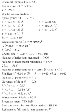
X-ray analysis. X-ray analysis of the colorless
crys-tal 3 (size; 0.20 × 0.30 × 0.50 mm) was performed on a Rigaku AFC5R diffractometer with graphite
monochromated MoKα radiation (λ= 0.71069 Å). The detailed measurement conditions and crystal data are listed in Table 1. The intensity data were collected at 296 K using theω-2θscan technique to a maximum 2θof value of 55.0゜.Of the 7096 reflections which were collected, 6779 were unique (Rint = 0.041).
The structure was solved by direct methods with SIR8814) and expanded using Fourier techniques.15) The non-hydrogen atoms were refined isotropically. All hydrogen atoms were placed at calculated posi-tions with their isotropic thermal parameters. The final cycle of the full-matrix least squares refinement was based on 2489 observed reflections [I >3.00 σ(I)] and 476 variable parameters. The final R1 and wR2 values were 0.043 and 0.043. The positional parame-ters are given in Table 2. The selected bond lengths, the bond angles, and torsion angles are shown in Ta-bles 3, 4, and 5. All calculations were carried out with the crystallographic software programe package TEX-SAN.16)
Table 1. Crystal and experimental data Chemical formula: C29H32N4O9
Formula weight = 580.59
T = 296 K
Crystal system: triclinic Space group: P 1- Z = 2 a = 12.171(5) Å α= 92.21(2)° b = 12.372(5) Å β= 109.75(2)° c = 10.431(2)Å γ= 89.19(3)° V = 1477.2(9) Å3 Dx = 1.305 g/cm3 Radiation: MoKα(λ= 0.71069 Å) μ(MoKα)= 0.98 cm-1 F(000)= 612 Crystal size = 0.20 × 0.30 × 0.50 mm Number of reflections measured = 7096 Number of independent reflections = 6779 2θmax= 55.0°
Number of reflections used = 2489[I >3.00 σ(I)]
R indices[I >3.00 σ(I)]:R1 = 0.043, wR 2 = 0.043 Number of parameters = 476 Goodness-of-fit on F2= 0.96 (△ /σ)max= 0.15 (△ρ)max= 0.14 e-/ Å3 (△ρ)min= -0.15 e-/ Å3
Measurement: Rigaku AFC5R Program system: TEXSAN
Structure determination: direct method (SIR88) Refinement: full-matrix least-squares on F2
Results and Discussion
An ORTEP drawing of 3 is illustrated in Fig.1 along with the atomic labeling scheme. The compound 3 has an intricate heterodiquinane framework containing a triazine, an indoline, and a pyrrolidine skeletons and these rings are cis-fused at axes of N(1)-C(9), N(3) -C(9), and C(9)-C(10). The torsion angles of C(7)-N (1)-C(9)-C(10), N(3)-C(9)-C(10)-C(4), and C (8)-N(3)-C(9)-C(10) are -154.8(3)゜, -130.0(2)゜, and
158.5(2)゜, respectively.
The stereochemistry of the ethoxycarbonylmethyl group at C-5 and the ethoxycarbonyl group at C-6 (Scheme 1) was determined as endo and exo
configu-rations with respect to the cis-fused indoline-pyrro-lidine ring on the basis of the ORTEP drawing. Further inspection of the ORTEP figure reveals that the ethox-yl group in the endo-position is just above the phenethox-yl ring of indoline. This special arrangement can be also deduced from 1H-NMR data of 3. The endo-ethoxyl protons on the 1H-NMR spectrum resonate at much higher field than exo-ethoxyl protons by diamagnetic anisotropy of the phenyl ring (δ1.17, 4.08 in endo-position and δ 1.36, 4.31 in exo-endo-position). The space distance between the methylene carbon C(22) of the
C(17) C(18) C(26) C(7) C(6) C(5) C(10) C(11) O(9) O(7) O(5) C(12) C(20) C(27) C(24) C(9) N(3) C(25) O(3) C(1) C(2) C(3) C(28) C(29) O(8) C(4) C(19) C(8) C(13) C(21) C(22) C(23) C(14) N(2) N(4) N(1) O(1) O(2) O(6) O(4) C(16) C(15)
Fig. 1. ORTEP drawing of the title compound (3) along with the atomic labeling scheme.
Table 2. Atomic coordinates and equivalent isotropic dis-placement (Å2) Atom x y z Beq O(1) 0.0711(2) 0.0803(2) 0.6629(2) 3.83(5) O(2) 0.3021(2) 0.3665(2) 0.8490(2) 3.95(6) O(3) 0.6780(2) 0.4724(2) 0.7614(3) 4.68(6) O(4) 0.3534(2) 0.0553(2) 0.7588(2) 4.57(6) O(5) 0.3528(2) - 0.1243(2) 0.7506(2) 4.52(6) O(6) -0.0693(2) 0.3819(2) 0.3428(3) 4.92(6) O(7) 0.1247(2) 0.0381(2) 0.3414(2) 3.10(5) O(8) 0.3417(2) 0.0908(2) 0.2299(3) 5.03(6) O(9) 0.3654(2) - 0.0576(2) 0.3536(2) 3.84(6) N(1) 0.2854(2) 0.3119(2) 0.6319(2) 2.69(5) N(2) 0.1569(2) 0.2461(2) 0.7309(3) 2.83(6) N(3) 0.1714(2) 0.1551(2) 0.5387(3) 2.59(5) N(4) 0.0101(2) 0.3007(2) 0.4100(3) 3.56(6) C(1) 0.5828(3) 0.4146(3) 0.6813(3) 3.24(7) C(2) 0.5933(3) 0.3704(3) 0.5617(4) 3.91(8) C(3) 0.5028(3) 0.3098(3) 0.4718(3) 3.62(8) C(4) 0.4029(3) 0.2938(2) 0.5023(3) 2.87(7) C(5) 0.3946(3) 0.3392(2) 0.6225(3) 2.69(7) C(6) 0.4830(3) 0.4014(3) 0.7150(3) 2.90(7) C(7) 0.2540(3) 0.3137(3) 0.7463(3) 2.81(7) C(8) 0.1276(3) 0.1537(3) 0.6422(3) 2.77(7) C(9) 0.2116(3) 0.2559(2) 0.5061(3) 2.57(6) C(10) 0.2967(3) 0.2259(3) 0.4289(3) 2.66(7) C(11) 0.3168(3) 0.1036(2) 0.4488(3) 2.56(6) C(12) 0.2065(3) 0.0594(2) 0.4718(3) 2.61(6) C(13) 0.1053(3) 0.2579(3) 0.8373(3) 2.90(7) C(14) 0.1340(3) 0.1886(3) 0.9431(4) 4.34(9) C(15) 0.0824(4) 0.2052(4) 1.0433(4) 5.5(1) C(16) 0.0072(4) 0.2878(4) 1.0368(4) 5.8(1) C(17) - 0.0206(4) 0.3567(4) 0.9312(5) 6.1(1) C(18) 0.0305(3) 0.3411(3) 0.8311(4) 4.73(10) C(19) 0.6757(4) 0.5215(4) 0.8856(5) 5.3(1) C(20) 0.2292(3) - 0.0416(3) 0.5572(3) 2.96(7) C(21) 0.3178(3) - 0.0280(3) 0.6981(3) 3.29(8) C(22) 0.4357(4) - 0.1229(4) 0.8906(4) 5.1(1) C(23) 0.4705(6) - 0.2321(5) 0.9288(6) 8.6(2) C(24) 0.1137(3) 0.3320(3) 0.4314(3) 2.86(7) C(25) 0.1469(3) 0.4376(3) 0.3920(5) 5.1(1) C(26) - 0.1843(4) 0.3413(5) 0.3096(6) 7.3(1) C(27) 0.3416(3) 0.0472(3) 0.3306(3) 3.18(7) C(28) 0.3720(3) - 0.1248(3) 0.2379(4) 4.44(9) C(29) 0.2547(5) - 0.1592(5) 0.1488(6) 8.5(2) Beq = (4/3) Σi Σj βij (ai. aj)
endo-ethoxyl group and least-squares plane of the
phe-nyl ring having the methoxyl group is 6.050 Å.
Acknowlegement
The author wishes to thank Dr. Satoko Tanaka for her valuable discussion.
References
1 ) Paquette, L. A., Top. Curr. Chem., 119, 1-158 (1984) 2 ) Mehta, G. and Srikrishna, A., Chem. Rev., 97, 671-720
(1997)
3 ) Tanaka, S., Seguchi, K., Itoh, and K., Sera, A., J. Chem. Soc., Perkin Trans. 1, 1994, 2335-2339
4 ) Tanaka, S. and Seguchi, K., Sera, A., Heterocycles, 38, 2581-2584 (1994)
5 ) Tanaka, S. and Seguchi, K., J. Chem. Soc., Perkin
Trans. 1, 1995, 519-520
6 ) Tanaka, S., Seguchi, K., Itoh, K., and Sera, A., Bull. Chem. Soc. Jpn., 69, 3533-3542 (1996)
7 ) Tanaka, S. and Seguchi, K., Chem. Lett., 1998, 1135-1136
8 ) Seguchi, K. and Tanaka, S., Heterocycles, 45, 707-713 (1997)
9 ) Seguchi, K. and Tanaka, S., Recent Res. Devel. in Org. & Biorg. Chem., 1, 15-24 (1997)
10) Tanaka, S., Kato, K., Kimoto H., and Seguchi, K., Anal. Sci., 15, 313-314 (1999)
11) Seguchi, K., Tanaka, S. and Kobayashi, A., Anal. Sci., 20, x147-x148 (2004)
12) Seguchi, K. and Tanaka, S., Bull. Mukogawa Women’s Univ. Nat. Sci., 58, 1-5 (2010)
13) Seguchi, K. and Tanaka, S., Bull. Chem. Soc. Jpn., 64, 3188-3190 (1991)
14) Burla, M. C., Camalli, M., Cascarano, G., Giacovazzo, Table 3. Selected bond lengths(Å)
Atom Atom Distance Atom Atom Distance O(7) C(12) 1.405(3) N(1) C(5) 1.412(4) N(1) C(7) 1.370(4) N(1) C(9) 1.470(4) N(2) C(7) 1.418(4) N(2) C(8) 1.413(4) N(3) C(8) 1.356(4) N(3) C(9) 1.440(4) N(3) C(12) 1.481(4) C(4) C(5) 1.388(4) C(4) C(10) 1.508(4) C(9) C(10) 1.546(4) C(9) C(24) 1.520(4) C(10) C(11) 1.541(4) C(11) C(12) 1.551(4) C(11) C(27) 1.509(5) C(12) C(20) 1.530(4) C(20) C(21) 1.505(4)
Table 4. Selected bond angles(°)
Atom Atom Atom Angle Atom Atom Atom Angle O(7) C(12)C(11)106.1(2) O(7) C(12)C(20)111.0(2) N(1) C(5) C(4) 109.0(3) N(1) C(9) C(10)104.1(2) N(1) C(9) C(24)108.3(2) N(3) C(12)C(11)100.8(2) N(3) C(12)C(20)113.6(3) C(4) C(10)C(9) 103.3(2) C(5) N(1) C(7) 127.7(2) C(5) C(4) C(10)110.5(3) C(8) N(3) C(9) 119.1(3) C(8) N(3) C(12)126.2(3) C(9) N(3) C(12)113.4(2) C(9) C(10)C(11)103.9(2) C(10)C(11)C(12)106.3(3) C(10)C(11)C(27)112.2(3) C(11)C(12)C(20)113.9(3) C(12)C(11)C(27)112.9(2) Table 5. Selected torsion angles(°)
Atom Atom Atom Atom Angle Atom Atom Atom Atom Angle O(1) C(8) N(3) C(9) 165.0(3) O(1) C(8) N(3) C(12) -29.0(5) O(2) C(7) N(1) C(5) 22.1(5) O(2) C(7) N(1) C(9) -172.0(3) O(7) C(12) N(3) C(8) 106.2(3) O(7) C(12) N(3) C(9) -87.1(3) O(7) C(12) C(11) C(10) 85.1(3) O(7) C(12) C(11) C(27) -38.3(3) N(1) C(9) N(3) C(8) 46.6(4) N(1) C(9) N(3) C(12) -121.1(3) N(1) C(9) C(10) C(4) -14.3(3) N(1) C(9) C(10) C(11) 104.7(3) N(3) C(9) N(1) C(5) 126.5(3) N(3) C(9) C(10) C(4) -130.0(2) N(3) C(9) C(10) C(11) -10.9(3) N(3) C(12) C(11) C(10) -30.6(3) N(3) C(12) C(11) C(27) -154.0(2) C(7) N(1) C(9) C(10) -154.8(3) C(8) N(3) C(9) C(10) 158.5(2) C(8) N(3) C(9) C(24) -74.8(3) C(8) N(3) C(12) C(11) -141.8(3) C(9) N(3) C(12) C(11) 24.9(3) C(9) C(10) C(11) C(12) 26.1(3) C(10) C(9) N(3) C(12) -9.3(3)
C., Polidori, G., Spagna, R., and Viterbo, D., J. Appl. Cryst., 22, 389-393 (1989)
15) Beurskens, P. T., Admiraal, G., Beurskens, G., Bosman, W. P., de Gelder, R., Israel, and R., Smits, J. M. M., DIRDIF94 program system, Technical Report of the Crystallography Laboratory, University of Nijmegen, Netherlands (1994)
16) TEXSAN:Crystal Structure Analysis Package, Molecu-lar Structure Corporation (1985)