Aetiological and Clinical Studies on Adenovirus Infections in Nagasaki, Japan 1964‑1966
Koichi FUTATSUKI
Abstract
During a period from June 1964 to February 1966 in Nagasaki, 676 patients with acute respiratory illness, diarrhea or skin rash were examined at the time of the initial visits, and in addition 145 of the cases were examined in approximately 2 to 4 weeks after onset of the illness. HeLa cells alone were employed throughout the study, and the total incubation period in each of the negative specimens was at least a total of 80 days through three passages, cultures being maintained in Hanks's B. S. S. containing 0.5% lact‑
albumin hydrolysate, 0.1% yeast extract, and usually 3 to 4% bovine serum. Forty‑nine adenovirus strains were isolated from 48 of the cases examined. Types 2, 3 and 5 were the commonest, and types 1, 6 and 11 were also recovered. Type 3 was mainly obtained from school children in a sharp outbreak in the summer of 1965. All the strains of type 3 caused upper respiratory disease and the recovery of this type was always accompanied by a rise in both CF and neutralizing antibody. Types 2 and 5 occurred chiefly in infants in both the summers of 1964 and 1965 and were associated with 3 cases of diarrhea and 6 cases of exanthema in addition to upper respiratory illness. There was a neutralizing antibody rise in 40% of each of the groups excreting type 2 and 5 agent respectively.
All strains of types 1,6 and 11, excepting one type 1 strain, were found in pre‑school children in the winter of 1965‑66, and in 3 cases of diarrhea besides upper respiratory disease. The 49 strains were obtained from 5.1% of 710 throat specimens collected, from 6.7% of 461 anal specimens, and from none of 117 eye specimens. A study of a limited number of the patients from whom both throat and anal specimen were collected indicates that adenoviruses were isolated with equal ease from throat and anal specimens. Those collected specimens were kept at 4℃ or at room temperature in six hospitals and five practitioner's offices from the time of collection before they were transported to the laboratory only once or twice a week without ice. Some of them spent more than 5 days
at 4℃ or at room temperature before transported. There was little influence of specimens spending longer than 6 hours at 4℃ or at room temperature not only on the isolation
Department of Virology, Institute for Tropical Medicine, Nagasaki University, (Director : Prof. Dr. Hideo FUKUMI)
(Received for publication November 25, 1967)
Contribution No. 517 fro:h the Institute for TropicA1 Medicine, Nagasaki Unilersitv
rate but on the time required for the first detection of the CPE. The optimal time for taking throat specimens for adenovirus isolation was the 4th day of illness, and that for taking anal specimens was the 4th day up to about 21st day. Serologically 18.4% of 136 patients were diagnosed as positive by the CFT. Although no adenovirus was isolated from any specimens of 206 adults, 13.5% of 37 adults were confirmed as adenovirus infection only by a rise in CF antibody titer. In addition to the adenovirus strains, enteroviruses
were isolated from 29 patients.
Aetiological and Clinical Studies on Adenovirus lnfections in Nagasal・i, Japan, 1964‑1966 2こ′7
Introduction
Since adenovirus was iso一ated for the first
time by Eowe et al. (1953)5i;i and Hilleman
and werner (1954)29), the etiologica一 ro一e of
adenoviruses in acute respiratory diseases has been studied by many investigators in the United States and other countries. In Japan,
since Fukiユmi et al. (1957)14) reported four
cases of pharyngoconjunctival fever due to
ad∈ノnovirus, many investigations of various
respiratory illnesses o.ue to adenovirus have
been carried out (Matumoto, 1963)42>. it
is now very clear that adenovirus is a causative agent of pharyngoconjunctival fever, which has been recognized to be accompanied b>
gastro‑intestina一 symptoms for some time
(Fukumi et al., 195816); Duncan and Hutchin‑
son, 196173; Kajietal., 196137)). Someinvesti‑
gators (Hirayama, 19593o;>3i ;) ; Kamiya et at., 196139>; Takatsu, 196154); have reported that adenoviruses were isolated from infants with diarrhea of white faeces, and the etiological relation of the adenoviruses to infantile diarrhea
especia】ly "pseudocnolera infantum"ミo is nowa―
days drawing attentions. On the other hand, recovery of a cytopathogenic agent, which was later classified as adenovirus (Bell et al.,
1956i;i)5 from faeces of an infant with rash resembling that of roseola infantum by Neva and Enders (1954)46 > gave rise to a suspicion about the relation between adenovirus and
exanthematous illness, and nowadays the relationship has become one of the most
important prob一ems in the adenovirus infec‑
tions. Recent一y Bell (1965〕3) has found that
5 per cent of the cases of aseptic meningitis and 10 per cent of those of encephalitis are associated only with an adenovirus infection, principa11y type 2, and has suggested that
the adenoviruses are imp一icated in infections
of the centra一 nervous system. Thus, many
researches have been made, but the role of the
adenoviruses in the etio一ogy of acute respira―
tory illnesses, d週arrlユea in chi一dren, and ex‑
ar〕tb.巳ma etc. is not yet fully understood, and
in addition some part of eco】ogical knowledge
of the ad.en―ovirus infection stil remains to
be studied. To make matters worse, in Kyushu
area, Japan, only a few of such detai一ed
studies have been carried out, and in Nagasaki any study has never been done.
The present report describes the findings in the studies carried out with a view to putting anything in requisition of prevention of an ill‑
ness or an epidemic, by investigating (1) the re」Nation between the types of aderiovirus and the clinical symptoms, (2) antibody responses to adenovirus infections according to the day of the川ness, and (3) the excretion period of the adenoviruses in various routes of the
nn十tentsー Thチire was clearly the need for
* The disease will be explained in Uiscussi(Hl.
to11ecting specimens as many as possible on different days after onset of the i1Iness from the patients of various age groups suffering from acute respiratory il1nesses, diarrhea or skin rashetc.. However, no effort was made to examine a control group of the general popu‑
lation. Between June, 1964, and February, 1966, in Nagasaki 1,288 specimens for virus isolation and 136 paired sera for serological tests from 676 patients wit'i so‑called "cold"
syndrome mainly in summer season and with diarrhea mainly in winter season were exam‑
ined for adenovirus. No particular atteitnpt was made to examineother viruses. To add to the resu1ts obtained in the subjects, this paper reports the findings in the problems of the de1ay in delivery of specimens to the labora‑
tory and the optimal time for taking the specimens, and also describes the detai1ed clmical features of 6 cases with exanthema.
Materials and Methods Condition of Patients
Specimens for virus isolation were obtained irom 676 infants, children and adults in Nagasaki during the period from June ll, 1964, up to February 20, 1966. Of these, 403 infants and children were seen in the outpatient
・clinics of six hospitals* and five practitioners**
mainly for acute raspiratory illness or diarrhea, and 190 adults were seen in the same out‑
patient climcs for acute respiratory illness.
0hIy 30 chi1dren and 16 adults were hospi‑
talized in 5 of the6hospitals and in 2 of the 5 practitioners wards for lower respiratory tract infection, and 37 infants were admitted to the ward of an asylum*** for acute respir‑
・atory disease or exanthema. Paired serums for serological studies were obtained from 136 of the 676 individua1s from whom the specimens
for virus isolation were collected.
Clinical Observations and Cases For the studies, detailed cl mical observations and interrogations of the outpatients were conducted at the time of the initia1 and the second visits, and those of the inpatients were
conductedeveryday.Theseweremadeby physiciansofeachhospitalandeachclinic.
A1lpatientswerec1assifledintothefo‖owing diseasecategories.
(A)Upperrespiratorytractinfection(U.R.T.I.) Thisisclassifiedin5syndromesaccording totheclassificationsdescribeddyDowling andLefkowitz(1963)6)andParrottetal.
(1963)47):
(a)coryzalsyndrome,whichischaracterized byincreasednasaldischargeandocculusion andrare1yaccompaniedwithfever;
(¥t
)pharyngealsyndrome,inwhichthe
prominentsymptomsaresorethroat,redness ofthepharynxandswel1ingofthetonsi1s, oftenwithfever,cough,coryzaandmild systemicsymptoms;
(c)pharyngocomunctivalfeverCP.C.F.^syndrome, inwhichthepharyngealsyndromeispresent inadditiontoconjunctivitis;
(d)influenzasyndrome,which,hasasits outstandingfeaturetheconstitutionalsymp‑
tomssuchaschi巾ness,headache,general aching,malaiseandanorexiainahighpercen・
* Mitsubishi Heavy Industries Ltd. Nagasaki Shipyard and Engineworks Hospital, Nagasaki Atomic Bomb Hospital, Nagasaki Municipal Citizen fIospital, Nagasaki Municipal Nagasaki Hospital, Nagasaki University Hospital, and Saisei‑Kai Hospital.
** Inoue Hospital, Satomi Pediatric Clinic, Takamori Clinic, Sumiyoshi‑Chuo Hospital, and Zeshinkai Takahara Hospital.
ネ** Nagasaki Municipal Suckling Asylum
A etiological and Clinical Studies on Adenovirus Infections in Nagasaki, Japan, 1964-1966 259
tage of coryzal or pharyngeal syndrome;
(e) herpangina syndrome, wich is charac- terized by vesicles in the pharynx and on the hard and soft palate.
(B) Lower respiratory tract infection (L.R.T.I.) This is also classified in 3 syndromes :
(f) pleurodynial syndrome, which is charac- terized by the sudden onset of severe pain in the thorax aggravated by breathing ;
(g) bronchitis, which includes croup ; (h) pneumonia, which is confirmed radi- ologicall y.
(CT) Fever of unknown origin (F.U.O.)
This is characterized by high fever alone, showing no symptom of acute respiratory disease, aseptic meningitis, encephalitis, or paralysis.
CD) Exanthema
This includes all exanthematous infections resembling rubella, measles, scarlet fever, exanthema subitum, urticaria, or roseola infantum, with or without fever, pharyngitis, or conjunctivitis.
(E) Diarrhea
This includes diarrhea associated with the coryzal or the pharyngeal syndrome, but when diarrhea is not so severe and the coryzal or the pharyngeal syndrome is prominent the patients are not included in this diarrhea group. Pseudocholera infantum is also included, which will be well explained in Discussion.
^F) Other diseases
This includes conjunctivitis, meningitis, myelitis, and the cases from whomno answer of clinical observation was received.
Collection of Specimens
All of serum, throat, anal and conjunctiva!
materials were tried to be taken from each patient during the acute phase of illness, but
throat swabs or pharyngeal garglings were mainly collected from the patients with acute
respiratory illness and anal swabs or faeces from the cases with diarrhea. Moreover approximately 2 to 4 weeks after onset of the illness, efforts were made to obtain the second those samples only to fail in almost
four-fifths of the cases, owing to the failure of the patients to return at the designated time. At the time of the initial visits of patients, though some specimens were taken more than 7 days after onset of the illness,, 584 throat, 364 ana], 94 eye and 370 serum specimens were collected from 676 patients.
The number of the specimens on the second visits were 126 throat, 97 anal, 23 eye and 136 serum specimens, which were collected from 145 among the 676 patients. Thus, 1,288 materials for virus isolation and 136 paired.
serums for serological tests were obtained.
Throat, anal and conjunctival swab speci- mens were collected by use of the cotton swabs which were placed in the test tubes with 3 ml of bonito infusion broth contain- ing 1,000 units of penicillin and 1,000 /zg of streptomycin per ml, and throat washings were obtained by gargling with 10 ml of the same transport medium. Faeces were put into the tubes with 10 ml of bonito infusion broth containing 25^ glycerin, 1,000 units of peni-
cillin and 1,000 jag of streptomycin per ml.
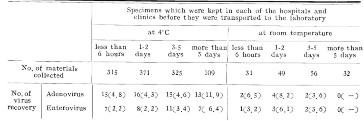
These specimens obtained in the hospitals and clinics were kept at 4°C or sometimes at roomtemperature from the time of collection before they were transported to the laboratory only once or twice a week without ice by road. Some of them spent more than 5 days at 4°C and several days at room temper-
ature before transported. In the laboratory
they were frozen at -20°C and. stored until
tested. Approximately 60^ of specimens
were inoculated into tissue cultures within
2 weeks after collection, but the others were
stored frozen at -20°C for several months until inoculated.
Specimens of blood were taken in the test tubes and kept at 4°C, until they were transported to the laboratory and separated from the clot. The serums were kept at -20°C until tested.
Tissue Culture
Throughout the study, a strain of HeLa cells was employed. HeLa cells were kindly supplied by Dr. M. Shingu, Department of Microbiology, Kurume University School of Medicine, Fukuoka, Japan. During the whole à"of the work, the cultures were grown at 36.5°C in Hanks's Balanced Salt Solution (B.S.
S.) containing 0.5^ lactalbumin hydrolysate and 10^ bovine serum. Test tube cultures for virus isolation were prepared by inoculating to culture tubes with0.6 to 0.8ml of a HeLa à"cell suspension containing approximately 200,000 cells per ml. The cultures ready for use, which were usually 3 days old, were washed once with 2 ml of Pack's B.S.S. and the washed cell sheets were then overlaid with 1.0 ml of Hanks's B.S.S. containing 0.5^
lactalbumin hydrolysate, 0.1^ yeast extract and 3 to 4 per cent bovine or calf serum.
For virus titration and neutralization test, test tube cultures were prepared by inoculating with 0.6 ml of growth medium containing 400,000 cells perml. After 24 to 36hours of incubation the tube cultures were washed twice with 2.0 ml of Pack's B.S.S., and 0.8 ml of Hanks's B.S.S. containing 0.5^ lact- albumin hydrolysate and 0.1^ yeast extract
<Y.L.H.) was added to each tube, and then the tubes were used. All mediums contained 200 units of penicillin and 200 fig of strep- tomycin per ml.
Virus Isolation Procedures
Faeces in transport medium were homoge-
nized with Hanks's B.S.S. to give a 10 to 20 per cent emulsion. They were then centri- fuged at 3,000 r.p.m. for 30 minutes 3 times.
To the supernatant fluid obtained from the third centrifugation, antibiotic solution was added to give the final concentration per ml of 1,000 units of penicillin, 1,000 ^tg"of strep- tomycin and 400 units of mycostatin, and then the samples were held at 4°C overnight.
The specimens were again centrifuged at 3,000 r.p.m. for 30 minutes, and 0.1 ml of the supernate was inoculated into 3-day-old tube cultures on maintenance medium which were prepaired. Two HeLa tube cultures were used for one specimen. The nutrient fluid was changed after incubation for 2 hours at 35°C.
Throat, anal and conjunctiva! swabs and pharyngeal washings in bonito infusion broth were centrifuged at 3,000 r.p.m. for 30 minutes, the supernate was removed, and antibiotic solution was added in the final concentration of 1,000 units of penicillin, 1,000 jug of strepto- mycin and 400 units of mycostatin per ml.
The sampleswere kept at 4°C for 4hours to overnight, and then centrifuged at 3,000 r.p.m.
for 15 minutes, and 0.2 ml of the supernate was inoculated into each of 2 HeLa tube cultures of 3-day-old on maintenance medium.
All tubes were incubated at35°C in a station-
ary position for at least 28days, and examined
microscopically 3 to 4 times a week for cyto-
pathic effect, changing the medium every 4
to 5 days. If any cytopathogenicity was
observed, at the height of cellular degener-
ation the fluid and cells were harvested by
freezing and thawing 6 times for next passage
and storage. If no cytopathogenicity was
noted for minimum of 28 days, two additional
passages were carried out by freezing and
thawing the cultures 6 times and by inocu-
lating 0.2m! of the fluid into fresh monolayers
Aetiological and Clinical Studies on Adenovirus Infections in Nagasaki, Japan, 1964-1966 261
to extend the incubation period to at least 80 days. The tubes that proved negative after 25 days of incubation in the third passage were discarded. When cytopathogenic changes
in the HeLa cells were not suggestive of adenovirus but rather of enterovirus occurred,
the cultures were harvested by freezing and thawing once and identified with polio- antiserums. In all critical and positive speci- mens the isolation procedure was repeated
from the original materials.
Identification of Adenoviruses
Neutralization tests were performed exclu- sively in 2-day-old HeLa cell cultures for typing any isolates. The technique for adeno- virus was a modification of those described
by Rowe et d. (1955)52), Grayston et al. (19- 56)20), Rafajko (1964)48>, Rose (1964)49)t Wigand et «/.(1965)57> and Ellis et al. (1966)8>.
The viruses isolated were propagated in HeLa cell cultures which were incubated, for a few days after the cells had shown a
4 + cytopathic changes. The infected cells and maintenance medium were harvested, frozen and thawed 6 times, and the cell debris were removed by centrifugation at 3,000 r.
p.m.for 15 minutes. Serial twofold dilutions of the isolates were prepared in Hanks's B.
S. S., and 0.2 ml of each dilution was inocu- lated into 2 cultures containing 0.8 ml of the serum free maintenance solution. The cultures were examined for CPE 48 hours after inocu- lation, and the endpoint was defined as the highest dilution of virus producing a 4+ CPE.
Some isolates produced very little cellular change and were allowed additional time for incubation. In these cases, the tubes were read for CPE on the 4th or the 5th day, or later.
The unknown viral agent was diluted in Hanks's B.S.S. to contain twice the virus
necessary to produce the endpoint CPE. 0.25 ml of the diluted virus was mixed in individual tubes with the same amount of each type- specific horse antiserum against adenovirus types 1 to 18, diluted 1:10. A virus control tube was prepared by mixing 0.3 ml of the diluted virus with equal volume of Hanks's B.S.S., and a 1Q'1 virus control was also pre- pared by mixing 0.1 ml of the first virus control suspension with 0.9 ml of Hanks's B.S.S.. After the serum-virus mixtures and the controls were held at 35°C for 2 hours and then at room temperature for 2 to 3 hours, 0.2ml of each of the mixtures and the controls was inoculated into 2 tube cultures containing 0.8 ml of the serum free maintenance medium.
The cultures were incubated at 35°C in a stationary position for 4 to 5 days and inspect- ed for CPE. Neutralization was considered as positive when antiserum-containing tubes showed a complete prevention of cytopath- ogenic change 48 hours after the virus control had reached a 4+ reading.
Identification of Polioviruses
The method was carried out according to a modification of the techniques described by
Kono et al. (1958)4l>, Melnick et d. (1964)43', Schmidt (1954)53>, and Ellis et al. a966)9>.
Specific typing antisera against 3 types of poliovirus were also kindly given by Dr. M.
Shingu. They were sera from monkeys immunized with type 1 (Mahoney), type 2 CSabin 2) and type 3 (Sabin 3), and from rabbits immunized with type 1 (Mahoney), type 2 (MEF 1) and type 3 (Saukett). All these sera except Anti-Mahoney monkey serum had neutralizing antibody titers of over 1:1,000 against 100 TCDso of homologous virus. 0.25 ml of each of antisera and pooled one of types
1 to 3 containing 20 antibody units per 0.1
ml was mixed with the same amount of tissue
culture fluid diluted to contain 1,000 TCDso per ml 4 days after inoculation. A virus control was also prepared. The mixtures and the virus control were incubated at 35° C for 90 minutes, and 0.2 ml of these was inoculated into each of 2 tubes of 2-day-old cultures.
This 1,000 TCD5o challenge virus was diluted
1:10, 1:100, 1:1,000, and 1:10,000 for the virus
titration, and 0.1 ml of each dilution was inoculated into 4 tubes. Results were read on the 4th day, and complete prevention of CPE was interpreted as a positive neutrali- zation.
Complement Fixation Procedure
The tests were simultaneously carried out with acute and convalescent sera of the same patient on plastic haemagglutination plates, according to the same method as that de- scribed by the National Institute of Health, Tokyo58). Isolated adenovirus types 3 and 5 were employed for complement-fixing anti- gen, which was prepared by propagating the viruses in bottle cultures of HeLa cells. The virus inoculum had been adjusted so that approximately 5 to 7 days were required for complete cytopathogenic changes. The bottles were held for 2 to 3 days after all the cells had become detached, the cells and medium were frozen and thawed 6 times, centrifuged, and the supernatant fluid was harvested. The antigen was made by pooling equal amounts of type 3 and 5 adenovirus supernatant fluid, which had been inactivated by heating to 56°C for 30 minutes. Serum was also inactivated at 56°C for 30 minutes, and twofold dilution was made.
On the plastic plates were dropped serum dilution in 0.02 ml (1 drop), 4 units of antigen in the same amount, and 2 full units of comle- ment in 0.04 ml (2 drops). After overnight incubation at 4°C the hemolytic system, which
consisted of equal volumes of a 2.5^ sus- pension of washed sheep cells and 20 units of sheep cell hemolysin, in 0.04ml (2 drops) was added. The plates were then reincu- bated at 37°Cfor 60 minutes, and the test was- read after additional incubation at 4°C for 2 to 4 hours. The endpoints were read as reciprocal of the highest original dilution of serum producing a 3+ fixation. The diluent used throughout was Veronal-buffered saline solution. The initial serum dilution used was- 1:4, except in some babies under one year.
A four-fold or greater rise in antibody be- tween the acute and the convalescent serum was interpreted to be a significant change.
Serum Neutralization Tests
Serum neutralization tests were carried out on paired sera against any virus isolated from that patient. Method for titrating neutralizing antibodies against adenoviruses was basically similar to that for typing adenoviruses. The sera were inactivated at 56°C for 30 minutes and twofold dilutions were made in Pack's B.S.S.. To 0.25 ml of each serum dilution was added 0.25 ml of virus dilution containing an amount of virus to produce 4+ cytopath- ogenic changes in 0.1 ml 48 hours after inocu- lation. The mixtures were allowed at 35° C for 2 hours and then 0.2 ml of each was inocu- lated into 2 tubes. Avirus control and a 10'1
virus control were included. The tubes were
incubated for 3 to 4 days and the test was
read 24 hours after the virus control had
shown a 4+reaction. The neutralizing titer of
serum was expressed as reciprocal of the
highest initial dilution showing greater than
a 2 + difference between the cytopathogenic
changes in the serum-containing tubes and the
virus control. In case of slowly growing
adenovirus, challenge virus was diluted to
produce a 4+ CPEin0.1 ml on the4th day or
Aetiological and Clinical Studies on Adenovirus Infections in Nagasaki, Japan, 1964-1966 265
later.
Serum neutralization procedure for entero- viruses was also the same as that for typing polioviruses. 0.25 ml of each of twofold dilu- tion of serum was mixed, with the same amount of virus diluted to contain 100 TCDso per 0.1 ml on the 4th day. The mixtures
were incubated at 35°C for 90 minutes and 0.2 ml of each of the mixtures was inoculated into 2 tubes. The neutralizing titer was taken as reciprocal of the highest original dilution of serum showing a 1 + CPE 4 days after inoculation.
Results
Distribution of Disease Categories October, 1964, 220 patients were included in Thedistributionofdiseasecategoriesthrough- the survey; 69^ of the cases were in the out the period of survey is shown in Table 1. U.R.T.I, group, 10^ in the L.R.T.I, group, During the summer season from June to ?>% in the exanthema group, <Q% in the
T able 1. The distribution of disease categories, June, 1964, to February, 1966.
Month of onset of illness of
patients
1964-- 6
7 8 9 10 ll 12
1965... 1
2 3 4
9
10 ll 12
1966... 1
2
Disease categories
U .R.T.I.* L.R.T.I.** F.U.O.*** Exanthema Diarrhea
1
71 50 29
13396 37 34
1 2
ll
Other
Diseases****
188
T otal
43
1
21
38
12 5 3 4 1 1
2 251
10 6
~2 1 2 1
1 221
18 4
2 4 ll
3 12
33 66 41 20
864
13
1112
64
* Upper respiratory tract infection
** Lower respiratory tract infection
*** Fever of unknown origin
**** Meningitis, myelitis, conjunctivitis, etc.
49
1
439
14 67 16 127
22141412
22 12 16
TT
45 75 71 34
~?A
22
75
34
39
diarrhea group, and VI% in the F.U.O. and the other diseases group. In the winter of 1964-65, the specimens were collected from only 5 patients, and besides in November,
1964, no case was examined. The number of the patients examined during the spring season from March to May, 1965, were 50;
46^ of the cases were in the L.R.T.I. group, 44^ in the exanthema group, and only k%
in the U.R.T.I, group. In the summer of 1985, 246 patients were examined ; similarly in the summer of 1964, 70^ of the cases were in the U.R.T.I. group; the other disease categories were responsible for 30^ of the cases. 155 patients were examined in the winter of 1965-66; 68^ of the cases were in the diarrhea group and only 20^ in the U.R.
T.I. group, while the patients with exanthema were not examined.
Virus Recovery
The number of specimens co/Jected and the number of isolates during the period of the survey are summarized in the first part of Table 2, and the number of virus strains
isolated and the number of patients having viruses in the latter part of Table 2. From 676 patients 710 throat, 461 anal and 117 eye specimens were collected. 36 adenoviruses and 17 enteroviruses were recovered from 710 throat swabs and garglings, 31 adenoviruses and 22 enteroviruses from 461 anal swabs and faeces, and none from 117 conjunctiva] swabs.
These 67 adenoviruses and 39 enteroviruses were isolated from 73 patients. 48 of the 73 patients had adenoviruses; 25 were the patients from whomspecimens were collected only once, and 23 were those from whom specimens were collected twice. Amongthese 23 patients, 3 had one or two adenoviruses and a poliovirus each on two different visits or from throat and anal specimens on the same day, 3 had a single adenovirus strain
each not only during acute phase of illness but during convalescence, 12 had an adeno- virus on the first visit only, and 5 had iton thesecond visit only. Among those 25 patients one had an adenovirus type 3 and
an unidentified enterovirus, and the others
T able 2. Summary of virus recovery from 676 patients, June ll,1964, to February 20, 1966.
T ested No.
Adenovirus positive No
Enterovirus
Adenovirus Poliovirus CB** E*3
l E Bi v vi Xi ?*1 ! it M m vi ?*2
Total
Specimens
Patients
Throat Anal Eye
710 461 117
Total 1. 288
Single virus
i nfectio )n
*3
Multiple virus infection
36 31 0
2 6 22 5
1 5 15 6 1 1
16 17
67 44 4
3 ll 37 ll 2 1 2 1 1
53 S3 0
1 1 33 106
3 6 22 8 2 1 2
- 3 1 1
1 - - 1 1 22
- 1 2 - - 1
69 4
Total 676 48 3 9 23 9 2 1 2 1 1 1 1 23 73
9 =Adenoviruses assumed to be types of higher number than 18.
9 =Viruses assumed to be coxsackieviruses or echoviruses.
Two adenovirus strains (type 2 and 5) and a poliovirus type 3 were obtained from one patient, and an a'denovims and an enterovirus from each of 3 other patients.
CB=Coxsackievirus B
E =Echovius
Aetiological and Clinical Studies on Adenovirus Infections in Nagasaki, Japan, 1964-1966
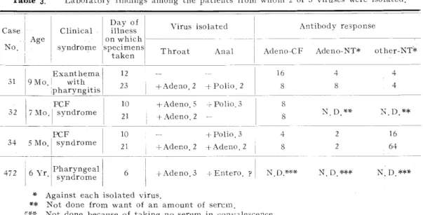
Table 3. Laboratory findings among the patients from whom 2 or 3 viruses were isolated.
C ase
No Age
31 I9Mo.
Clinical syndrome
Day of illness on which
E xanthema with pharyngitis
32 7Mo.
Virus isolated
Throat Anal
+Adeno.2 +Polio.2 PCF
syndrome
5Mo.
PCF syndrome
472 I6Yr. Pharyngeal syndrome
10 +Adeno.5 -rPolio.3
21 i +Adeno.2 -
10 - +Polio.3
21 ; +Adeno.2 +Adeno.2
Antibody response
Adeno-CF Adeno-NT* other-NT'*
16 4 4
8 8 . 4
8N D ** D.**
4 2 16
8 2 64
Adeno 3 -i-Entero. ? N.D.*** N.D.*** N.D.***
* Against each isolated virus.
** Not done from want of an amount of serum.
«** Not done because of taking no serum in convalescence.
h ad an adenovirus strain each. Thus, four patients were excreting two or three different viruses; one had two strains of adenovirus type 2 and 5 with a poliovirus type 3, and the others had an adenovirus and an entero- virus each. The details of these cases, which couldnot be confirmed serologically in all of the cases, are given in Table 3. A total of 49 adenovirus strains were therefore recovered from the 48patients. Of these strains, adeno- virusesof types 2, 3 and 5 were 84^', and over half of these were type 3. The remainder of the adenovirus strains were types 1,6 and
ll; in addition 2 strains were recovered, which have not yet been identified and are assumed
to be adenoviruses of the higher numbered designations than 18.
39 isolates which were assumed to be enteroviruses were isolated from 29 patients.
Four isolates which were obtained with adeno- viruses from four patients were identified as polioviruses of type 1,2 or 3. Two isolates were identified as coxsackievirus type B3 and
echovirus type 6, which were kindly per- formed by Dr. K. Ishii, National Institute of
H ealth, Tokyo. The remainder could not be identified as polioviruses and are assumed to be coxsackieviruses or echoviruses, owing to their characteristics of CPE, resistance to temperature, and sensitivity to HeLa and monkey kidney cells and newborn, mice..
Thus, adenoviruses and enteroviruses were recovered from 73 of the G76 patients exam-
ined.
The Relation of Recovery of Adeno- viruses to Season
Table 4 shows the distribution of the patients
having viruses according to the month when
the patients had their onsets of illness. It
would be dangerous to discuss on the seasonal
distribution of the viruses from such a small
number of isolates and moreover from lack
of isolations during the winter and the spring
from October, 1964, to May, 1965, because of
very few patients examined and most of them
suffering from L.R.T.I, or exanthema, but
some interesting points in this respect are ob-
served from the study. Only ll of 220 patients
examined (5.0^) had adenoviruses in the
summerof 1964 and 10 of 155 patients (6.5^>
T able 4. The frequency distribution of the patients having adenoviruses, according to the month of onset of the illness.
Month of onset of illness of
patients
No.of
patients tested
1964- 6 7 8 9 10
No.of
patients
having adeno-
Adenovirus
Enterovirus
Cox.
Poliovirus B Echo
1 II IE V VI XI ? i n m BE vi ?
Total
No. of
positive patients
23
83 89 43
00
9*
2 0
1965.
ll 12
. 1
2
3
4
5
6
7
8
9
10 ll
4 1 4
_ 2
1 - 1 2
22 00
1 1**
4 0
00000023
6
ll
45 | i
2 1 -
12
1966... 1
2
22 75 34
0000002
10 14 13**
5
~5~
3 4 2 2 -
1 1
- - 7
1 - 7
- - 3
1 1
2 1
1 - 1
1 1
1 1Total -676 48s 3 9 23 1 23 73**
* Two adenovirus strains (type 2 and 5) with a poliovirus type3 were obtained from one patient.
** One ortwo adenovirus strains and an enterovirus were obtained from each of 4 patients.
Table 5. Age distribution of the patients having adenoviruses.
Age
No. of
No. of patients patients having tested adeno-
viruses
0 -ll months 1- 4 years 5-14 years 15-81 years
Adenovirus Enterovirus
Cox.
Poliovirus B Echo
i E IK v vi xi ? i H n n vi ?
T No.of otal positive patients
107 13*(12. 1)
191 12( 6.3)
172 23 (13.4)
206 0( 0.0)
8 - 6 - - - 1 1 2 1 1 3
2 - 5 - 2 1 2
1 1 18 3 _ _ _
10
19**
22
30**
2
T otal 676 48*^7.1) 9 23 1 23 73*1*
* Two adenovirus strains (type 2 and 5) with a poliovirus type 3 were obtained from one patient.
** One or two adenovirus strains and an enterovirus were obtained from eachof4patients.
Aetiological and Clinical Studies on Adenovirus lnfections in Nagasaki, Japan, 1964‑1966
had them in the winter of 1965‑66, as com‑
pared with 27 of 246 patients (ll.0^) having them in the summer of 1965. Adenoviruses of type 3 appeared most significantly in a sharp outbreak which took p1ace during the summer season from June to October, 1965, but only one recovery of this type was made out of 220 patients examined in the summer of 1964. Adenoviruses of types 2 and 5 were predominantly observed for a brief period in both the summers of 1964and 1965. Although the number was sma11, all adenovirus types 1,6 and ll were obtained only m winter season of 1965‑66, except one type 6 in October,
1966, and some strains of types 2,3 and 5 were also found in this winter season. Two unidentified adenovirus recoveries were made
週n August of 1964 and in that of 1965.
′The majority of enteroviruses occurred in
the summer of 1965, and some of them in the summer of 1964 and in the winter of
1965‑66.
The Relation of Recovery of Adeno‑
viruses to Age
when the different types of adenovirus are compared with the age of the patients some marked differences are observed, as indicated in Tab1e 5. Of the 676 patients examined,.
Table 6. The relation between the types of adenovirus isolated and the defined disease categories, in 470 infants and children.
No.
Disuseti
categories,*ァるof :nts
ed coryzal syndrome Pharyngeal― l
′′
P.C.F.***
′′
Influenza
′′
Herpangina
′′
40
98
23
4
25
L.R.T.I.
F U.O.***** ― 39
Exanthema ― 48
Di arrhea*** ***
pseudocnolera infantum
Other diseases 28
Total ― 470
No.of patients
having adeno‑
viruses
Adenovirus
Enterovirus Cox,
polioいTirus BEcho
l iL 皿 V VI XI '? ⊥ 1L l止Ill vl ?
1
20 2 13
―. 1 ‑‑―, ‑― ‑― ‑ ‑ 122 10 2 ‑ ‑ 1 ‑ ‑ 2 ‑ ‑ ‑
6 ‑ 4 ‑ 2
3 : 1 1 ― 1
1―
2―
48* ― 3 9 23 1 1 1 21― 1**
2adenovirusstrains(type2and5)withapoliovirustype3wereobtainedfrom onepatient.
*株1or2adenovirusstrainsandanenteroviruswereobtainedfromeachof4patients‑
・
*#* ;*Ph
*Fe'い?erぎngo‑conjunctn
。funknown。い.と1fever
gin約*一 **ォ
***話oh一ceこrespirat except重orytract
fr。mpseuるnfec l。ch三ion
lerainfantu‑
Total No. of positive patients
IEE
O
O
‑ 2 10 2 ‑ ‑ 1 ‑ ‑ 2 ‑ ‑ ‑
5 6
31**
14**
〔)
1
7. 7.
107 were infants under 1 year of age, 191 pre-school children of 1-4 years, 172 school children of 5-14 years, and 206 adults of 15
years and over. During the preiod of the survey, adenoviruses were recovered from
12.1^ of the infants, 6.3^ of the pre-school à"children, 13.4^ of the school children, and none of the adults. All strains of type 3 were found in children between the ages of 1 and 14, and the school children had the highest incidence of this type. Only one strain of adenovirus type 2 was isolated from the school
children, and none from the pre-school chil- dren, while there were 8 from the infants under 1 yearof age. As intype2, no adeno- virus type 5 occurred in the pre-school chil- dren and most of them came from the infants.
In contrast with adenovirus types 2 and 5, it will be noted that all strains of types 6 and ll and two of three strains of type 1 were from the pre-school children, 1-4 years, and that both the unidentified adenoviruses
were also from these children.
All of enteroviruses were isolated from infants and children, except that only 2 adults shed unidentified enteroviruses of which one was isolated from a man of 17 years of age with the pharyngeal syndrome and the other irom that of 31 years with pharyngitis. Six
strains identified as poliovirus, coxsackievirus type B3 and echovirus type 6 were recovered from the infants under 1 year.
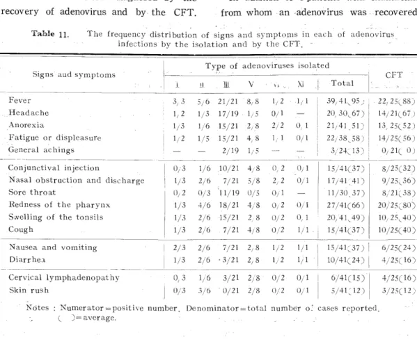
The Relation of Recovery of Adeno- viruses to Disease Category
The disease categories defined and the results of recovery of viruses in infants and children are recorded in Table 6. Among
470 infants and children examined throughout the period of the survey, 190 were in the
U. R.T. I.group, 45inthe L. R.T. I. group, -39 in the F. U. O. group, 48 in the exanthema
group, and 126 in the diarrhea group. Adeno- viruses occurred in 35 of the 190 cases (18.4^)
intheU. R. T. I. group, 1 of the 39(2.6^9 in the F. U. O. group, 6 of the 48 (12.5^) in the exanthema group, 6 of the 126 (4.8^9 in the diarrhea group, and none of the L. R. T. I. and the other diseases group.
In the U.R.T.I, group, adenoviruses were isolated from 60.9^ of 23 cases with the P. C. F. syndrome, 20.4^ of 98 cases with the pharyngeal syndrome, 2.5^ of 40 cases with the coryza) syndrome, and none of 29 cases with the influenza or the herp- angina syndrome. Of the 126 cases examined in the diarrhea group, 28 were the patients with pseudochoJera infantum and 98 were those with diarrhea associated with U.R.T.I. ; adenoviruses were recovered from 10.7^
of the former and 3.1^ of the latter.
All adenoviruses of type 3 were isolated from the cases with the pharyngeal or the P.C.F. syndrome. There were recoveries of adenovirus types 2 and 5 in the cases with the pharyngeal or the P. C. F. syndrome, the fever of unknown origin, exanthema, or diarrhea. A strain each of adenovirus types 1,6 and ll were observed in the diarrhea group and the other strains of types 1 and 6 were in the U.R.T.I. group. The two strains of unidentified adenovirus were found in the cases with the pharyngeal or the P. C. F.
sy ndrome.
Approximately half of enterovirus. isol ations were made from the cases with pharyngeal syndrome, and the others occurred in all the disease categories except the influenza syndrome, the L. R.T.I. group, and the other diseases group.
The Relation of Age and Disease Cate- gory in Recovery of Adenoviruses
The influence of age and disease category
Aetiological and Clinical Studies on Adenovirus Infections in Nagasaki, Japan, 1964-1966 269 Fig- !à" The relation of age and disease category in the patients from whom
adenovirus was recovered. Numbers indicate type of the viruses isolated.
Disease categories
C oryzal svn.
: 5: o
oPharyngeal syn.
l^l
ED ° CDEDED ° 'à"5^°
0 |1| ° ED ° [1||3|I3I o I3Io
oo
P .C.F.
[Tj= P3
=ra
H
H erpangina
svn. B :-!
F .U.0.
E xanthema
o m
pi 6 ll
Diarrhea
Pseudocholera infantum
E6
K ey [ \ -Adenovirus recovery on the first visit j^ | =Adenovirus recovery on the second visit
° =Unidentified enterovirus recovery
P =Poliovirus B= Coxsackievirus B E = Echovirus
= =Virus recovery from the same patient
on the patients from whom viruses were recovered may be seen in Fig.1. Of 23 adenovirus type 3 recoveries 13 were from patients with the pharyngeal syndrome, ll -of whom were school children of 5-9 years.
The other types observed in the pharyngeal syndrome were adenovirus types 1,2 and 5, and an unidentified adenovirus and entero- viruses were also found in this syndrome.
The remaining adenovirus type 3 recoveries
were made from children with the P. C. F.
syndrome whose ages were 2 to 14 years.
Three infants under 1 year old with the P.C.F. syndrome had adenovirus type 2 or 5, orpoliovirustype 3. This type2 and an unidentified adenovirus were recovered from the specimens which were collected when the patients revisited during convales- cence. In children of 1-7 years with the coryzal syndrome were found an adenovirus type 6, which was obtained from a throat swab on the second visit, and unidentified enteroviruses.
Only one coxsackievirus type B3 occurred in an infant aged 9 months with herpangina.
There were an adenovirus type 5 and an identified enterovirus recovery in the F.U.
O. group ; the former was isolated from an infant and the latter from a school child.
8 cases of exanthema were associated with the recovery of adenovirus type 2 or 5, polio- virus type 2, or unidentified enterovirus ; all patients were under 1 year of age. Adenovirus types 1,2 and 5, echovirus type 6, and uniden- tified enterovirus were isolated from infants and children with diarrhea associated with U.R.T.I., except pseudocholera infantum ; thier ages were under 9 years. From infants, 6 to 23 months of age, with pseudo- cholera infantum adenovirus types 5,6 and
ll, and poliovirus type 1 were recovered.
The Recovery of Adenoviruses Related to the Type of Specimens and to the Day of Illness Specimens Were Collected As had already been mentioned in Table 2, eye swabs yielded no virus and all the iso- lates were obtained from throat and anal speci- mens. Pharyngeal garglings, all of which were taken from adults of 15 years and
over, yielded no adenovirus and yielded only 2 unidentified enteroviruses. Adenoviruses and enteroviruses were isolated more frequently
f romfaeces than from anal swabs.
Table 7 gives the relation of frequency of recovery of viruses from throat and anal specimens in the patients from whomboth throat and anal specimen were collected. The patients who revisited hospitals or clinics with
no apparent symptom during convalescent phase were regarded here as control group and compared with the patients from whom specimens were collected at the time of illness. Adenoviruses were recovered with equal ease from throat and anal specimens not only in case of the patients with evident symptoms but also in case of those with no apparent symptom. It may be noted that 2 strains of unidentified adenovirus were isolated only from anal specimens and that no enterovirus was recovered from throat specimen alone.
The findings relating the isolation rate of viruses from throat and anal specimens of a total of the 676 patients to the day of illness on which the specimens were collected are presented in Table 8. The day of illness was employed, i. e. if the specimens were taken on the same day as the onset of symptoms, this was the first day of illness ; if the following day, this was the 2nd day.
Neither throat nor anal specimens which
were collected on the first day of illness
yielded adenovirus and the throat specimens
collected on the second day gave a low
isolation rate (2.7^0, although 60^ of the
60 throat specimens collected on the first
day and lk% of the 183 throat specimens
collected on the second day were from the
patients with U. R. T. I. as compared with
75^ of the 60 anal specimens collected on
the first day being from the cases with
diarrhea. The day which showed the
highest isolation rate for adenovirus from
Aetiological and Clinical Studies on Adenovirus Infections in Nagasaki, Japan, 1964-19&6 271
T able 7. The comparison of recovery of adenoviruses from throat and anal specimens of 350 cases from whom both the specimens were taken.
No of cases from whom adenovirus was isolated
Type
The patients from whom the speci- mens were collected during acute phase
(^tested No. =272) Throat
only
Throat and Anal
anal only
The patients fromwhom the speci- mens were collected during convalescence (^tested No. =78)
Throat and Anal
anal only
Adenovirus
HEV
VI
?
T otal Entero- Poliovir*s
Total
1
236 1
12 1
14
1211
5 _ 2 - 7 5 0 7 7
14 2
1
1 4
- 1
1 2
0 1 3
Table 8. The isolation rate for adenovirus from throat and anal specimens of a total of the 676 patients examined, according to the day of illness on which the specimens were collected.
Specimens Type
Throat
Day of illness
1 2 3 4
5 - 7
8 - 14
15 - 21
22+
Subtotal
No. of No.of
speci- adeno-
mens viruses
tested isolated
0( 0.0) 5( 2-7) 9( 7.5) 9( 9.9) 6C 8.1) 1C 1-4) 4( 5.7) 2( 4.9) 36( 5.1)
Viruses isolated
Adenovirus Enterovirus
Poliovirus C.B E.
i E n v vi xi i n m n vi
I 4
2 7
1 - 5 3
- - 6 -
- 2
2 6
1 -
1 1
9 i
1
10 1 2 1
Total
22 1 0 0 0 0 0 1 0 16
A nal
1234
5 - 7
8 -14
15 - 21
22+
0( o.o)
5( 6.5) 3( 5.6) 6(ll.3) 5( 6.7) 3( 5-9) 6(ll.5) 3C 7.7) S ubtotalI 461 31(6.7)
11
- 2 1 1
2 1 - -
IS
1 1 1 2 1
1 -
1 2 1
Total ll,171 67C5.7) ] 3 ll 37 ll
25212311
17 S3
3
10 5 7 8 8 7 5 53
1 1 1 33 106
throat specimens was the 4th day (9.9^), and the isolation rate was reduced after that day. In case of isolation from ana]
specimens, adenoviruses were recovered most frequently on the 4th day (ll.3^) and on the 15th to 21st day(ll.5^). The mean isolation rates of adenovirus for all days irrespective of the day of illness were 5.1^
and Q.796 for the 710 throat and the 461 ana] specimens, and those for all days greater
than 7 were 7/182 (3.8^0 and 12/142 (8.5^9
for the throat and the anal specimens.
Thus, ad.enoviruses were recovered a little more frequently from anal specimens than from throat specimens in case of all the 676 patients examined, as compared with what were seen in the patients from whom both throat and anal specimen were taken.
The throat specimens collected on the 2nd day (5.5^; and the anal specimens collected
Fig. 2-a. The distribution of adenoviruses isolated from throat swabs, according to the day of illness on which the specimens were collected.
Type 6
204086 X D
x_ X X
Type 5
«
Type 3
M
C3
Z
491
nn
B X
X
X XX
XType 2
X
X X
Type 1
2t n
n n
X X
PM \*\ HI
10
8
14
12 16
22 26 30
24 28
B
Day of illness
Virus isolation with specific antibody rise Virus isolation alone without specific antibody rise The same virus isolation
from different specimen uion imen alone
D
=This virus isolation from the specimen
i n case of no collection of serum.
X =Novirus isolation from the patient who had the
virus isolated from different specimen.
Aetiological and Clinical Studies on 1Adeho<4TuisiIfrf^t(tfens in Nagasaki, Japan, 1964-1966 Fig. 2-b. The distribution of adenoviruses isolated from anal swabs and faeces, according
to the day o: illness on which the specimens were collected.
Tyi
0900
oi 4
8 2
" 0
d
? :^4
9
0
4
2
0
90 X
Type ll
Type 6
Type 5
nx
M Type 3
Type 2
nx _ x
Type 1
à"
X X
X K&5] Ewvi Y V
X
f «"|^ X|«
2 -3 4
Day of illness
K ey = The same as'that of Fig. 2-a
on the 8th to 14th day (9.8^) yielded enteroviruses most frequently.
The, distribution of adenoyiruses isolated from throat and anal specimens according to the day of illness the specimens were taken are depicted in Fig. 2-a and Fig. 2-b.
All 22 patients who shed adenovirus type 3 from the oropharynx were within 8 days of illness. From 7 of these 22 patients throat specimens were again taken without yielding any viruses during convalescence after the 16th day of illness, and from any
of them no specimen taken during the 8th to the 16th day. From the intestine, 13 patients shed adenovirus type 3 by the llth
day of illness, and 2 patients, who had- had
the same type which was recovered from
throat specimens collected during acute
phase and showed an antibody rise to the
virus isolated, shed on the 17th and the 21st
day. Specimens were collected after the
llth day of illness for the first time from
3 of the 9 patients excreting adenovirus
type 5; one shed this type from both the
oropharynx and the intestine on the 17th day, the second did from the intestine on both the 12th and the 26th day, and the third did from the intestine only on the 17th day. All these 3 patients produced neutral- izing antibody rises in their convalescent serums which were collected after the 25th day of illness. There was one type 5 isolation from a faeces on the 18th day without recovery of this type during acute phase and without a specific antibody rise.
From 4 other patients adenovirus type 5 wasrecovered from throat or anal specimens within 5 days of illness. One patient had this type recovered from a throat swab collected on the 10th day of illness, a polio- virus type 3 from a faeces on the same day,
and an adenovirus type 2 from a throat swab on the 21st day. To add to this patient,
4 patients shed adenoviruses of type 2 during the 21st to 24th day of illness, not having this type during acute phase ; one excreted this type not only from a throat swab but from a faeces on 21st day without an antibody rise and a poliovirus type 3 from a faeces
during acute phase, the second excreted an adenovirus type 2 from a throat swab on the 23rd day with a specific antibody rise and a poliovirus type 2 from a faeces on the same day, the third excreted an adenovirus type 2 from a throat swab alone on the 23rd day with an antibody rise, and the fourth excreted this type from a faeces alone on the 24th day without an antibody rise. Four other patients had type 2 by the 6th day of illness; one of them shed from the oropharynx as well as from the intestine and he showed a rise in antibody. Without recovery during acute phase of illness, a type 6 was recovered from a throat swab and an unidentified adenovirus from a faeces during conva-
lescence.
Antibody Response
The cases confirmed as adenovirus infection by the complement-fixation test are shown in Table 9, by means of age and disease category. Of 136 infants, children and adults from whomblood samples in the acute and the convalescent stages were obtained in addition to materials for virus isolation, 25
T able 9. Adenovirus infections diagnosed by the complement-fixation test*, by age and disease category, June, 1964 to February, 1966.
Age
0 -ll Mo.
Disease category Coryzal syndrome Pharyngeal syndrome
P. C. F. syndrome
L.R.T.I.
F.U.O.
Exant hema Diarrhea Other diseases
12
1-4 Yr 5 Yr. -14
15-64 Yr.
6211
2
T otal Total .
positive teggg- Average
41 4851
232
0
32 40 13 12 15 ll
5 8
12.5 20.0 38.5 8.3 13.3 27.3 40.0 0
Total positive Total tested Average
3
17 17.6
5
31 16 1
12 51 23 5
5
37 13 5
Against the pooled antigen of types 3 and 5 isolated
25
136
à"18.4
A etiological and Clinical Studies on Adenovirus Infections in Nagasaki, Japan, 1964-1966 275
T able 10. The CF and the neutralizing antibody response of the patients from whom adenovirus was recovered from throat or anal specimen, or both.
Adenovirus recovered from Type
Oi
adeno- virus
Throat specimen only
A ntibody rise*
Throat and anal specimen
A ntibody rise
Anal specimen only
N o.
CF** NT***
n
IT v
321
No
CF NT
No.
A ntibody rise
CF NT
0 67
100 100
0 0
261
so
100 100
50 100 100
T otal 33 67
* A four fold or greater rise
** Against the pooled antigen of types 3 and 5
*** Against the virus isolated
89
2035 %0
33
%0
33
20 20
C18.4^)showed a fourfold or greater antibody rise to the CF antigen pooled types 3 and 5
isolated. ll of them occurred in patients from whomadenovirus was isolated and 14 in those from whom no virus was recovered.
The percentage of patients with a four fold or greater CF antibody response in the different age groups was 17.6^ in the infants under 1 year of age, Vo.\% in the pre-school children between 1 and 4 years, and 23.5^ in the school children of 5-14 years. As compaired with no recovery of adenovirus from the adults over 14 years old and only one recovery from the children with the coryzal syndrome, 5 adults (13.5^) were positive by the CFT; 4 of them were suffering from the coryzal syndrome and the other from the pharyngeal syndrome.
Seven children with the pharyngeal syn- drome, from 5 of whom adenovirus type 3 was isolated, showed a CF antibody rise; 6 of them were the school children. In infants and children with the PCF syndrome there were 5 with a rise in antibody titer ; all but one were associated with the recovery of
adenovirus. Although no adenovirus was recovered from the patients in the L. R. T. I.
group, only one child with bronchitis produced anantibodyrise. Of 7 patients in the F. U.
O. , the exanthema and the diarrhea group, 2 were infants with exanthema from whom adenovirus was recovered and the others were children from whomno virus was isolated.
Paired serums were obtained from only 22 of the 48 patients excreting adenoviruses.
A four fold or greater rise both in complement
fixing and in neutralizing antibody titer occurred
in 9 of the 22 cases, it in complement fixing
antibody titeralone occurred in 2, and it in
neutralizing antibody titer alone occurred in
4. Thus, of the 22 patients ll showed a four-
fold or greater rise in CF antibody and
13 showed it in neutralizing antibody. From
the case who shed adenovirus type 2 and
5 with poliovirus type 3 a pair of sera was
taken ; no complement fixing antibody rise
was detected and a neutralizing antibody
response to each type isolated could not be
demonstrated from want of an amount of
serum. A serum pair which was obtained
from one of the 2 cases excreting uniden- tified type gave a twofold rise in each of complement fixing and neutralizing antibody.
Excluding these 2 patients, in Table 10 the CF and the neutralizing antibody response
of 20 cases from whomadenoviruses of types 2, 3 and 5 were recovered are analyzed to
show the response according to each of the types and to indicate the response according to whether adenovirus was recovered from
a throat or an anal specimen, or both. A fourfold or greater rise in CF and neutralizing antibody each occurred in 89^' of the patients when the same virus was isolated from both
throat and anal specimen. In contrast to this, only 33^ of patients in whomadenovirus was recovered from the throat specimen alone.produced a CF antibody rise and only 20^ of those in, whom adenovirus was recovered from the anal specimen alone did it. Neutralizing antibody responses were found in-§1% of the cases shedding adenovirus from the throat alone and 20.& of those
shedding it from the intestine alone. Adeno- virus type 3 isolations were always accompa-
nied by both CF and neutralizing antibody rise, but only 1/7 (14^) of the cases from whomtype 2 was recovered developed a CF antibody response and 3/7 (43^) of those a neutralizing antibody response. There were a CF and a neutralizing antibody rise in 40^ of 5 patients from whomthe recovery of type 5 was made.
The CF and the neutralizing antibody response to adenovir.us infections according
to the day of illness are shown in Fig. 3. A curved line for CF antibody response to ad'enovirus was drawn by making an entry of the antibody titers in the acute and the convalescent serums of 25 cases who showed a fourfold or:greater CF antibody rise. As will be seen,, the CF antibody began to rise from about the 8th to 10th day of illness.and got to the peak by the 20th day. Neutralizing antibody titers to the isolated viruses in the acute and the convalescent serums of the
lij. 3. The CF and the neutralizing antibody response to adenovirus infections, according to.the day of illness.
A A
à"
A «, -
à">1t' -I- ? à" /:à"-'':à"$à"à"
§à"à"
.: .: .
.. . . .
ot-is:
à" A
NT'Adono 3 ]
à" à"
A A
CK and XT(Ade
5 ,--'"""NT'A(!eno 2 ;
à" - -