Acta Med. Nagasaki 35:189-193
PERIPHERAL AMELOBLASTOMA OF THE BUCCAL MUCOSA : WITH A REVIEW
OF THE LITERATURE
Takashi SHINZATO 1, Takayoshi TODA 1, Masaya KIYUNA 1 Yushi HAMADA 1, Seitetsu HOKAMA 1 and Katsuji AKAMINE 2
Department of Clinical Laboratory and Laboratory Medicine 1 and Department of Otolaryngology 2, School of Medicine, University of the Ryukyus, Okinawa, Japan
Received for publication, June 25, 1990
SUMMARY : A peripheral ameloblastoma arising on the buccal mucosa is exceedingly rare. Only three document cases have been reported, and we present one additional case of this tumor. In this case, the lesion repeatedly occurred in the same location in spite of twice local excisions. The first lesion was diagnosed as basal cell carcinoma with ameloblastomatous features. The pathological findings of the lesion were palisading of the peripheral cells of epithelial masses and structures resembling stellate reticulum, which could also be observed also in peripheral ameloblastomas. The second lesion was diagnosed as basal cell adenoma, and the third as peripheral ameloblastoma.
The histogenesis of peripheral ameloblastomas and intraoral basal cell carcinomas are discussed, especially with reference to the lesions of the buccal mucosa.
INTRODUCTION
Ameloblastoma is a odontogenic neoplasm that most often occurs in bone, and its origin has been accepted to be one of the following sources : (1) the developing enamel organ, (2) cell rests of the enamel organ, (3) epithelium of odontogenic cysts, (4) basal cells of thr surface epithelium, or (5) heterotopic epithelium, as in lesions that occur at extraoral sites (10).
The histogenesis of the peripheral amelobla- stoma is controversial. The peripheral ame- loblastoma is described as a neoplasm arising in soft tissue overlying tooth bearing region, not including the buccal mucosa, the lips, the craniopharyngeal duct, or the tibia (1). On the other hand, Shiba emphasized (12) that ameloblastomas arising in buccal mucosa or
labial mucosa should come within the same category of peripheral ameloblastoma, and indeed three cases of ameloblastoma arising in the buccal mucosa has been described (2, 7, 13).
And it is generally accepted that peripheral ameloblastoma arises from the basal cells of the oral mucosa of from remnants of the dental lamina (3).
We herein report a fourth case of a peripheral ameloblastoma arising in the buccal mucosa of which histological feature was confused with
basal cell carcinoma, and review literatures concerning the histogenesis of the peripheral ameloblastoma and the differentiation from basal cell carcinoma.
CASE REPORT
A 46-year-old Japanese man was referred to
the Otolaryngology Clinic at our hospital for the evaluation of a raised, tender, friable mass on the right buccal mucosa. The lesion had been present for a few months, and except for the tenderness it was asymptomatic. The clinical diagnosis was basal cell carcinoma. After surgical excision, the microscopic examination revealed that the lesion was mainly composed of a superficial proliferation of epithelial islands arising from overlying surface epithelium (Fig.
1), and individual islands were sometimes attached to the basilar layer of the overlying epithelium (Fig. 2). The outer layers of the islands consisted of palisaded columnar cells with their nuclei polarized away from the basement membrane. In the central area of the lesion, there was a stellate reticulum arrangement with occasional acanthomatous areas. Based on these histological findings, a diagnosis of
basal cell carcinoma with ameloblastomatous
features, was made.
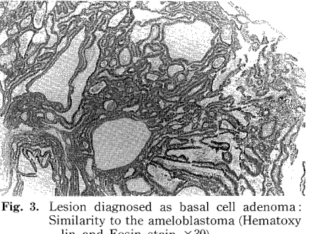
Seven years after the initial treatment, a tumor occurred at the right buccal mucosa near the maxilla. The second surgical excision was performad, and the lesion was diagnosed as basal cell adenoma (Fig. 3) by another pa- thologist.
Six years after the second operation, a third lesion was noted at the same location as the previous second lesion. The third surgical excision of the tumor was done. A diagnosis of peripheral ameloblastoma, follicular type, was made (Fig. 4).
These tumors were immunohistochemically examined in paraffin-embedded tissues by the ABC methods previously described (6). Lectins such as Con A, UEA-I, WGA, PNA, and SBA (EY Lab. INC) and antibody for keratin (DAKO) were used. Immunohistochemical results are shown in Table 1. The presence of Lectin
Fig. 1. Islands and nest of tumor epithelium lying below the surface epithelium (Hematoxylin
and Eosin stain x 100).
Fig. 3. Lesion diagnosed as basal cell adenoma:
Similarity to the ameloblastoma (Hematoxy
lin and Eosin stain X30).
Fig. 2. Island of ameloblastomatous tissue in continuity with the surface mucosa
(Hematoxylin and Eosin stain X300).
Fig. 4. Peripheral ameloblastoma, follicular type.
The epithelium is arranged in discrete islets
(Hematoxylin and Eosin stain X30).
Fig. 5. Immunohistochemical staining for Con A binding. Inner cells of the lesion are more
positive than outer layer (ABC Method x
300).
binding and keratin was slightly more remark- able in the inner layers than in the outer layers of the lesion (Fig. 5).
DISCUSSION
The peripheral ameloblastoma is the rare tumor which is usually found in the mandibular molar area (14) and in the maxillary gingiva (11).
Only three cases of the peripheral amelobla- stoma arising from buccal mucosa have been reported (2, 10,13) (Table 1).
The histologic apperance of the peripheral ameloblastoma _is almost the same as that of the central ameloblastoma (12, 14). The basal cell adenoma may resemble the ameloblastoma (13). The histologic features of the second lesion of the present case were similar to that of peripheral ameloblastoma, follicular type. such as the third lesion. The first pathological diagnosis of the present case was reported as basal cell carcinoma with ameloblastomatous features. We reviewed the histology of the first tumor and we could diagnose it as peripheral ameloblastoma, basal cell type. Simpson re- ported (11) a case of basal cell carcinoma with
ameloblastomatous lesion as our case, describ- ing that the histologic appearance was not exclusive to ameloblastoma. Thus, the differ- entiation between the peripheral amelobla- stoma, especially acanthomatous or basal type, and the intraoral basal cell carcinoma is sometimes confused.
Greer and Hammond (5) believed that the peripheral ameloblastoma exhibits central polarization of the nuclei of the peripheral cells, while the true intraoral basal cell carcinoma shows a sharp demarcation' between the peripheral cells and the central reticular cells.
On the other hand, Gardner (4) considered these two lesions to be indistinguishable. The ultra- structural findings of previously reported cases (5) are rather nonspecific. The immunohisto- chemical examination of this present case showed almost the same findings as those of the case that was reported by Yamamoto et al.
(14). They stated that immunohistochemical study disclosed no significant differences between peripheral ameloblastoma and basal cell carcinoma. We suppose that most of the authors (4, 9, 11, 13) consider the peripheral ameloblastoma and the oral basal cell carcinoma as the same lesion.
Lucas indicated that the possible origin of the tumor was the oral mucosa or cell rests (8).
In many cases including our case (Fig, 2), there is an epithelial continuity between the tumor cells and the overlying basal epithelial cells.
Therefore, it is not surprising that peripheral ameloblastoma or intraoral basal cell carcinoma arise from the basal cell layer of the oral mucosa overlying the alveolar processes (3, 8, 13).
However, it is difficult to explain the origin of peripheral ameloblastoma arising in the buccal mucosa on the basis of dental lamina rest proliferation, because of the rare occurrence of dental lamina rest in such a location.
Although the possibility of salivary gland (rests
Table 1. Lectin binding and keratin in peripheral ameloblastoma
Con A UEA-I WGA PNA SBA Keratin
Outer layer - ± - - - +
Inner layer + ± ± ± ± ±