血流感染を伴う Mycobacterium haemophilum による 播種性皮膚感染症の一例
1)大阪大学医学部附属病院臨床検査部,2)同 感染制御部
木村 圭吾
1)萩谷 英大
2)満井 友美
1)西 功
1)朝野 和典
2)(令和元年5月7日受付)
(令和2年1月17日受理)
Key words : Mycobacterium haemophilum,Mycobacterium leprae, 16SrDNA sequence
序 文
Mycobacterium haemophilum
は免疫不全患者や健康 な小児に対し,皮膚・軟部組織感染症や肺感染症,リ ンパ節炎,骨・関節感染症等,種々の感染症の原因と なることが知られており,近年その報告数が増加して いる
1).しかし,微生物検査室で本菌を検出するため には,培養には血液成分を要すること,菌種同定可能 な試薬は市販されていないため正確な同定には分子生 物学的手法が必要であることなどにより,依然,検査 室での検出・同定は十分とは言い難い状況にある.本 症例は
M. haemophilumによる血流感染を伴う播種性 皮膚感染症という貴重な症例と考え報告する.
症 例
患者:41 歳,日本人女性.
主訴:不明熱,手背・下腿を中心とした有痛性の浸 潤性紅斑(特に手背は腫脹強く,圧痛あり).
既往歴:30 歳〜原因不明の慢性腎障害のため維持 透析導入,36 歳時に生体腎移植
内服薬:プレドニゾロン
20mg/日,タクロリムス2mg/日,ミコフェノール酸モフェチル1,000mg/日,エ
ベロリムス
1.5mg/日,ラベプラゾール10mg/日環境への暴露歴:特記すべき事項なし
現病歴:来院
3カ月ほど前より
38℃ の発熱と有痛性の浸潤性紅斑が出現した.前医にて皮膚生検で白血 球破砕性血管炎と診断され,プレドニゾロン,ヨウ化 カリウムで加療するも発熱持続し,下腿を中心に皮下 硬結が出現したため,精査加療目的に当院皮膚科に紹 介となった.
入院時現症:意識清明.血圧
122/80mmHg,脈拍整
93/分,体温36.6℃.体表の視診では,手指関節・手背の腫脹・圧痛(Fig. 1A),両眼瞼の腫脹(Fig. 1
B),両下腿に手拳大の皮下硬結を伴う有痛性紅斑を認めた.その他,頭頸部および体幹部に所見は認めな かった.
検査所見:入院時血液検査所見を
Table 1に示す(ただし,血中薬物濃度は入院
15日目に測定).入院
2週間前の皮膚生検の病理検査では,皮下脂肪組織を 中心とした好中球を主体とする炎症細胞浸潤を認め た.
入院後経過:鑑別疾患として,結節性紅斑,血管炎 症候群,ベーチェット病,Sweet 病,ハンセン病など が挙げられた.両手単純レントゲンでは両手関節に変 形や関節腔狭小化など認めず,全身
CTでも頭蓋内お よび体幹臓器に特記所見を認めなかった.眼科・耳鼻 咽喉科・婦人科診察,また上下部内視鏡でも悪性所見 を含めた特記所見は認められなかった.入院後,発熱 は持続的に続き,血液培養を複数回提出するも陰性で あった.入院
2週間後に顔面・頸部に浮腫の強い有痛 性紅斑が出現し,これまでの浸潤性紅斑とは異なる皮 疹を呈したため(Fig. 1C)再度,頸部の皮膚生検を 実施した.組織学的には,表皮直下に著明な浮腫と帯 状の蜜な好中球主体の炎症細胞浸潤を認めた
2).微生 物検査では,グラム染色にて抗酸菌の存在が疑われ
(Fig. 2A),チール・ネルゼン染色陽性(Fig. 2B)で あったことより抗酸菌感染症であることが判明した
(その後,病理検査室でもチール・ネルゼン染色が実 施され,抗酸菌が確認された).その後に提出された 左下腿および後頸部の皮膚生検からも多数の抗酸菌を 認め,この時点で免疫抑制状態に発症した播種性抗酸
症 例別刷請求先:(〒565―0871)大阪府吹田市山田丘2―15 大阪大学医学部附属病院臨床検査部感染微生物
検査室 木村 圭吾
Fig. 1 Erythematous rashes emerging on back of the hand (A), palpebra (B), and neck (C).
A B
C
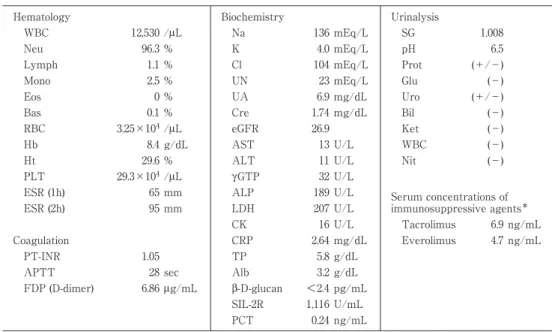
Table 1 Laboratory findings on admission
Hematology Biochemistry Urinalysis
WBC 12,530 /μL Na 136 mEq/L SG 1.008
Neu 96.3 % K 4.0 mEq/L pH 6.5
Lymph 1.1 % Cl 104 mEq/L Prot (+/−)
Mono 2.5 % UN 23 mEq/L Glu (−)
Eos 0 % UA 6.9 mg/dL Uro (+/−)
Bas 0.1 % Cre 1.74 mg/dL Bil (−)
RBC 3.25×10⁴/μL eGFR 26.9 Ket (−)
Hb 8.4 g/dL AST 13 U/L WBC (−)
Ht 29.6 % ALT 11 U/L Nit (−)
PLT 29.3×10⁴/μL γGTP 32 U/L
ESR (1h) 65 mm ALP 189 U/L Serum concentrations of
immunosuppressive agents*
ESR (2h) 95 mm LDH 207 U/L
CK 16 U/L Tacrolimus 6.9 ng/mL
Coagulation CRP 2.64 mg/dL Everolimus 4.7 ng/mL
PT-INR 1.05 TP 5.8 g/dL
APTT 28 sec Alb 3.2 g/dL
FDP (D-dimer) 6.86μg/mL β-D-glucan <2.4 pg/mL SIL-2R 1,116 U/mL
PCT 0.24 ng/mL
*Tested on Day 15 of admission
菌感染症と考えられた.
菌種同定:菌体には,結核菌を疑わせるコード形成 がわずかに認められたため(Fig. 2B),Loopamp 結 核菌群検出試薬キット(栄研化学)を用いて遺伝子検 査を実施したが陰性であり,抗原特異的インターフェ ロン
γ遊離検査(T―スポット.TB Oxford Immu-
notec)も陰性であったことより結核菌である可能性は低いと判断された.そこで,皮膚生検材料から遺伝 子を抽出し,直接,抗酸菌に特異的な
2領域(rpoB,
hsp65)のシークエンス解析を実施した(rpoB:For- ward primer;MF 5ʼ―CGA CCA CTT CGG CAA
CCG-3ʼ,Reverse primer;MR 5ʼ―TCG ATC GGG CAC ATC CGG-3ʼ,hsp65:Forward primer;Tb11 5ʼ―ACC AAC GAT GGT GTG TCC AT-3ʼ,Reverse primer;Tb12 5ʼ―CTT GTC GAA CCG CAT ACC CT-3ʼ)2)3)
.PCR 条件は両領域ともに
95℃3分,(95℃
30
秒,63℃
30秒,72℃
30秒)×37 サイクル,72℃
5
分とした.その結果,両領域ともに
M. haemophilum ATCC29548(Type strain)と100% の相同性を認め(rpoB:328bp/328bp,hsp65:422bp/422bp),塗
抹検査で認められた抗酸菌は
M. haemophilumと同定
された.
Fig. 2 Microscopic examination of the dermatologic lesion.
A, Gram stain; arrows indicate so-called ghost bacteria. B, Ziehl-Neelsen stain.
B A
Fig. 3 Mycobacterium haemophilum colonies on agar plates after 14 days.
A, chocolate agar in 5% CO2 at 35℃. B, blood agar in ambient air at 30℃.
A B
抗酸菌の分離培養:提出された検体はすべて,固形 培地:極東
2% ビット培地SP(極東製薬),液体培地:
ミジット分離培養剤(BD),平板培地:バイタルメディ ア 血液寒天培地(ウマ)(極東製薬)およびバイタル メディア チョコレート寒天培地(極東製薬)に接種 した.固形培地と液体培地は
37℃ で,血液寒天培地は
30℃ 好気,チョコレート寒天培地は35℃ 5%CO2で培養を実施した.この中で,もっとも良好な発育を 示した条件は「チョコレート寒天培地,35℃,5%CO
2」 であった(Fig. 3A).提出検体のうち血液検体に関し ては,十分量の滅菌精製水を加え混和した後,3,000
rpm 20min
遠心,その沈渣に再度,滅菌精製水を加
え混和し,十分溶血させた後に各培地に接種するよう 注意した.最終的に,穿刺吸引した膿および血液から も
M. haemophilumが分離された.
診断:病理検査に提出された後頸部や両下腿,また 右前腕の皮膚生検材料すべてから抗酸菌が検出され た.全身の
CT画像検査ではリンパ節腫大等の異常所 見を認めず,皮膚病変以外の感染巣はあきらかではな かったことから,本症例は
M. haemophilumによる血
流感染を伴う播種性皮膚感染症と診断された.
抗 菌 薬 治 療:入 院
23日 目 よ り
clarithromycin(
CAM 400 mg)+
minocycline(
MINO 200 mg)+
ciprofloxacin(CPFX 400mg)の3
剤の経口抗菌薬に よる治療を開始した.20 日目に
MINOによる嘔気お よび色素沈着が出現したため
rifabutin(RBT 150mg)へ変更した.その後,新たな紅斑の出現も認めず改善 を認めている.腎機能の悪化にも注意し免疫抑制剤の 投与量を適宜調整しながら治療継続.最終的に
CAMは
800mg/日に増量して軽快退院となったが,2カ月
後,発熱と四肢の紅班出現を認め再入院となった.培 養検査は陰性であったが依然,塗抹検査では抗酸菌を 認めており,meropenem(MEPM 1g)が追加された ことで症状は改善し,最終的に
faropenem(FRPM 600
mg)に変更され退院となった.その後,発熱と紅班の出現は認めるもののその頻度は低下しており,1 年 以上治療が継続されている.
考 察
本症例は,腎移植後の患者に発症した
M. haemophi- lumによる播種性非結核性抗酸菌症である.臓器への
Table 2 Summary of Mycobacterium haemophilum infections in transplant patients CaseYearAge (yr) /sexaUnderlying diseasebTransplant- ed organcClinical presentationCulture sourceCultureTreatmentdOutcomeRefer- ence 1201062/Mischemic heart diseaseHeartfever, warm and tender erythematous plaques with undefined borders observed on both shinsbiopsy of skinpositiveCAM+CPFX+RFPimproved11 2201159/Mdiabetic nephropathyKidneycutaneous and subcutaneous nodules on the up- per and lower limbsneedle aspirationpositiveCAM+CPFXimproved12 3201159/Fhepatitis B virus-related cirrhosis, diabetes mellitusLivergeneralized and nodular skin lesionsbiopsy of skinpositiveCAM+MFLX+EB+ AMKresolved13 4201122/Mdysplastic kidneyKidneyprogressive swelling and erythema developed in forearm and palm with paintissue samplespositiveCAM+MFLX+EBimproved14 5201258/Mchronic renal failureKidneyarthralgia, swelling of finger, skin rash, erythema, papules and subcutaneous nodulebiopsy of skinpositiveCAM+RFP+EB+INHresolved15 6201367/Mtype 2 diabetes mellitusKidneyasymptomatic rash and swelling over right thighbiopsy of skinpositiveCAM+CPFXresolved16 7201466/F
hepatocellular carcinoma, ITP, type 2 diabetes mellitus, chronic kidney disease, vitamin B12 deficiency, hysterectomy for menorrhagia, osteoporosis
Livertender and raised pustular skin lesions over face, arms, chest, and legssynovial fluid from the kneepositiveCAM+IPMimproved17 8201454/MAMLStem cellnodules on limbs, thighs, and abdomenbiopsy of skinpositiveCAM+RBT+EBimproved18 9201432/MIgA nephropathyKidney
multiple episodes of epistaxis and sensation of nasal fullnessintranasal mass −CAM+CPFX+RFP ↓ CLDM+CPFX+RFPrelapse19 multiple, painless, non-discharging erythema- tous nodulesbiopsy of skin 10201627/MANCA-associated necrotizing glomerulonephritisKidneyfever, arthralgia, multiple subcutaneous tumors, edema in left knee, lower leg, and handsbiopsy of skin, bonepositiveCAM+CPFX+RBT+ antitubercular therapyresolved20 11201657/FAMLStem cellthigh rashbiopsy of skinpositiveCAM+CPFX+RBTresolved21 12201761/FCrohnʼs disease, chronic venous stasis dermatitisKidneytender erythematous nodule on left forearmbiopsy of tissuepositiveAZM+CPFX+RBTresolved22 13201771/Mhypertension, gouty arthritisKidneyfever, right palm swelling, multiple erythema- tous subcutaneous nodulesfluid aspiratepositiveCAM+CPFX+RFPimproved23 14201714/FPTLDHeartleft arm and lower leg swelling with painbiopsy of tissuepositiveCAM+CPFX+RFPresolved24 15201865/Frenal disease, hypertension, EB virus-positive PTLDKidneyfever, rashbiopsy of skinpositiveCAM+CPFX+RBTdied due to respiratory failure25 16201832/Mendocarditis, gastrointestinal bleeding, hypertensionKidneymultiple painful nodular lesionsbiopsy of tissue−CAM+CPFX+RBTresolved26 17201941/Fchronic renal failureKidneyfever, arthralgia, multiple tender erythema on face, neck, and extremitiesbiopsy for tissue, bloodpositiveCAM+CPFX+RBT+ FRPMpersistedThis case a M, male; F, female b ITP, idiopathic thrombocytopenic purpura; AML, acute myelogenous leukemia; ANCA, antineutrophil cytoplasmic antibody; PTLD, posttransplant lymphoproliferative disorder c Included Stem cell d CAM, clarithromycin; CPFX, ciprofloxacin; RFP, rifampicin; MFLX, moxifloxacin; EB, ethambutol; AMK, amikacin; IPM, imipenem; INH, Isoniazid; RBT, rifabutin; CLDM, clindamycin; AZM, azithromycin; FRPM, faropenem
感染は認めないものの,血液および全身の体表から本 菌が検出された.微生物検査室で実施したグラム染色 が本症例の診断に大きな役割を果たした.微生物検査 に最初に提出された検体は綿棒で採取されており,得 られた検体量も微量で細胞成分もわずかにしか認めな い状況であったが,そのわずかな細胞成分上に,グラ ム染色では染色されない,所謂
ghost状態の菌体 を認め抗酸菌が疑われた(Fig. 2A).病理検査室では 通常チール・ネルゼン染色は実施されているが,抗酸 菌の存在が確認されているかもしくは疑われている検 体に限定されている.同様に,当微生物検査室でもチー ル・ネルゼン染色は通常,抗酸菌検査依頼時にのみ実 施する.本症例は,特徴的なグラム染色所見を見逃さ なかった微生物検査技師による検鏡が診断に大きく寄 与した症例と考える.
M. haemophilum
の細菌学的特徴は,発育に
X因子
(ヘミン)やクエン酸鉄第二アンモニウム等の鉄成分 を要求する点と,至適発育温度が
30〜32℃ である点である
1)4)5).従って,通常,抗酸菌培養に用いられる 固形培地の小川培地や液体培地の
MGIT(Mycobacte-
rium Growth Indicator Tube,BD)やMP抗酸菌培養 ボトル(SYSMEX bioMérieux)等での発育は期待で きないため,Middlebrook7H10 寒天培地等の培地に ヘミン含有のストリップを設置するか,血液寒天培地 等を用いる工夫が必要となる
1)4)5).至適発育温度が
30〜32℃ で あ る 抗 酸 菌 と し て はMycobacterium marinumや
Mycobacterium ulceransが 知 ら れ て い る が
1)4)6),いずれも血液要求性はなく,患者背景や臨床 像が特徴的であることから,両者と
M. haemophilumとの鑑別は比較的容易であると考える.
本症例から分離された
M. haemophilumは
30℃ 血液寒天培地にて発育を認めたが,もっとも発育が良好 であった条件は「チョコレート寒天培地,35℃,5%
CO2
」であった(Fig. 3).チョコレート寒天培地には 血液寒天培地に比し多くのヘミンが含まれているた め,M. haemophilum がより発育し易い環境であった と 考 え ら れ た.さ ら に,M. haemophilum は
5〜10%の
CO2が発育を支持することが知られており
7)8),通 常,微生物検査室で使用している
CO2フラン器を利 用することができる.当院で過去に経験した症例にお いても当該条件がもっとも良好な発育を認めたことか ら,本菌を疑う場合は,30℃ 血液寒天培地での培養 に加え,「チョコレート寒天培地,35℃,5%CO
2」と いう条件で培養を実施することにより,検出感度の向 上が期待できる.また本症例では,血液を接種した
37℃ 培養のMGIT(BD バクテック
MGlT―320(BD)
により培養)が培養
18日で陽転した.これは,患者 血液中に含まれる鉄成分が発育を支持したと推測され
る.M. haemophilum は
30〜32℃ 培養が推奨されているが,37℃ でも発育したとの報告
5)もあることから,
種々の培養温度条件で発育可能であると推測される.
本症例は血流感染を伴う播種性皮膚感染症と診断さ れたが,皮膚生検病理検査により好中球主体の結節性 紅斑を示唆する所見を得たことより,当初,ハンセン 病の可能性も考慮された.実際,
Mycobacterium. lepraeとの鑑別を要した症例
6)や,稀ではあるが混合感染の 症例も報告されており
9),M. haemohilum と
M. lepraeとの鑑別は重要であると考える.M. leprae は人工培 地では発育せず微生物学的診断が困難であることか ら,ハンセン病の診断は臨床像・病理検査所見・末梢 神経所見・細菌学的検査を総合して行われる.しかし 本症例は,患者が高齢ではない
41歳の日本人であっ た点,皮膚組織中の抗酸菌が培地上で検出され,且つ 遺伝子学的に
M. haemophilumと同定された点から考 慮すると,患者がハンセン病である,もしくは
M.leprae
との混合感染の可能性は非常に低いと考えられ
た.実際,菌種同定には
M. lepraeとの鑑別が十分に 可能な遺伝子領域を利用しており
2),誤同定の可能性 は極めて低い.
M. haemophilum
に対しては標準的な薬剤感受性試
験の方法が存在しないため本症例では実施せず,過去 の文献
1)を参考に薬剤を選択し投与量を決定した.そ の後,MINO から
RBTに変更,MEPM が 追 加 さ れ た後に
FRPMに変更され,1 年以上の抗菌薬治療が 継続されている.
原因が明らかとなり抗菌薬治療により改善を認めた が,臨床所見と病理検査所見から当初は
Sweet病が もっとも疑われており
10),診断に時間を要した.病理 検査では好中球主体の結節性紅斑との所見が得られ,
さらに患者は腎移植後で
4剤の免疫抑制剤服薬中の易 感染性状態であるため,より早期に細菌感染を鑑別疾 患として考慮する必要があったと思われる.
M. haemo-philum
による感染症は世界的に症例数が増加してお
り
1),国内も例外ではないと思われる.HIV 陽性患者 や移植患者等の免疫抑制患者が症例の大半を占めてい るが
1),移植患者に限定すると,2010 年以降で本症例 を含め少なくとも
17例が報告されている(Table 2).
中でも腎移植患者の感染症症例が多い印象を受け,臨 床症状としては結節性紅斑や腫脹,発疹が多く共通し ており,出現部位は顔や四肢,腹部など多様である.
培養に関しては,チョコレート寒天培地を用いた
30℃培養という同じ条件であっても,コロニー発育に要し た期間が
3日の症例
14)もあれば
44日の症例
25)も報告さ れているため,培養条件の工夫が重要であり,より早 期に微生物学的診断を行うためには,検体から直接,
遺伝子同定を実施することが有用であると考えられ
る.腎移植後であり,既報と類似した症状を呈してい たことを考慮すると,本患者への抗酸菌感染も早期に 想 定 す べ き で あ っ た と 言 え る.治 療 に は
CAMや
CPFX,RFP
や
RBTが多用されているが,治療困難
例もあり
19),今後の症例報告のさらなる増加と治療成 績の蓄積により,より適切な抗菌薬治療の確立と,薬 剤感受性試験の標準法の開発を期待したい.
謝辞:皮膚所見に関してご助言を頂きました当院皮 膚科 中川幸延先生,清水友理先生,M. leprae との鑑 別に関してご助言を頂きました国立感染症研究所ハン セン病研究センター センター長 石井則久先生に深謝 申し上げます.
利益相反自己申告:著者朝野和典は
MSD(株)から講演料を受けている.
文 献
1)Kelley CF, Armstrong WS, Eaton ME:Dissemi- natedMycobacterium haemophiluminfection. Lan- cet Infect Dis. 2011;11:571―8.
2)Shimizu Y, Nakagawa Y, Kotobuki Y, Kimura K, Hagiya H, Katayama I:Case of Mycobacte- rium haemophilum infection presenting with Sweetʼs syndrome-like reactions. J Dermatol.
2019;46:e319―20.
3)McNabb A, Eisler D, Adie K, Amos M, Ro- drigues M, Stephens G,et al.:Assessment of partial sequencing of the 65-kilodalton heat shock protein gene (hsp65) for routine identifica- tion ofMycobacteriumspecies isolated from clini- cal sources. J Clin Microbiol. 2004;42:3000―11.
4)Itoh S, Kazumi Y, Abe C, Takahashi M:Het- erogeneity of RNA polymerase gene (rpoB) se- quences of Mycobacterium gordonae clinical iso- lates identified with a DNA probe kit and by conventional methods. J Clin Microbiol. 2003;
41:1656―63.
5)Saubolle MA, Kiehn TE, White MH, Rudinsky MF, Armstrong D:Mycobacterium haemophilum: microbiology and expanding clinical and geo- graphic spectra of disease in humans. Clin Mi- crobiol Rev. 1996;9:435―47.
6)斎藤 肇,戸田憲一,松本いづみ,松尾清光,中 永和枝,石井則久:免疫不全患者の皮膚病巣か ら分離されたMycobacterium haemophilumの細菌 学的特性.感染症誌 2004;78:389―97.
7)Ishii K, Ishii N, Nakanaga K, Nakano K, Saito I, Asahina A:Mycobacterium haemophilum infec- tion with prominent facial manifestation mim- icking leprosy. J Dermatol. 2015;42:992―5.
8)Sompolinsky D, Lagziel A, Rosenberg I:Fur- ther studies of a new pathogenic mycobacte- rium (M. haemophilumsp. nov.). Can J Microbiol.
1979;25:217―26.
9)Males BM, West TE, Bartholomew WR:Myco- bacterium haemophiluminfection in a patient with
acquired immune deficiency syndrome. J Clin Microbiol. 1987;25:186―90.
10)SoRelle JA, Beal SG, Scollard DM, Gander RM, Cohen J, Nuara A,et al.:Mycobacterium leprae and Mycobacterium haemophilum co-infection in an iatrogenically immunosuppressed patient. Di- agn Microbiol Infect Dis. 2014;78:494―6.
11)Sagi L, Leshem E, Barzilai A, Baum S, Har- Zahav Y, Rahav G:Mycobacterium haemophilum infection presenting as bilateral cellulitis and an- nular lesion in a heart transplant recipient. Isr Med Assoc J. 2010;12:57―8.
12)Castro-Silva AN, Freire AO, Grinbaum RS, El- mor de Araújo MR, Abensur H, Araújo MR,et al.:CutaneousMycobacterium haemophiluminfec- tion in a kidney transplant recipient after acu- puncture treatment. Transpl Infect Dis. 2011;
13:33―7.
13)Lau SK, Curreem SO, Ngan AH, Yeung CK, Yuen KY, Woo PC:First report of dissemi- natedMycobacterium skin infections in two liver transplant recipients and rapid diagnosis by hsp 65 gene sequencing. J Clin Microbiol. 2011;
49:3733―8.
14)Clement CG, Loeffelholz MJ, Eltorky MA, Tang YW, Williams-Bouyer N:Mycobacterium haemo- philumandHistoplasma capsulatumcoinfection in a renal transplant patient. J Clin Microbiol.
2011;49:3425―8.
15)Takeo N, Hatano Y, Okamoto O, Saruwatari K, Nakanaga K, Ishii N,et al.:Case of Mycobacte- rium haemophilum infection in a Japanese renal transplant patient and a review of Japanese cases. J Dermatol. 2012;39:968―9.
16)Copeland NK, Arora NS, Ferguson TM:Myco- bacterium haemophilumMasquerading as Leprosy in a Renal Transplant Patient. Case Rep Derma- tol Med. 2013;2013:793127.
17)Doherty T, Lynn M, Cavazza A, Sames E, Hughes R:Mycobacterium haemophilum as the Initial Presentation of a B-Cell Lymphoma in a Liver Transplant Patient. Case Rep Rheumatol.
2014;2014:742978.
18)Brissot E, Gomez A, Aline-Fardin A, Lalande V, Lapusan S, Isnard F,et al.:Report of dissemi- nated Mycobacterium haemophiluminfection after double cord blood allo-SCT. Bone Marrow Transplant. 2014;49:1347―8.
19)Ducharlet K, Murphy C, Tan SJ, Dwyer KM, Goodman D, Aboltins C,et al.:RecurrentMyco- bacterium haemophilum in a renal transplant re- cipient. Nephrology (Carlton). 2014;Apr;19
(Suppl 1):14―7.
20)Brix SR, Iking-Konert C, Stahl RA, Wenzel U:
Disseminated Mycobacterium haemophilum infec- tion in a renal transplant recipient. BMJ Case Rep. 2016;bcr2016216042.
21)Baluch A, Pasikhova Y, Snyder M:Successful management of Mycobacterium haemophilum lower extremity cutaneous infection in a matched-unrelated donor stem cell transplant recipient. Transpl Infect Dis. 2017;19: doi:
10.111/tid.12627.
22)Krueger S, Rork J, Lee J, Cornejo K, Gibson L, Daly JS,et al.:Mycobacterium haemophiluminfec- tion in a renal transplant patient with inflamma- tory bowel disease. Dermatol Online J. 2017;
23:pii:13030/qt8vs8509k.
23)Anandh U, Jayanna K:Nontubercular Myco- bacterial Infection in a Renal Allograft Recipi- ent. Indian J Nephrol. 2017;27:478―81.
24)Takeyari S, Hashii Y, Yoshida H, Kogaki S, Ozono K:Mycobacterium haemophilumosteomye- litis in the immunocompromised host. Pediatr Int. 2017;59:1279―81.
25)Jacobs SE, Zhong E, Hartono C, Satlin MJ, Ma- gro CM, Jenkins SG,et al.:Closing the Brief Case : Disseminated Mycobacterium haemophilum Infection in a Kidney Transplant Recipient. J Clin Microbiol. 2018;56:e00562―17.
26)Abolghasemi S, Abbasi F, Tehrani S, Nafar M, Nasiri MJ:Cutaneous Infection WithMycobacte- rium haemophilum in an Immunocompromised Patient. Iran J Kidney Dis. 2018;12:312―4.
A Case of Cutaneously DisseminatedMycobacterium haemophilumInfection Accompanying Blood Stream Infection
Keigo KIMURA1), Hideharu HAGIYA2), Tomomi MITSUI1), Isao NISHI1)& Kazunori TOMONO2)
1)Laboratory for Clinical Investigation and2)Division of Infection Control and Prevention, Osaka University Hospital
Mycobacterium haemophilum is a non-tuberculous mycobacterium that causes systemic infections involv- ing the skin and soft tissue, pulmonary system, bones and joints, particularly in immunocompromised pa- tients. We report herein on a rare case of cutaneously disseminatedM. haemophiluminfection accompanying blood stream infection in a patient following renal transplantation. A 41-year-old Japanese woman who had undergone a kidney transplant 5 years previously was referred to us due to prolonged undetermined fever.
She was taking prednisolone, tacrolimus, everolimus, and mycophenolate mofetil at the time of hospitaliza- tion. In addition to the high fever, the patient suffered from chronic erythema emerging over her body. The results of Gram staining were suggestive of mycobacterium infection, and a genetic analysis based on se- quencing of thehsp65and rpoBgenes finally identified this case to be a disseminatedM. haemophiluminfec- tion. After we initiated combination therapy including clarithromycin, ciprofloxacin, and rifabutin, the pa- tientʼs dermatological condition ameliorated and she was discharged 3 months later in remission.
Diagnosis of non-tuberculous mycobacterial infection is challenging from the aspect of both the clinical and laboratory approaches. In this case, careful observation of Gram staining in laboratory was a clue to the diagnosis of the infection. Among non-tuberculous mycobacteria, M. haemophilum is clinically a rare patho- gen. Difficulty in identifying the pathogen may be a reason for its lower prevalence.M. haemophilum prefers a lower temperature (30-32℃) and requires iron or hemin (e.g. a blood agar plate) for proliferation, although we noticed that a chocolate agar plate in 5% CO2 at 35℃gave the best culture conditions in this case. In case of refractory and unidentified dermatologic diseases, attention should be paid to the possibility of myco- bacterial infection.