Acta Med. Nagasaki 44 : 33-36
Papillary Adenoma of Type 2 Pneumocytes in the Lung
Masataka INADA 1), Naoko YAMASAKI 1), Yoshikazu HIGAMI 1), Yuji SHIGEOKA 2), Jin YAMAIE 2), Isao SHIMOKAWA 1),
1) First Department of Pathology, Nagasaki University School of Medicine 2) Division of Surgery, Obase Hospital
Recent developments in radiologic imaging and thoracoscopic excision techniques have increased the possi- bility of encountering small, benign or premalignant tumors in the lung. In this report, we describe a rare case of papil- lary adenoma of type 2 pneumocytes. A 51-year-old Japanese woman was hospitalized following a traffic acci- dent. Helical CT scan of the chest incidentally detected a nodular ground-glass opacity measuring 8 mm in diameter at the subpleural region of left lung. The nodule was thoracoscopically resected out. Light microscopic examina- tion demonstrated a noninfiltrative tumor consisting of cuboidal cells covering fibrovascular cores; thus, the tumor exhibited a branching papillary appearance. The cuboidal cells showed little nuclear atypia. Mitotic figures, necrosis, and intercellular mucin were absent. The cytoplasm was immunohistochemically stained for surfactant apoprotein A and cytokeratin, though not for carcinoembryonic antigen (CEA), epithelial membrane antigen (EMA), or S100 pro- tein. The morphologic and immunohistochemical findings fulfilled the criteria of papillary adenoma of type 2 pneumocytes.
Key Words: papillary adenoma of type 2 pneumocytes, surfactant apoprotein A
ments in radiologic imaging and thoracoscopic exci- sion techniques have increased the possibility of en- countering such benign or premalignant tumors in the lung. In this report, we describe a case of papillary adenoma of type 2 pneumocytes in the lung. We also discuss the pathologic differentiation among those be-
nign or premalignant tumors.
Case Report
A 51-year-old Japanese woman was hospitalized in July 1998 following a traffic accident. The patient was generally bruised. Radiologic examination was per- formed to evaluate possible internal injuries. Helical computed tomography (CT) of the chest incidentally found a nodular ground-glass opacity measuring 8 mm in diameter at the subpleural region of the left lung
(B'+Z ); no other lesion was noted in the lung itself.
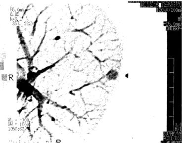
After recovering from the bruise, the patient under- went CT of the chest on December 1998, which dem- onstrated that the size of the lesion remained un- changed (Fig. 1). Physical and laboratory examinations
Introduction
Although most bronchioloalveolar tumors are malig- nant, apparently benign or potentially malignant forms have been described as the following types; papillary adenoma, mucinous cystadenoma, bronchioloalveolar adenoma, also referred to as atypical alveolar hyperplasia, and alveolar adenoma'). Recent develop-
Address Correspondence: Isao Shimokawa, M.D.
First Department of Pathology, Nagasaki University School of Medicine 1-12-4 Sakamoto, Nagasaki City 852-8523 TEL: +81-95-849-7050, FAX: +81-95-849-7052
E-mail: [email protected]
Figure 1. Computed tomography of the chest. Note a nodular ground-glass opacity in the subpleural region of left lung(B'+2, arrowhead).
were within normal limits. The patient underwent thoracoscopic excision biopsy of the nodule. She has been well for 4 months after the operation. She has no history of smoking or exposure to known occupational or environmental toxins.
Pathologic Examination
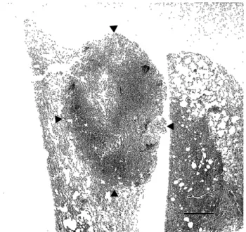
The biopsy specimen was fixed in 10%-buffered formalin. The tumor, 5 x 3 mm in size at the cut surface and located at the subpleural region without any pleural indentation, was circumscribed, but nonencapsulated
Figure 4. The nuclei of cuboidal cells contain delicate chromatin and nuclear membranes No cellular atypia or mitoses is present (Hematoxyline Eosin Stain, bar: 25 p m)
Figure 2. Low power view of the nodule. The tumor is cir- cumscribed but not encapsulated (arrowhead. bar; 1 mm).
(Fig. 2). These sectioned specimens were embedded in paraffin and cut into 4 u m-thick sections for routine stains and immunohistochemistry. The antibodies used for immunohistochemistry were as follows: anti- sur- factant apoprotein A (DAKO JAPAN, Co. Ltd., Tokyo), cytokeratin (DAKO Corporation, Carpinteria, CA), carcinoembryonic antigen (CEA; Immuno-Biological Laboratories, Fujioka, Gunma, Japan), epithelial mem- brane antigen (EMA; Nichirei Co. Ltd., Tokyo), and
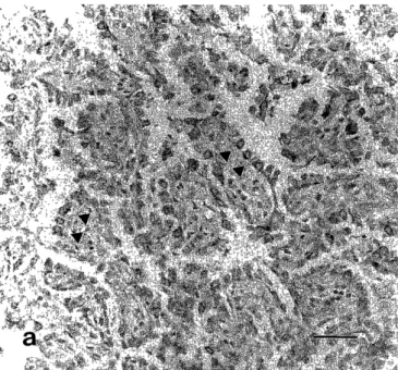
S100 protein (DAKO A/ S, Glostrup, Denmark). Light microscopic examination revealed papillary structures in the tumor (Fig. 3). Cuboidal cells with round to oval nuclei containing delicate chromatin and nuclear membranes covered fibrous cores (Fig. 4). We noted no histologic features of carcinoma such as cellular atypia, necrosis, or mitoses. The cytoplasm was immuno- histochemically stained for surfactant apoprotein A and cytokeratin, though not for CEA, EMA or S 100 pro- tein (Figure 5). The histologic and immunohistochemical findings fulfilled the diagnostic criteria of papillary adenoma of type 2 pneumocytes23).
Figure 3. Cuboidal cells cover the papillae containing fibrovascular cores (Hematoxyline Eosin Stain, bar: 100 u m).
Figure 5. a) Tumor cells are positive for surfactant apoprotein A (arrowhead, bar; 50 fi m).
studies have also shown that the tumor cells exhibit a Clara cell-specific antigens' or surfactant apoproteins in the cytoplasm"). Although most authors have regarded this type of tumor as benign, Mori et al.') report one case in which tumor cells invaded into the surround- ing lung tissues and a small vessel.
The differential diagnosis of papillary adenoma of type 2 pneumocytes includes bronchioloalveolar ade- noma, bronchioloalveolar carcinoma, alveolar adenoma, and sclerosing hemangioma3). Bronchioloalveolar ade- noma, a possible precursor lesion of bronchioloalveolar carcinoma, exhibits a lepidic growth rather than a pap- illary pattern, and greater cytologic atypia than papil- lary adenoma'). Most cases are found in the lung adja- cent to bronchioloalveolar carcinoma or bronchogenic adenocarcinoma'). Alveolar adenoma, first identified by Yousem and Hochholzer9), presents as a solitary subpleural nodule. Microscopically, it is composed of multiple cystic spaces filled with serous or floccular material or by blood. The lining of the cysts is largely inapparent or consisting of flattened cells that are immunohistochemically uncertain in the cell origin.
Sclerosing hemangioma, originally described by Liebow and Hubbell"), has been thought to represent epithelial neoplasms derived from bronchiolar epithe- lial cells, type 2 pneumocytes, or Clara cells. It shows a greater diversity of histopathologic patterns that Katzenstein et al.") have categorized as solid, hemorrhagic, papillary, and sclerotic patterns.
The histologic and immunohistochemical findings in the present case favor the diagnosis of papillary ade- noma of type 2 pneumocytes, although some parts of the tumor might mimic the pattern of bronchioloa- lveolar adenoma.
Acknowledgements
Figure 5. b) Tumor cells were negative for CEA (bar; 50 u m).
The authors wish to thank Ms. Yuko Moriyama for her technical assistance. This case study was con- ducted as a part of the research seminar of the 4th gear medical students of Nagasaki University School of Medicine.
Comments
Spencer et al.') have noted unusual, benign, non- invasive papillary tumors in which the lining cells re- semble Clara cells. An ultrastructural study') demon- strates concentric lamellar inclusions in the cytoplasm of tumor cells, indicating their similarity to Clara cells or type 2 pneumocytes. Subsequent immunohistochemical
Reference
1) Saldana MJ, Mones JM, Garcia-Moliner M, Frable WJ. Localized diseases in the bronchi and lung. In Principles and Practice of
Surgical Pathology and Cytopathology. 3rd. ed. (Silverberg SG,
Delellis RA, Frable WJ eds.; Churchill Livingstone Inc., New York)
pp. 1200-1203, 1997
2) Noguchi M, Kodama, T, Shimosato Y, Koide T, Naruke T, Singh G, Katyal SL. Papillary adenoma of type 2 pneumocytes Am J Surg
Pathol 10: 134-139, 1986
3) Colby TV, Koss MN, Travis WD. Tumor of the Lower Respiratory
Tract. Fascicle 13. In Atlas of Tumor Pathology. Third Series. pp.
57-63, 1994
4) Spencer H, Dail DH, Arneaud A. Non-invasive bronchial epithelial papillary tumors. Cancer 45: 1486-1497, 1980
5) Fantone JC, Geisinger KR, Appelman HD. Papillary adenoma of the lung with lamellar and electron dense granules: An ultrastructural
study. Cancer 50: 2839-2844, 1982
6) Hegg CA, Flint A, Singh G. Papillary adenoma of the lung. Am J Clin Pathol 97: 393-397, 1992
7) Fine G, Chang C-H. Adenoma of type 2 pneumocytes with
oncocytic features. Arch Pathol Lab Med 115: 797-801, 1991 8) Mori M, Chiba R, Tezuka F, et al. Papillary adenoma of type II
pneumocytes might have malignant potential. Virchows Arch 428:
195-200, 1996
9) Yousem SA, Hochholzer L. Alveolar adenoma. Hum Pathol 17:
1066-1071, 1986
10) Liebow AA, Hubbell DS. Sclerosing hemangioma (histiocytoma,
xanthoma) of the lung. Cancer 9: 53-75, 1956
11) Katzenstein AL, Gmelich JT, Carrington CB. Sclerosing hemangioma of the lung. a clinicopathologic study of 51 cases. Am
J Surg Pathol 4: 343-356, 1980