TUMSAT-OACIS Repository - Tokyo University of Marine Science and Technology (東京海洋大学)
Purification and characterization of
metal-binding proteins from the digestive
gland of the Japanese scallop Mizuhopecten
yessoensis
著者
Gao Jialong, Ishizaki Shoichiro, Nagashima
Yuji
journal or
publication title
Fisheries Science
volume
82
number
2
page range
337-345
year
2015-12-23
権利
(c) 2016 Japanese Society of Fisheries Science
and Springer Japan. This is the author's
version of the work. It is posted here for
your personal use. To
cite/redistribute/reproduce this work, the
Publisher's version in
https://doi.org/10.1007/s12562-015-0950-z
should be used, and obtain permission from
Publishers, if required.
URL
http://id.nii.ac.jp/1342/00001939/
ホタテガイの中腸腺由来金属結合タンパク質の分離 高加龍ab,石崎松一郎b*, 長島裕二b a広東海洋大学食品科技学院,b東京海洋大学大学院海洋科学技術研究科 ホタテガイをカドミウム、銅または鉛を含む人工海水に暴露した後、金属の蓄積能およ び金属結合タンパク質の存在を調べた。いずれの金属も中腸腺に顕著に蓄積され、その蓄積 量はカドミウム、銅、鉛の順に高かった。中腸腺から分子量約 28、37 および 42kDa の金属結 合タンパク質が精製され、これらのアミノ酸部分配列解析により、Coccidioides immitisの
calcium-binding protein またはPleurocapsa sp.の ion-transporter 類似タンパク質と高い 相同性を示すことを明らかにした、これらのタンパク質はホタテガイの金属蓄積または解毒 メカニズムに関与していることが示唆された。
Purification and characterization of metal-binding proteins from the digestive gland of
1
the Japanese scallop Mizuhopecten yessoensis
2
3
Jialong Gaoa,b, Shoichiro Ishizakib*, and Yuji Nagashimab
4
5
aCollege of Food Science and Technology, Guangdong Ocean University, Haida Road 1,
6
Mazhang, Zhanjiang 524088, China
7
bGraduate School of Marine Science and Technology, Tokyo University of Marine Science and
8
Technology, 4-5-7 Konan, Minato, Tokyo 108-8477, Japan
9 10 *Corresponding author: 11 Dr. Shoichiro Ishizaki 12
Graduate School of Marine Science and Technology, Tokyo University of Marine Science and 13
Technology 14
Konan, Minato, Tokyo 108-8477, Japan 15
Tel & Fax: +81-3-5463-0614 16
17
Jialong Gao, [email protected]
18
Shoichiro Ishizaki, [email protected]
19
Yuji Nagashima, [email protected]
20 21 22
Manuscript Click here to download Manuscript Manuscript for Fish Sci.doc
Click here to view linked References
1 2 3 4 5 6 7 8 9 10 11 12 13 14 15 16 17 18 19 20 21 22 23 24 25 26 27 28 29 30 31 32 33 34 35 36 37 38 39 40 41 42 43 44 45 46 47 48 49 50 51 52 53 54 55 56 57 58 59
Abstract
23
24
Marine bivalves accumulate high concentrations of potentially toxic heavy metals in their 25
tissues. We investigated accumulation patterns of cadmium (Cd), copper (Cu), and lead (Pb) 26
in tissues of the Japanese scallop Mizuhopecten yessoensis and clarified that their metal 27
accumulations were associated with certain intracellular metal-binding proteins, after 28
exposure to artificial seawater containing Cd, Cu, or Pb. The scallop was demonstrated to 29
accumulate higher concentrations of Cd than Cu and Pb, and most of the metals were detected 30
in the digestive gland. We purified metal-binding proteins from the digestive gland and 31
performed a preliminary characterization. Three proteins with molecular masses of 32
approximately 28, 37, and 42 kDa were isolated by gel-filtration and anion-exchange column 33
chromatography. Partial amino acid sequences show high sequence similarity to 34
metal-binding proteins and ion-transporters. Metalloprotein profiles in the digestive gland 35
indicated that some proteins were upregulated after metal exposure. We suggest that these 36
proteins are involved in mechanisms of metal accumulation and detoxification in M. 37
yessoensis.
38
39
Key words: Scallop; heavy metals; bioaccumulation; purification; metal-binding protein
40 41 42 43 1 2 3 4 5 6 7 8 9 10 11 12 13 14 15 16 17 18 19 20 21 22 23 24 25 26 27 28 29 30 31 32 33 34 35 36 37 38 39 40 41 42 43 44 45 46 47 48 49 50 51 52 53 54 55 56 57 58 59
44
Introduction
45
46
Heavy metals are natural components of the Earth’s crust, and heavy metals from both natural 47
and anthropogenic sources readily accumulate in marine sediment. This is especially true in 48
coastal zones, which often receive chemical input from many diverse sources of 49
contamination [1]. Heavy metals can enter the aquatic food chain through direct consumption 50
of water or biota and can potentially accumulate at each level of the food chain, which may 51
cause humans to amass a quantity of heavy metals through diet [2]. Therefore, heavy metals 52
are considered a serious environmental threat. 53
Although marine bivalves have no clear evidence of adaptive immunity [3, 4], they can 54
survive and reproduce in severely contaminated environments for decades and bioaccumulate 55
several metals [5, 6]. Previous research proved that metal-binding proteins called 56
metallothioneins (MTs) play a key role in biochemical detoxification of potentially toxic 57
metals [7, 8]. MTs are well known as low molecular mass (6–7 kDa) cytoplasmic 58
metal-binding proteins that are ubiquitous in eukaryotes [9]. They are also known to play 59
important biological roles such as essential metal homeostasis, detoxification of trace metal 60
ions, and protection against free radicals and intracellular oxidative damage [10, 11]. Recently, 61
considerable research has focused on diversity in inducibility of MTs by metals in bivalves 62
such as oysters Crassostrea corteziensis [12] and Crassostrea gigas [13], clams Meretrix 63
meretrix [14], Mactra veneriformis [15], Scapharca inaequivalvis [16] and Cerastoderma 64 1 2 3 4 5 6 7 8 9 10 11 12 13 14 15 16 17 18 19 20 21 22 23 24 25 26 27 28 29 30 31 32 33 34 35 36 37 38 39 40 41 42 43 44 45 46 47 48 49 50 51 52 53 54 55 56 57 58 59
edule [11], and mussels Mytilus galloprovincialis [10] and Mytilus sp. [17]. However, until
65
now, the only report on MTs in scallops has been on the bay scallop Argopecten irradians 66
[18]. 67
The Japanese scallop, Mizuhopecten yessoensis (Jay, 1857), is one of the most 68
economically important marine mollusks living in the cold seas along the coasts of the 69
northern islands of Japan, the northern part of the Korean Peninsula, and the Sakhalin and 70
Kuril islands [19, 20]. Previous studies of M. yessoensis focused on metal accumulation and 71
metal bioaccumulation patterns suggested that scallops have evolved a natural capacity to 72
accumulate, detoxify, and store metals in their tissues [21, 22]. Hence, it is necessary to study 73
the ability to bioaccumulate different heavy metals and corresponding metal-binding proteins 74
including MTs. 75
In the present work, bioaccumulation and tissue distribution of heavy metals in the scallop 76
M. yessoensis were examined by using laboratory experiments under controlled conditions.
77
Accumulation of metals was monitored using living shells exposed to seawater containing 78
cadmium (Cd), copper (Cu), and lead (Pb). Furthermore, to clarify the function of the 79
metal-rich proteins in M. yessoensis after acute metal exposure, proteins in the digestive gland 80
were purified by gel-filtration and ion-exchange chromatography, and then separated by 81
electrophoresis. Finally, the isolated proteins were characterized by partial amino acid 82
sequence analysis. 83
84
Materials and methods
85 1 2 3 4 5 6 7 8 9 10 11 12 13 14 15 16 17 18 19 20 21 22 23 24 25 26 27 28 29 30 31 32 33 34 35 36 37 38 39 40 41 42 43 44 45 46 47 48 49 50 51 52 53 54 55 56 57 58 59
86
Scallops and metal exposure
87
Adult Japanese scallops M. yessoensis were collected from Tokyo central wholesale fish 88
market, Japan. Scallops (shell length: 11.6 ± 0.3 cm, weight: 193.7 ± 31.0 g) were maintained 89
in aquariums for 7 days prior to experiments. They were fed a commercial diet of Ultra Clam 90
(Fauna Marin, Holzgerlingen, Germany), which is a special food for filter feeders without 91
heavy metals and was added every day at a concentration of 0.1 g per 100 l seawater during 92
both acclimatization and exposure periods. Glass tanks with dimensions 90 × 45 × 45 cm 93
were filled with 40 l of synthetic seawater (salinity = 3.3%) and aerated by a diffuser system. 94
Temperature was set at 18 ± 1oC and the photoperiod was fixed at 12 h using artificial light 95
sources. After the acclimation period, 60 scallops were used for metal exposure experiment, 96
and randomly divided into 6 groups, each group with 10 individuals. Cd was added as 97
CdCl2·2.5H2O at the concentrations of 200 µg/l and 400 µg/l. Cu and Pb were added as
98
CuCl2·2H2O and Pb(NO3)2 at the concentrations of 100 µg/l and 200 µg/l, respectively. The
99
water in each tank was changed every two days to ensure no accumulation of toxic materials 100
from the scallops, which could change water quality. As a control, 10 scallops without metals 101
were maintained under similar conditions. 102
After each sampling time (7 and 10 days), 5 scallops were randomly selected from both 103
the control and metal-treated groups. Tissues of the digestive gland, gill, mantle, gonad, and 104
adductor muscle were collected separately from individual scallop. Tissues for investigation 105
of metal accumulation were completely dried at 80oC for more than 24 h until a constant 106 1 2 3 4 5 6 7 8 9 10 11 12 13 14 15 16 17 18 19 20 21 22 23 24 25 26 27 28 29 30 31 32 33 34 35 36 37 38 39 40 41 42 43 44 45 46 47 48 49 50 51 52 53 54 55 56 57 58 59
weight was reached, and then the dried tissues were stored in a desiccator at room temperature 107
until they were processed. The digestive glands used for protein extraction were stored at 108
–80oC for further use.
109
110
Quantification of metals in the tissues of scallops
111
Approximately 50 mg aliquots of dried tissue was digested with 14 mol/l nitric acid (HNO3)
112
for 24 h at room temperature and then heated at 80oC for 6 h until totally digested [23]. 113
Milli-Q water was added to the digested samples to dilute the HNO3 to 2.0% (v/v). Finally,
114
the samples were filtrated with 0.22 µm membranes before atomic absorption 115
spectrophotometry (AAS) measurement. In control and metal-exposed scallops, Cd, Cu, and 116
Pb levels were determined using a Hitachi Z-2000 AAS (Hitachi, Japan). All metal 117
concentrations are given on a dry weight basis (µg/g dry wt), the values are the mean ± SD of 118
five individual experiments performed in triplicate. 119
120
Extraction and separation of metalloproteins
121
Approximately 30 g of the digestive glands from scallops exposed to metals and non-exposed 122
scallops were pooled separately and homogenized in 3 volumes of ice-cold 10 mM Tris-HCl 123
buffer (pH 8.6), which contained 1 mM dithiothreitol (DTT) and 0.1 mM 124
phenylemethylsulfonyl fluoride (PMSF) as an antioxidant and antiproteolytic mixture. The 125
homogenate was centrifuged at 30,000 × g for 40 min (4oC). The supernatant (subcellular 126
fraction) containing metalloproteins was partially purified by acetone fractionation (50–80%) 127 1 2 3 4 5 6 7 8 9 10 11 12 13 14 15 16 17 18 19 20 21 22 23 24 25 26 27 28 29 30 31 32 33 34 35 36 37 38 39 40 41 42 43 44 45 46 47 48 49 50 51 52 53 54 55 56 57 58 59
as previously described [24, 25]. Briefly, cold acetone (–20oC) was added to the supernatant 128
to a final concentration of 50%. The sample was maintained at 4oC for 30 min with magnetic
129
stirring and then centrifuged at 14,500 × g for 10 min (4oC). The pellet was discarded and the 130
acetone concentration of the supernatant was raised to 80%. The preparation was maintained 131
at 4°C for 40 min with magnetic stirring and then centrifuged again at 14,500 × g for 10 min 132
(4oC). The 80% acetone-precipitated pellet was resuspended in 10 mM Tris-HCl buffer (pH 133
8.6), which contained 1 mM DTT and 0.1 mM PMSF. Protein concentrations of all samples 134
were measured in triplicate by the Bradford method (Quick Protein Assay, BioRad, Hercules, 135
CA, USA). Metal concentrations in proteins were determined in triplicate by AAS as 136
previously described. Fifty mg of dissolved 50-80% acetone pellet from scallops in the 137
non-exposed group and those exposed to 200 µg/l of CdCl2, CuCl2, or Pb(NO3)2 was loaded
138
onto a Sephadex G-50 gel-filtration column (2.6 × 100 cm) equilibrated with 10 mM Tris-HCl 139
buffer (pH 8.6), that contained 1 mM DTT and 0.1 mM PMSF. The samples were eluted with 140
the same buffer at a flow rate of 0.5 ml/min after sample application. To detect proteins and 141
thiolate-metal linkage in protein, absorbance of the eluted fractions at 280 and 254 nm was 142
monitored continuously with an ultraviolet spectrometry (Shimadzu UV-1800, Shimadzu, 143
Japan) [26]. Cd, Cu or Pb levels were also continuously monitored with AAS. The column 144
was calibrated for molecular weight estimation with bovine serum albumin (66 kDa), bovine 145
erythrocyte carbonic anhydrase (29 kDa), horse cytochrome C (12.4 kDa), and aprotinin from 146
bovine lung (6.5 kDa) (Sigma-Aldrich, St. Louis, MO, USA). Cd-containing fractions were 147
pooled and applied directly onto a Mono QTM 5/50 GL column (GE Healthcare, Pittsburgh, 148 1 2 3 4 5 6 7 8 9 10 11 12 13 14 15 16 17 18 19 20 21 22 23 24 25 26 27 28 29 30 31 32 33 34 35 36 37 38 39 40 41 42 43 44 45 46 47 48 49 50 51 52 53 54 55 56 57 58 59
PA, USA). The column was equilibrated with buffer A (10 mM Tris-HCl, pH8.6). Following a 149
wash with buffer A, proteins were eluted with a linear gradient of buffer B (250 mM Tris-HCl, 150
pH 8.6) in buffer A at a flow rate of 0.5 ml/min. UV intensity of proteins on 254 nm was 151
monitored by a MD-2010 UV detector (JASCO, Tokyo, Japan). Cd contents of the eluted 152
peaks were determined by AAS as described above. Purified Cd-binding protein samples were 153
analyzed by sodium dodecyl sulfate-polyacrylamide gel electrophoresis (SDS-PAGE). 154
155
SDS-PAGE and 2-Dimensional (2-D) electrophoresis
156
Proteins extracted from the digestive glands of scallops were subjected to SDS-PAGE (12.5% 157
acrylamide). For further analysis, 2-D electrophoresis was also performed. Fifty µg aliquot of 158
each protein sample was diluted with 125 µl of rehydration buffer (7.0 M urea, 2.0 M thiourea, 159
4.0% CHAPS (w/v), 20 mM DTT, 2 mM tributylphosphine and 0.4% (w/v) pharmalyte 3–10), 160
and applied to a 7 cm linear IPG strip (pH 3–10, GE Healthcare). Isoelectric focusing (IEF) 161
was conducted using an Ettan IPGphor Ⅱ system (GE Healthcare) at 300 V for 45 min, 300 to 162
1000 V for 30 min, 1000 to 5000 V for 1.2 h, and 500 V for 25 min, successively. The focused 163
IPG strips were reduced for 25 min with 1.0% DTT (w/v) in equilibration buffer (50 mM 164
Tris-HCl, 6.0 M urea, 30% (v/v) glycerol, 2% (w/v) SDS, 0.001% (w/v) bromophenol blue, 165
pH 8.8), followed with alkylation for 25 min with 2.5% (w/v) iodoacetamide in the 166
equilibration buffer. After equilibration, the strips were subjected to SDS-PAGE (5–20% 167
acrylamide) at 20 mA per gel. Finally, the proteins were visualized by staining with a Rapid 168
CBB Staining kit (Kanto Chemicals, Tokyo, Japan). 169 1 2 3 4 5 6 7 8 9 10 11 12 13 14 15 16 17 18 19 20 21 22 23 24 25 26 27 28 29 30 31 32 33 34 35 36 37 38 39 40 41 42 43 44 45 46 47 48 49 50 51 52 53 54 55 56 57 58 59
170
Measurement of ultraviolet absorption spectrum
171
The Cd-containing fraction obtained from Sephadex G-50 was assessed by ultraviolet 172
absorption spectroscopy from 200 to 400 nm (Shimadzu UV-1800, Shimadzu, Japan). In 173
addition, the Cd-containing fraction was acid-hydrolyzed with 0.1 M HCl and 0.5 M EDTA at 174
room temperature for 20 min, and then its UV spectrum was also examined as described 175
above. 176
177
Amino acid sequencing
178
Following SDS-PAGE, purified protein was transferred to a PVDF membrane, and then 179
stained with CBB. The target bands were cut out and analyzed in a protein sequencer (ABI 180
Procise 491HT, Applied Biosystems, Nippi Research Institute of Biomatrix, Japan). 181
182
Data analysis
183
Values are expressed as mean ± SD. SPSS software (version 20, IBM, Armonl, NY, USA) was 184
used for statistical analysis. Data from control scallops and metal-exposed scallops were 185
compared using one-way analysis of variance (ANOVA) and statistically different treatments 186
were identified using Duncan’s test. All differences were considered significant at p < 0.05. 187
The letters a, b, c, and d indicate significant differences between groups. 188 189 Results 190 1 2 3 4 5 6 7 8 9 10 11 12 13 14 15 16 17 18 19 20 21 22 23 24 25 26 27 28 29 30 31 32 33 34 35 36 37 38 39 40 41 42 43 44 45 46 47 48 49 50 51 52 53 54 55 56 57 58 59
191
Concentrations of Cd, Cu, and Pb in different tissues of scallops
192
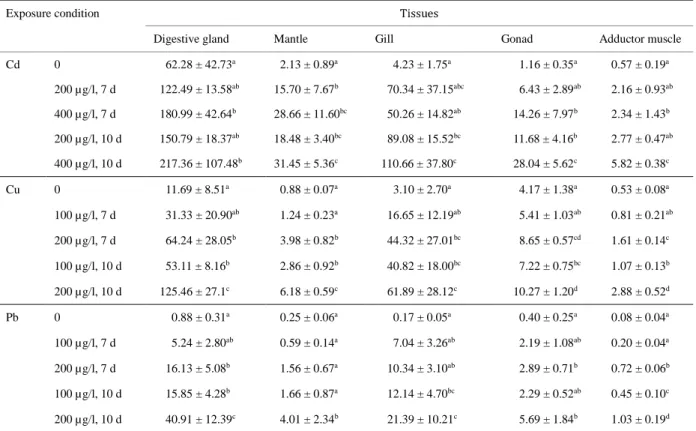
Concentrations of metal in tissues of control and exposed scallops M. yessoensis were 193
measured by AAS (Table 1). In tissues from control scallops, the highest Cd concentration 194
(62.28 ± 42.73 µg/g dry wt; p < 0.05) was observed in the digestive gland. This was 14 times 195
higher than that observed in gills (4.23 ± 1.75 µg/g dry wt), which had the second highest 196
concentration. Similarly, the digestive gland displayed the highest Cu (11.69 ± 8.51 µg/g dry 197
wt; p < 0.05) and Pb (0.88 ± 0.31 µg/g dry wt; p < 0.05) concentrations among tissues, Cu and 198
Pb levels in the digestive gland were 2 times higher than that in gonads, which had the second 199
highest concentrations.Following exposure to metals, metal concentrations in different tissues 200
increased globally. Moreover, the digestive gland displayed highest concentrations for all 201
metals, with the exception of scallops exposed to 100 µg/l of Pb for 7 d. Although a 202
significant increase in Cd concentration in the digestive gland of scallops was observed with 203
400 µg/l of CdCl2 exposure (p < 0.05), there was no significant difference with exposure time
204
(p > 0.05). In contrast, Cu and Pb concentrations in the digestive glands of the exposed 205
scallops presented significant differences (p < 0.05) and both showed significant differences 206
with exposure time (p < 0.05), with the exception of scallops exposed to 100 µg/l of CuCl2
207
and Pb(NO3)2 for 7 days. Given that the digestive gland is considered the major tissue that
208
accumulates trace elements in Japanese scallops, the digestive glands of scallops were 209
dissected and homogenized to investigate protein profiles. 210 211 1 2 3 4 5 6 7 8 9 10 11 12 13 14 15 16 17 18 19 20 21 22 23 24 25 26 27 28 29 30 31 32 33 34 35 36 37 38 39 40 41 42 43 44 45 46 47 48 49 50 51 52 53 54 55 56 57 58 59
2-D electrophoretic analyses
212
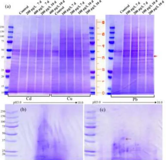
Figure 1 presents electrophoretic analysis of extracts obtained from the digestive glands. 213
SDS-PAGE analysis revealed that some proteins with molecular weights of approximately 214
100, 50, 42, 37, and 28 kDa were differentially expressed during exposure to heavy metals. 215
The most marked difference among different exposure groups was a band at approximately 42 216
kDa, which was upregulated when exposed to all three metals. On the other hand, some bands 217
(approximately 100 kDa) were significantly appeared when Cu and Pb ions were exist (Fig.
218
1a). Further analysis by 2-D electrophoresis also showed a distinctive difference between 219
control scallops and scallops exposed to 200 µg/l of CdCl2 for 7 days, with a protein spot of
220
approximately 42 kDa that had an acidic pI (red arrow in Fig. 1c). 221
222
Protein purification
223
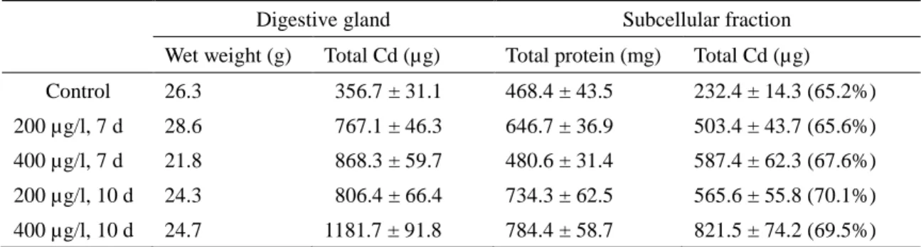
Results of AAS analyses of the digestive gland and its extracts indicated that more than 65% 224
of the total Cd accumulated in the digestive gland was detected in the subcellular fractions, 225
irrespective of metal exposure conditions (Table 2). To purify metal-binding proteins, extracts 226
from the digestive glands of control and metal-exposed scallops were subjected to 227
gel-filtration. Figure 2 shows the Sephadex G-50 column elution profiles of resuspended 228
pellets obtained from the 50–80% acetone precipitations. The profiles at 280 nm indicated 229
two major peaks and a minor peak, the first with relatively high molecular weight (> 25 kDa), 230
corresponding to void volume of the column, the second with molecular weight less than 10 231
kDa, and the minor peak with low molecular weight (< 1.5 kDa) at around fraction number 232 1 2 3 4 5 6 7 8 9 10 11 12 13 14 15 16 17 18 19 20 21 22 23 24 25 26 27 28 29 30 31 32 33 34 35 36 37 38 39 40 41 42 43 44 45 46 47 48 49 50 51 52 53 54 55 56 57 58 59
100 in Fig. 2c and d, corresponding to bed volume of the column. However, metals Cd, Cu, 233
and Pb were detected at only the first peak, which indicated that these metals were bound to 234
high molecular weight substances.
235
The Cd-containing fractions were subjected to further purification on Mono QTM 5/50 GL 236
ion exchange HPLC. Eluted fractions were monitored by absorbance at 254 nm. As shown in 237
Fig. 3a, at least four peaks were eluted at the retention times of 8, 28, 65, and 93 min, and 238
then the unabsorbed fraction, fraction 1, and fraction 2 were pooled separately. The 239
concentrations of Cd in three fractions were 42, 28, and 55 µg/mg protein of Cd, and the Cd 240
amounts of these fractions were 5.7, 7.3, and 33.1 µg, respectively. Therefore, it is indicated 241
that fraction 2 mainly contained Cd-binding proteins. Results of SDS-PAGE analysis revealed 242
that fraction 2 consisted of two clear bands with molecular weights of approximately 42 and 243
37 kDa, in unabsorbed fraction and fraction 1, there were one clear band with a molecular 244
weight of approximately 37 kDa and some faint bands with molecular weights of 245
approximately 28 and 22 kDa (Fig. 3b). 246
247
Characterization of Cd-binding proteins
248
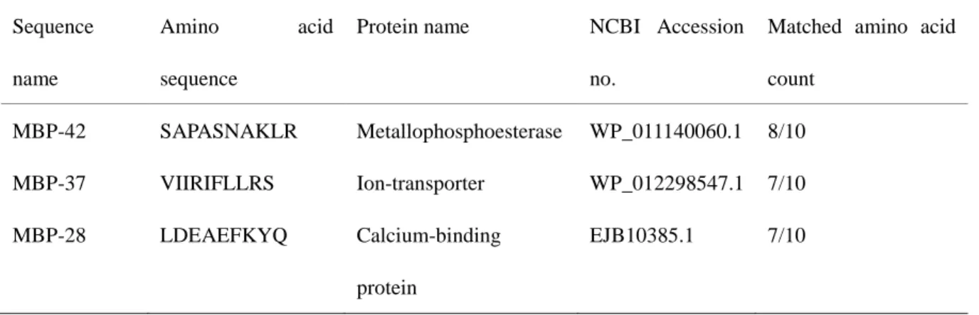
Two components with relative high molecular weights of approximately 42 and 37 kDa were 249
detected in the main Cd-containing fraction 2, which designated to metal-binding protein 250
(MBP)-42 and MBP-37, respectively. To confirm whether these proteins are related to 251
metal-binding, amino acid sequence analysis was carried out. N-terminal amino acid 252
sequences of MBP-42 and MBP-37 were determined as follows: SAPASNAKLR and 253 1 2 3 4 5 6 7 8 9 10 11 12 13 14 15 16 17 18 19 20 21 22 23 24 25 26 27 28 29 30 31 32 33 34 35 36 37 38 39 40 41 42 43 44 45 46 47 48 49 50 51 52 53 54 55 56 57 58 59
VIIRIFLLRS, respectively. Additionally, MBP-28 from the unabsorbed fraction was 254
determined to be LDEAEFKYQ. A database similarity search using these partial amino acid 255
sequences revealed high identities with some proteins of metallophosphoesterase 256
(WP_011140060.1), metal-binding proteins (EJB10385.1) or ion-transporter 257
(WP_012298547.1) (Table 3). 258
All of metal-containing fractions eluted from Sephadex G-50 exhibited relatively high UV 259
absorption at 254 nm (Fig. 2), which indicated that metals were linked to the metal-binding 260
protein fractions with high thiol content. The OD254/280 ratio of Cd-exposed scallops was 261
1.61, which was higher than that of Cu (1.40) and Pb (1.26) (Fig. 2b, c, and d). An UV 262
absorbance spectrum of pooled Cd-containing fractions showed a characteristic pattern in the 263
200–400 nm range, with a broad peak at 256 nm that is similar to the typical pattern of Cd 264
thiolate cluster [24]. The procedure of treating Cd-rich fractions with HCl at pH 1 removed 265
metal ions from metal-binding proteins, which then led to a red shift of the absorption 266
maximum of its retinal from 256 nm to 267 nm (Fig. 4). 267
268
Discussion
269
270
The present work confirmed the high metal bioaccumulation potential of the Japanese scallop 271
M. yessoensis. The digestive gland is the organ to accumulate metals at the highest levels in
272
the scallop not only in natural but also metal-exposed conditions. In this study, to assess the 273
contamination status and bioaccumulation ability of the Japanese scallop M. yessoensis, 274 1 2 3 4 5 6 7 8 9 10 11 12 13 14 15 16 17 18 19 20 21 22 23 24 25 26 27 28 29 30 31 32 33 34 35 36 37 38 39 40 41 42 43 44 45 46 47 48 49 50 51 52 53 54 55 56 57 58 59
concentrations of Cd, Cu, and Pb were analyzed in relative to metal exposure (Table 1). 275
Interestingly, in the normal state without metal exposure, the digestive gland accumulated 276
most of the elements. In contrast, other tissues seemed to play a minor role in the storage of 277
Cd, Cu, and Pb, although they may play a major role in uptake and transfer of trace elements 278
[27]. Previous studies have pointed out the ability of various scallop species to accumulate 279
high trace elements in their tissues, even in remote areas such as Antarctica or Arctic Oceans 280
where are not subjected to direct anthropogenic inputs [25, 28, 29]. Furthermore, it is reported 281
that Cd in the digestive glands of 1–8 year-old M. yessoensis collected from the Sea of Japan 282
increased from 39 to 400 µg/g dry wt, but in the muscle, mantle and gill did not exceed 6 µg/g 283
dry wt in the oldest scallop [30]. This suggests either a higher Cd bioaccumulation capacity of 284
this scallop species and/or lower bioavailability of Cu and Pb for M. yessoensis in fields of 285
cultured scallop. Therefore, we compared bioaccumulation ability of the scallops by exposure 286
to Cd, Cu, and Pb at the concentration of 200 µg/l. Following exposure, metal concentrations 287
in all scallop tissues increased globally and most of metals were detected in the digestive 288
gland where the concentration of Cd was significantly higher than that of Cu and Pb (p < 0.05, 289
Table 1). This indicates that M. yessoensis can accumulate different metals such as Cd, Cu, 290
and Pb, but that the bioaccumulation ability differs such that: Cd > Cu > Pb. 291
The concentration of Cd in the digestive gland of M. yessoensis is age-specific [21]. Cd in 292
the cytoplasmic fraction of the digestive gland is also age-specific; 71.7% of this metal was 293
detected in subcellular fractions of the digestive glands from l-year-old scallops, compared 294
with 98.8% in 8-year-olds [30]. In our study, more than 65% of metals were found in the 295 1 2 3 4 5 6 7 8 9 10 11 12 13 14 15 16 17 18 19 20 21 22 23 24 25 26 27 28 29 30 31 32 33 34 35 36 37 38 39 40 41 42 43 44 45 46 47 48 49 50 51 52 53 54 55 56 57 58 59
cytoplasmic protein fraction of the digestive gland, and the metals were likely bound with 296
high molecular weight substances like proteins. Previous studies reported that Cd was bound 297
with different kinds of proteins in the hepatopancreas of M. yessoensis, including 298
metallothionein (MT)-like proteins and high molecular weight proteins (HMWP) [8, 30, 31]. 299
Nakayama and co-workers (1995) noted that most of the accumulated Cd existed in the 300
soluble fractions of hepatopancreas, with molecular weights of approximately 40 and 30 kDa. 301
More recent work reported the existance of two Cd-binding proteins with molecular weights 302
of 72 and 43 kDa in M. yessoensis, which are thermally stable [8]. Although the 40 and 30 303
kDa materials were not purified and the characteristics of 43 kDa Cd-binding protein were not 304
fully elucidated, it seems that some of them probably are identical to the purified proteins 305
found in this study. In the digestive gland of the Antarctic scallop Adamussium colbecki, Cd 306
was associated with MT only (about 10 kDa) [25]. However, our results showed that in the 307
digestive glands of Japanese scallop M. yessoensis, not only Cd but also Cu and Pb were 308
bound to only HMWPs (> 25 kDa), but not in MT fraction. 309
In our study, three proteinaceous components with molecular weights of approximately 42, 310
37, and 28 kDa were isolated as possible metal-binding proteins. Database similarity search 311
using their partial amino acid sequences revealed identities with several proteins related 312
metal-binding and ion-transport proteins, such as metallophosphoesterase which contains two 313
ions in a typical dinuclear center [32] and heavy metal ion-transporters play roles in metal 314
uptake, regulation of metals, and export of metals [33]. Although information on these 315
proteins does not appear to be available for bivalves, it is possible that these proteins isolated 316 1 2 3 4 5 6 7 8 9 10 11 12 13 14 15 16 17 18 19 20 21 22 23 24 25 26 27 28 29 30 31 32 33 34 35 36 37 38 39 40 41 42 43 44 45 46 47 48 49 50 51 52 53 54 55 56 57 58 59
newly are involved in the metal accumulation and detoxification in M. yessoensis, since MTs 317
were not found in the digestive gland of scallop. Further investigations on the primary 318
structures and characteristics of these proteins are in progress to elucidate the underlying 319 mechanisms. 320 321 322 323 Acknowledgments 324 325
This work was partially funded by the Sasakawa Scientific Research Grant from The Japan 326 Science Society (26-325). 327 328 References 329 330 331
1. Meng X, Tian X, Liu M, Nie G, Jiang K, Wang B, Wang L (2014) The transcriptomic 332
response to copper exposure by the gill tissue of Japanese scallops (Mizuhopecten 333
yessoensis) using deep-sequencing technology. Fish Shellfish Immunol 38:287-293
334
2. Zhu B, Gao KS, Wang KJ, Ke CH, Huang HQ (2012) Gonad differential proteins 335
revealed with proteomics in oyster (Saccostrea cucullata) using alga as food 336
contaminated with cadmium. Chemosphere 87:397-403 337 1 2 3 4 5 6 7 8 9 10 11 12 13 14 15 16 17 18 19 20 21 22 23 24 25 26 27 28 29 30 31 32 33 34 35 36 37 38 39 40 41 42 43 44 45 46 47 48 49 50 51 52 53 54 55 56 57 58 59
3. Bachère E, Gueguen Y, Gonzalez M, De Lorgeril J, Garnier J, Romestand B (2004) 338
Insights into the anti-microbial defense of marine invertebrates: the penaeid shrimps and 339
the oyster Crassostrea gigas. Immunol Rev 198:149-168 340
4. Shan Z, Li H, Bao X, He C, Yu H, Liu W, Hou L, Wang J, Zhu D, Sui L (2011) A 341
selenium-dependent glutathione peroxidase in the Japanese scallop, Mizuhopecten 342
yessoensis: cDNA cloning, promoter sequence analysis and mRNA expression. Comp
343
Biochem Physiol B 159:1-9 344
5. Metian M, Bustamante P, Hedouin L, Warnau M (2008) Accumulation of nine metals 345
and one metalloid in the tropical scallop Comptopallium radula from coral reefs in New 346
Caledonia. Environ Pollut 152:543-552 347
6. Shulkina VM, Presley BJ, Kavun VI (2003) Metal concentrations in mussel 348
Crenomytilus grayanus and oyster Crassostrea gigas in relation to contamination of 349
ambient sediments. Environ Int 29:493-502 350
7. Choi YK, Jo PG, Choi CY (2008) Cadmium affects the expression of heat shock protein 351
90 and metallothionein mRNA in the Pacific oyster, Crassostrea gigas. Comp Biochem 352
Physiol C 147:286-292 353
8. Zhukovskaya AF, Belcheva NN, Slobodskova VS, Chelomin VP (2012) 354
Metallothionein-like proteins induced by cadmium stress in the scallop Mizuhopecten 355
yessoensis. Ocean Sci J 47:189-195
356
9. Amiard JC, Amiard-Triquet C, Barka S, Pellerin J, Rainbow PS (2006) Metallothioneins 357
in aquatic invertebrates: their role in metal detoxification and their use as biomarkers. 358 1 2 3 4 5 6 7 8 9 10 11 12 13 14 15 16 17 18 19 20 21 22 23 24 25 26 27 28 29 30 31 32 33 34 35 36 37 38 39 40 41 42 43 44 45 46 47 48 49 50 51 52 53 54 55 56 57 58 59
Aquat Toxicol 76:160-202 359
10. Grattarola M, Carloni M, Dondero F, Viarengo A, Vergani L (2006) Expression, 360
purification and preliminary characterization of mussel (Mytilus galloprovincialis) 361
metallothionein MT20. Mol Biol Rep 33:265-272 362
11. Paul-Pont I, Gonzalez P, Montero N, Montaudouin X, Baudrimont M (2012) Cloning, 363
characterization and gene expression of a metallothionein isoform in the edible cockle 364
Cerastoderma edule after cadmium or mercury exposure. Ecotoxicol Environ Saf
365
75:119-126 366
12. Bernal-Hernandez YY, Medina-Diaz IM, Robledo-Marenco, Velazquez-Fernandez JB, 367
Giron-Perez MI, Ortega-Cervantes L, Maldonado-Vazquez WA, Rojas-Garcia AE (2010) 368
Acetylcholinesterase and metallothionein in oysters (Crassostrea corteziensis) from a 369
subtropical Mexican Pacific estuary. Ecotoxicology 19:819-825 370
13. Tanguy A, Moraga D (2001) Cloning and characterization of a gene coding for a novel 371
metallothionein in the Pacific oyster Crassostrea gigas (CgMT2): a case of adaptive 372
response to metal-induced stress? Gene 273:123-130 373
14. Wang Q, Wang X, Wang X, Yang H, Liu B (2010) Analysis of metallotionein expression 374
and antioxidant enzyme activities in Meretrix meretrix larvae under sublethal cadmium 375
exposure. Aquat Toxicol 100:321-328 376
15. Fang Y, Yang H, Wang T, Liu B, Zhao H, Chen M (2010) Metallothionein and 377
superoxide dismutase responses to sublethal cadmium exposure in the clam Mactra 378
veneriformis. Comp Biochem Physiol C 151:325-333
379 1 2 3 4 5 6 7 8 9 10 11 12 13 14 15 16 17 18 19 20 21 22 23 24 25 26 27 28 29 30 31 32 33 34 35 36 37 38 39 40 41 42 43 44 45 46 47 48 49 50 51 52 53 54 55 56 57 58 59
16. Andreani G, Carpene E, Capranico G, Isani G (2011) Metallothionein cDNA cloning, 380
metallothionein expression and heavy metals in Scapharca inaequivalvis along the 381
Northern Adriatic coast of Italy. Ecotoxicol Environ Saf 74:366-372 382
17. Amiard JC, Journel R, Bacheley H (2008) Influence of field and experimental exposure 383
of mussels (Mytilus sp.) to nickel and vanadium on metallothionein concentration. Comp 384
Biochem Physiol C 147:378-385 385
18. Wang L, Song L, Ni D, Zhang H, Liu W (2009) Alteration of metallothionein mRNA in 386
bay scallop Argopecten irradians under cadmium exposure and bacteria challenge. 387
Comp Biochem Physiol C 149:50-57 388
19. He C, Yu H, Liu W, Su H, Shan Z, Bao X, Li Y, Fu L, Gao X (2012) A goose-type 389
lysozyme gene in Japanese scallop (Mizuhopecten yessoensis): cDNA cloning, mRNA 390
expression and promoter sequence analysis. Comp Biochem Physiol B 162:34-43 391
20. Nagashima K, Sato M, Kawamata K, Nakamura A, Ohta T (2005) Genetic structure of 392
Japanese scallop population in Hokkaido, analyzed by mitochondrial haplotype 393
distribution. Mar Biotechnol (NY) 7:1-10 394
21. Belcheva NN, Zakhartsev M, Silina AV, Slinko EN, Chelomin VP (2006) Relationship 395
between shell weight and cadmium content in whole digestive gland of the Japanese 396
scallop Patinopecten yessoensis (Jay). Mar Environ Res 61:396-409 397
22. Santoso J, Ishizuka Y, Yoshie-Stark Y (2012) Characteristics of minerals extracted from 398
the mid-gut gland of Japanese scallop Patinopecten yessoensis at various pH values. 399 Fisheries Sci 78:675-682 400 1 2 3 4 5 6 7 8 9 10 11 12 13 14 15 16 17 18 19 20 21 22 23 24 25 26 27 28 29 30 31 32 33 34 35 36 37 38 39 40 41 42 43 44 45 46 47 48 49 50 51 52 53 54 55 56 57 58 59
23. Luo L, Ke C, Guo X, Shi B, Huang M (2014) Metal accumulation and differentially 401
expressed proteins in gill of oyster (Crassostrea hongkongensis) exposed to long-term 402
heavy metal-contaminated estuary. Fish Shellfish Immunol 38:318-329 403
24. Ma WL, Yan T, He Y, Wang L (2009) Purification and cDNA cloning of a 404
cadmium-binding metallothionein from the freshwater crab Sinopotamon henanense. 405
Arch Environ Contam Toxicol 56:747-753 406
25. Ponzano E, Dondero F, Bouquegneau JM, Sack R, Hunziker P, Viarengo A (2001) 407
Purification and biochemical characterization of a cadmium metallothionein from the 408
digestive gland of the Antarctic scallop Adamussium colbecki (Smith, 1902). Polar Biol 409
24:147-153 410
26. Marcano L, Nusetti O, Rodriguez-Grau J, Vilas J (1996) Uptake and Depuration of 411
Copper and Zinc in Relation to Metal Binding Protein in the Polychaete Eurythoe 412
complanatu. Comp Biochem Physiol 114C:179-184
413
27. Bustamante P, Miramand P (2005) Evaluation of the variegated scallop Chlamys varia as 414
a biomonitor of temporal trends of Cd, Cu, and Zn in the field. Environ Pollut 415
138:109-120 416
28. Bustamante P, Miramand P (2005) Subcellular and body distributions of 17 trace 417
elements in the variegated scallop Chlamys varia from the French coast of the Bay of 418
Biscay. Sci Total Environ 337:59-73 419
29. Metian M, Warnau M, Hédouin L, Bustamante P (2009) Bioaccumulation of essential 420
metals (Co, Mn and Zn) in the king scallop Pecten maximus: seawater, food and 421 1 2 3 4 5 6 7 8 9 10 11 12 13 14 15 16 17 18 19 20 21 22 23 24 25 26 27 28 29 30 31 32 33 34 35 36 37 38 39 40 41 42 43 44 45 46 47 48 49 50 51 52 53 54 55 56 57 58 59
sediment exposures. Mar Biol 156:2063-2075 422
30. Evtushenko ZS, Lukyanova ON, Belcheva NN (1990) Cadmium bioaccumulation in 423
organs of the scallop Mizuhopecten yessoensis. Mar Biol 104:247-250 424
31. Nakayama K, Jin K, Tsuzuki T (1995) Studies on cadmium accumulated in 425
hepatopancreas of scallop, Mizuhopecten yessoensis. Rep Hokkaido Inst Pub Health 426
45:13-20 427
32. Canales J, Fernandez A, Ribeiro J, Cabezas A, Rodrigues J, Cameselle J, Costas M 428
(2008) Mn2+-dependent ADP-ribose/CDP-alcohol pyrophosphatase: a novel 429
metallophosphoesterase family preferentially expressed in rodent immune cells. 430
Biochem J 413:103-113 431
33. Paulsen IT, Saier MHJ (1997) A novel family of ubiquitous heavy metal ion transport 432
proteins. J Membrane Biol 156:99-103 433
434
Figure captions
435
436
Fig. 1 Electrophoretic analyses of metalloproteins extracted from the digestive glands of
437
scallops. a: Ten µg of protein analyzed by 12.5% sodium dodecyl sulfate-polyacrylamide gel 438
electrophoresis (SDS-PAGE). b and c: Isoelectric focusing (IEF) analysis of 50 µg of protein 439
from control scallops (b) and 50 µg of protein from scallops exposed to 200 µg/l of Cd for 7 440
days (c). Gels were stained using CBB staining kit. Red arrows indicate a protein with a 441
molecular weight of approximately 42 kDa. 442 1 2 3 4 5 6 7 8 9 10 11 12 13 14 15 16 17 18 19 20 21 22 23 24 25 26 27 28 29 30 31 32 33 34 35 36 37 38 39 40 41 42 43 44 45 46 47 48 49 50 51 52 53 54 55 56 57 58 59
443
Fig. 2 Chromatographic profiles (Sephadex G-50) of resuspended acetone precipitate from the
444
digestive gland of scallops in which gel-filtration chromatography was monitored at 280 and 445
254 nm. a: control, b: 200 µg/l Cd for 7 days, c: 200 µg/l Cu for 7 days, d: 200 µg/l Pb for 7 446
days. 447
448
Fig. 3 Anion-exchange chromatogram on a Mono Q column detected by UV detector at 254
449
nm (a) and 12.5% sodium dodecyl sulfate-polyacrylamide gel electrophoresis (SDS-PAGE) 450
analysis of the purified proteins (b). The samples were obtained using a Sephadex G-50 451
column. M: marker, Un: Unabsorbed fraction from anion-exchange chromatography, 1 and 2: 452
fractions 1 and 2 collected from anion-exchange chromatography. Red arrows indicate target 453
proteins of MBP-42, MBP-37, and MBP-28. 454
455
Fig. 4 Absorbance spectrum of Cd-binding proteins purified using a Sephadex G-50 column.
456
Black line: Cd-binding proteins, red line: Cd-binding proteins treated with HCl. 457 458 1 2 3 4 5 6 7 8 9 10 11 12 13 14 15 16 17 18 19 20 21 22 23 24 25 26 27 28 29 30 31 32 33 34 35 36 37 38 39 40 41 42 43 44 45 46 47 48 49 50 51 52 53 54 55 56 57 58 59
Table 1 Metal concentrations (µg/gdry weight) in different tissues of scallops.
Exposure condition Tissues
Digestive gland Mantle Gill Gonad Adductor muscle
Cd 0 62.28 ± 42.73a 2.13 ± 0.89a 4.23 ± 1.75a 1.16 ± 0.35a 0.57 ± 0.19a 200 µg/l, 7 d 122.49 ± 13.58ab 15.70 ± 7.67b 70.34 ± 37.15abc 6.43 ± 2.89ab 2.16 ± 0.93ab 400 µg/l, 7 d 180.99 ± 42.64b 28.66 ± 11.60bc 50.26 ± 14.82ab 14.26 ± 7.97b 2.34 ± 1.43b 200 µg/l, 10 d 150.79 ± 18.37ab 18.48 ± 3.40bc 89.08 ± 15.52bc 11.68 ± 4.16b 2.77 ± 0.47ab 400 µg/l, 10 d 217.36 ± 107.48b 31.45 ± 5.36c 110.66 ± 37.80c 28.04 ± 5.62c 5.82 ± 0.38c Cu 0 11.69 ± 8.51a 0.88 ± 0.07a 3.10 ± 2.70a 4.17 ± 1.38a 0.53 ± 0.08a 100 µg/l, 7 d 31.33 ± 20.90ab 1.24 ± 0.23a 16.65 ± 12.19ab 5.41 ± 1.03ab 0.81 ± 0.21ab 200 µg/l, 7 d 64.24 ± 28.05b 3.98 ± 0.82b 44.32 ± 27.01bc 8.65 ± 0.57cd 1.61 ± 0.14c 100 µg/l, 10 d 53.11 ± 8.16b 2.86 ± 0.92b 40.82 ± 18.00bc 7.22 ± 0.75bc 1.07 ± 0.13b 200 µg/l, 10 d 125.46 ± 27.1c 6.18 ± 0.59c 61.89 ± 28.12c 10.27 ± 1.20d 2.88 ± 0.52d Pb 0 0.88 ± 0.31a 0.25 ± 0.06a 0.17 ± 0.05a 0.40 ± 0.25a 0.08 ± 0.04a 100 µg/l, 7 d 5.24 ± 2.80ab 0.59 ± 0.14a 7.04 ± 3.26ab 2.19 ± 1.08ab 0.20 ± 0.04a 200 µg/l, 7 d 16.13 ± 5.08b 1.56 ± 0.67a 10.34 ± 3.10ab 2.89 ± 0.71b 0.72 ± 0.06b 100 µg/l, 10 d 15.85 ± 4.28b 1.66 ± 0.87a 12.14 ± 4.70bc 2.29 ± 0.52ab 0.45 ± 0.10c 200 µg/l, 10 d 40.91 ± 12.39c 4.01 ± 2.34b 21.39 ± 10.21c 5.69 ± 1.84b 1.03 ± 0.19d
Cadmium (Cd), Copper (Cu) and Lead (Pb) concentrations are the mean ± SD of five individual experiments performed in triplicate. Significant differences (p < 0.05) in metal concentration of each tissue among different exposure conditions are indicated with letters (a, b, c, or d).
Table 2 Cadmium (Cd) distributions in subcellular fractions from the digestive glands of the
scallops.
Digestive gland Subcellular fraction
Wet weight (g) Total Cd (µg) Total protein (mg) Total Cd (µg) Control 26.3 356.7 ± 31.1 468.4 ± 43.5 232.4 ± 14.3 (65.2%) 200 µg/l, 7 d 28.6 767.1 ± 46.3 646.7 ± 36.9 503.4 ± 43.7 (65.6%) 400 µg/l, 7 d 21.8 868.3 ± 59.7 480.6 ± 31.4 587.4 ± 62.3 (67.6%) 200 µg/l, 10 d 24.3 806.4 ± 66.4 734.3 ± 62.5 565.6 ± 55.8 (70.1%) 400 µg/l, 10 d 24.7 1181.7 ± 91.8 784.4 ± 58.7 821.5 ± 74.2 (69.5%)
Cd and protein contents are mean ± SD of pooled sample performed in triplicate, numbers in ()indicate mean of Cd content as percent total Cd in the digestive gland.
Table 3 Database similarity search usingN-terminal protein sequences.
Sequence name
Amino acid
sequence
Protein name NCBI Accession no.
Matched amino acid count MBP-42 SAPASNAKLR Metallophosphoesterase WP_011140060.1 8/10 MBP-37 VIIRIFLLRS Ion-transporter WP_012298547.1 7/10 MBP-28 LDEAEFKYQ Calcium-binding protein EJB10385.1 7/10