症例報告
中脳下部病変により眼球運動障害と尿閉をきたした 1 例
齊ノ内 信
1)2)* 中村 道三
1)3)増田 裕一
1)4)大谷 良
1)要旨:症例は 86 歳女性.調理中に突然出現した複視を主訴に来院した.両眼性複視,両側眼球内転障害と輻湊 障害,両側側方注視時に外転眼のみに粗大な単眼性眼振を認めた.入院翌日の MRI で中脳下部背側に拡散低下域 を認めた.入院 1 日半後に尿閉,尿意の消失に気付かれた.脳梗塞を念頭に抗血小板薬で治療を行い,発症 2 ヶ 月後には眼球運動は正常になり,複視も消失.尿閉も消失した.眼球運動障害については動眼神経の内転筋亜核 から両側 medial longitudinal fasciculus にかけての病変を想定している.尿閉については排尿中枢の一つである 中脳水道周囲灰白質への障害が原因と考えている.中脳の微小な梗塞で尿閉を呈する例は稀であるため報告する. (臨床神経 2021;61:24-28) Key words:尿閉,眼球運動障害,MLF 症候群,中脳水道周囲灰白質,中脳梗塞 はじめに 脳卒中の合併症として排尿障害を呈する症例はしばしば 経験されるが,その正確な責任病巣については議論があると ころである.これまで Functional MRI や positron emission tomography を使った研究により,排尿に関与する領域が同 定されてきた.橋や延髄,大脳辺縁系,前頭葉が関与するこ とが以前から提唱されており,特に橋排尿中枢(pontine micturition center,以下 PMC と略記)がよく知られている が,中脳水道周囲灰白質(periaqueductal gray,以下 PAG と 略記)も蓄尿と排尿の調節に極めて重要な働きをしているこ とがわかってきている1).蓄尿と排尿は末梢と中枢の自律神 経による調節を受けており,脊髄と脳幹の連絡により排尿反 射が起こり,さらに大脳からの支配を受けることで,随意的 な排尿調節が可能になっている.PAG は上行する感覚情報と 上位中枢からの入力が交わる場所に位置しており,尿意の感 覚や蓄尿から排尿への切り替えにかかわるとされる2).また, PAG は排尿以外にも痛覚の調節や恐怖,防衛行動,発声,生 殖行動にも関わるとされる3). 今回我々は中脳の微小な梗塞により尿閉と眼球運動障害を きたした症例を経験した .ヒトの排尿コントロールに関与す る構造を考察する上で貴重な症例と考えられ,文献的考察を 交えて報告する. 症 例 症例:86 歳女性 主訴:複視 既往歴:十二指腸潰瘍,内臓逆位. 生活歴:独居,喫煙なし,機会飲酒. 現病歴:入院当日の夜,調理中に突然複視が出現した.そ の後ふらつき,嘔気も出現したため救急要請した.頭痛はな かった. 一般身体所見:体温 36.1°C,血圧 181/68 mmHg,脈拍 89 回/分,整.意識清明. 神経所見:視野障害なし.眼瞼下垂はなく,瞳孔径は両側 ともに 3 mm 大で,対光反射は敏速であった.両眼性複視あ り.両側眼球は軽度外転位で,両側眼球の内転障害を認めた. 上下方向への眼球運動は正常であった.両側への側方注視時 に外転眼のみに粗大な単眼性眼振を生じた.両側で輻輳はで きなかった(Fig. 1).顔面感覚は正常.顔面筋は正常で,構 音障害はなかった.舌運動は正常であった.四肢に麻痺およ び失調はなかった.指鼻指試験はやや拙劣であったが,複視 が影響していると考えられた.四肢に感覚障害はなかった. 血液生化学的所見:Hb8.9 g/dl と貧血を認めた以外に血 算,凝固,生化学的検査に異常は認めなかった.甲状腺機 能,ビタミン B1 は正常範囲であった.抗 AQP4 抗体は陰性 であった. *Corresponding author: 独立行政法人国立病院機構京都医療センター脳神経内科〔〒 612-0861 京都市伏見区深草向畑町 1-1〕 1) 独立行政法人国立病院機構京都医療センター脳神経内科 2) 現:新潟大学脳研究所病理学分野 3) 現:社会医療法人愛仁会尼崎だいもつ病院脳神経内科 4) 現:大阪医科大学内科学 IV 教室脳神経内科
(Received May 29, 2020; Accepted July 27, 2020; Published online in J-STAGE on December 15, 2020) doi: 10.5692/clinicalneurol.cn-001489
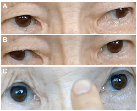
来院時画像所見:明らかな拡散低下域は認めず,FLAIR で 異常信号は認めなかった.MRA で主幹動脈に狭窄病変は認め なかった. 入院後経過:画像上病変は指摘できなかったものの,突然 発症の病歴から脳梗塞が疑われたため入院とし,当日よりオ ザグレルで治療を開始した.入院翌日の MRI で中脳下部に新 たに拡散低下域が確認された(Fig. 2).入院 1 日半後に下腹 部の緊満に気づかれ,導尿で 1 リットル近くの残尿を認め た.尿意が消失しており,生理食塩水を膀胱に注入し,自尿 の有無を確認するも,自尿は出なかった.脊髄 MRI では異常 所見を認めなかった.発症約 3 週間後の転院時まで両側眼球 の内転障害と尿閉は残存し,尿道カテーテル留置を要したが, クロピドグレルによる治療を続け,その後は経時的に症状は 改善した.発症約 2 ヶ月後には眼球運動はほぼ正常化し複視 も消失していた.排尿についても 3 時間以上の間隔で自尿が 出るようになり,入院前と同じ状態に戻った.発症 20 日後 Fig. 1 Representative images of the oculomotor disorder.
The patient presented bilateral adduction weakness (A, B) and convergence disorder (C). Fig. 1 is published with patient’s permission.
Fig. 2 Brain MRI diffusion-weighted image on the second day after onset.
A small abnormal signal is evident in the dorsal part of the lower midbrain. The lesion is markedly hyperintense. The mildly hyperintense areas in the ventral part of the lower midbrain are artifacts. A: Diffusion-weighted image (Axial, 1.5 T; TR 3,172.0 ms, TE 92.3 ms), B: Diffusion-weighted image (Sagittal, 1.5 T; TR 2,990.3 ms, TE 93.5 ms).
の MRI では FLAIR で同部位に淡い高信号域を認め,半年後 の撮影では異常信号は消失していた. 考 察 本症例では両側眼球の内転障害と両側への側方注視の際に 外転眼の単眼性眼振を認めることから,一見両側 medial longitudinal fasciculus(MLF)への障害が示唆された.しか し,輻輳がみられないことと正面視で両眼がやや外転してい る点は MLF 症候群のみでは説明できない.PAG と動眼神経 核,MLF の位置関係を Fig. 3 に示す.動眼神経核の尾側極は MLF の背内側に位置している.動眼神経核の中でも内直筋を 支配する亜核には三つのサブグループ A,B,C があり,A は 動眼神経核の腹側,B は背側,C は背内側に位置しており, 特に B と C はともに動眼神経核の尾側極まで延びているとさ れる4).特に C は輻輳への関与が示唆されており興味深い4). 本症例には内転以外の外眼筋麻痺はないため,これらの内直 筋を支配する亜核から MLF に入る経路が限局的に障害され たと推測している. 脳血管障害発症後に排尿障害を呈する症例はしばしば経験 される.様々な病理学的研究や動物実験から延髄,橋,中脳, 大脳辺縁系や前頭葉が排尿コントロールに関わることが示唆 されているが,ヒトの尿閉に関わる正確な領域については議 論がなされているところである.脳幹の排尿中枢として PMC がよく知られているが,中脳の PAG も排尿中枢として提唱さ
Fig. 3 Location of the lesion. Transverse section of the midbrain. Red areas indicate the oculomotor nucleus (OMN). A, B and C indicate the ventral, dorsal and dorsomedial parts of the medial rectus subnuclei in the OMN, respectively. Blue areas represent the medial longitudinal fasciculus (MLF). The area bounded by the bold outline is the periaqueductal gray (PAG). The yellow areas are the ventrolateral columns in the PAG (vlPAG). The area bounded by the narrow outline is the cerebral aqueduct (CA). The translucent circular area overlapping the vlPAG, OMN and MLF represents the lesion. れている.PAG は約 14 mm 長で 4~5 mm 幅の中脳水道を取 り囲む低分化な灰白質であり,吻側は後交連から尾側は青班 核の高さまで延びており5),前述の動眼神経核に対して背側 に位置している.PAG は機能的に別れた円柱(column)から なる6).すなわちそれぞれ左右一対の ventrolateral column
(vlPAG),lateral column, dorsolateral column と背側にある一つ の dorsomedial column である1).この中で vlPAG は副交感神経
系の機能を,lateral column と dorsolateral column は交感神経 系の機能を持つ5)7).vlPAG は脊髄と直接の連絡があり,膀胱 機能の調節に重要な働きを果たしていると考えられている1)8). 実際に実験動物で vlPAG への電気的刺激や化学的刺激によ り,膀胱収縮が起こることが確かめられており9),vlPAG へ 抑制因子(cobalt chloride)と刺激因子(L-glutamate)の注射 を行った実験では,抑制因子の注射により,排尿が抑制され, また刺激因子の注射により排尿が誘発されることが報告され ている10).また,ヒトでも排尿中に橋から中脳を含む領域が 活性化されると報告されており,実験動物と同様の排尿回路 の存在が提唱されるなど,画像研究によりヒトの正常な排尿 に中脳や PAG が関わることが示されてきた11)~13).さらに, 尿意を感じる際,PAG の血流が増加し14),膀胱に尿が溜まる ほどに PAG の血流が増加するという報告がある他15),排尿 の最中にも PAG の血流が増加することが報告されている16). 上記の報告などを背景に,現在 PAG は排尿中枢の一つであ り,大脳と脊髄の間の中継地点として蓄尿から排尿への切り 替えや,尿意の感覚に関わっていると考えられている.これ までに排尿に関わる大脳皮質領域が幾つか明らかになってお り,PAG は前頭前皮質や前帯状皮質,島皮質,視床下部,大 脳基底核や小脳など多数の領域よりコントロールされている ことがわかっている2).現在提唱されている排尿中枢モデル を Fig. 4 に示す.排尿の際,膀胱壁の一次感覚ニューロンが 膀胱の充満を検出し,その情報を仙髄背側角の二次感覚 ニューロンを介して上位中枢に伝える.PMC は脊髄からの求 心性線維を直接受け取ることはなく,排尿反射の主な引き金 である膀胱の充満についての情報は,PAG を介して PMC に 達する.このことは脊髄から PAG へ濃密な投射があることと 矛盾がない3).遠心性ニューロンは下降して仙髄に連絡し, そこから膀胱収縮を起こす経路を興奮させる一方,膀胱括約 筋を弛緩させるオヌフ核へ連絡する間接経路を興奮させる17). この様にして PAG は排尿反射の感覚と運動の橋渡し役を担 う.それゆえに PAG の障害により膀胱からの情報が PMC や 大脳皮質に伝わらず,尿意の消失,排尿反射の障害が起こる と推測される.本症例の病巣が PAG を含んでいると見れば尿 意の消失と尿閉を矛盾なく説明できる. 中脳の脳血管障害で意識障害と尿閉をきたした既報告では, 中脳下部の左傍正中部の出血性梗塞で,第四脳室内への出血 も伴っていた18).PAG も病巣に含まれていたと考えられる. 脳梗塞ではないが,PAG 病変で尿閉と膀胱感覚の消失を呈し た症例報告がある.同症例においては各種検査で唯一指摘さ れた異常が微小な PAG の病変のみであった.病態は不明だ が,ステロイドが奏効したことから何らかの自己免疫性機序 61:26 臨床神経学 61 巻 1 号(2021:1)
が想定されている19).本症例も画像検査や血液学的検査にお いて中脳病変以外に有意な所見を認めず,同病変が眼球運動 障害と尿閉を引き起こしたものと考えている.今回複視が突 然発症したことから,病態は脳血管障害を想定する.抗血小 板薬による治療のみで一相性の経過で症状が軽快したことも 脳梗塞の経過として矛盾がない.中脳下部の前内側領域は脳 底動脈由来の傍正中枝による灌流を受けており,同血管の末 梢が今回の責任病巣と考えられる20).今回尿閉に加え,MLF +動眼神経亜核の障害を呈していることから,PAG の中で排 尿に関与する部分がこれらの構造の近傍である事が示唆され る.特に前述した vlPAG は,中脳水道より腹側に位置してお り,動眼神経核に近く(Fig. 3),今回の尿閉に関与していた と推測する. 本報告の要旨は,第 108 回日本神経学会近畿地方会で発表し,会長 推薦演題に選ばれた. ※著者全員に本論文に関連し,開示すべき COI 状態にある企業, 組織,団体はいずれも有りません. 文 献
1)Zare A, Jahanshahi A, Rahnama’i MS, et al. The role of the periaqueductal gray matter in lower urinary tract function. Mol Neurobiol 2019;56:920-934.
2)de Groat WC, Griffiths D, Yoshimura N. Neural control of the lower urinary tract. Compr Physiol 2015;5:327-396.
3)Carrive P, Morgan MM. Periaqueductal gray. The human
nervous system. 2012;3:368-401.
4)Büttner-Ennever JA, Akert K. Medial rectus subgroups of the oculomotor nucleus ant their abducens internuclear input in the monkey. J Comp Neurol 1981;197:17-27.
5)Linnman C, Moulton EA, Barmettler G, et al. Neuroimaging of the periaqueductal gray: state of the field. Neuroimage 2012;60:505-522.
6)Bandler R, Shipley MT. Columnar organization in the midbrain periaqueductal gray: modules for emotional expression? Trends Neurosci 1994;17:379-389.
7)Benarroch EE. Periaqueductal gray: an interface for behavioral control. Neurology 2012;78:210-217.
8)Blok BF, De Weerd H, Holstege G. Ultrastructural evidence for a paucity of projections from the lumbosacral cord to the pontine micturition center or M-region in the cat: a new concept for the organization of the micturition reflex with the periaqueductal gray as central relay. J Comp Neurol 1995;359:300-309.
9)Taniguchi N, Miyata M, Yachiku S, et al. A study of micturition inducing sites in the periaqueductal gray of the mesencephalon. J Urol 2002;168:1626-1631.
10)Matsuura S, Downie JW, Allen GV. Micturition evoked by glutamate microinjection in the ventrolateral periaqueductal gray is mediated through Barrington’s nucleus in the rat. Neuroscience 2000;101:1053-1061.
11)Blok BF, Holstege G. The central nervous system control of micturition in cats and humans. Behav Brain Res Fig. 4 Schematic diagram of the micturition center.
The PAG receives information from the bladder, and then sends a signal to the PMC, which provides descending output to the sacrum. This results in contraction of the bladder and relaxation of the urethral sphincter. Therefore, the PAG plays a key role in the micturition reflex and occupies a pivotal location between the brain and the bladder. It also passes information about the bladder to the higher regions of the brain and receives information back from the cerebral control system in order to suppress or enhance storage or voiding. PAG: periaqueductal gray, PMC: pontine micturition center
1998;92:119-125.
12)Fukuyama H, Matsuzaki S, Ouchi Y, et al. Neural control of micturition in man examined with single photon emission computed tomography using 99mTc-HMPAO. Neuroreport 1996;7:3009-3012.
13)Blok BF, Sturms LM, Holstege G. Brain activation during micturition in women. Brain 1998;121:2033-2042.
14)Takao T, Tsujimura A, Miyagawa Y, et al. Brain responses during the first desire to void: a positron emission tomography study. Int J Urol 2008;15:724-728.
15)Athwal BS, Berkley KJ, Hussain I, et al. Brain responses to changes in bladder volume and urge to void in healthy men. Brain 2001;124:369-377.
16)Blok BF, Willemsen AT, Holstege G. A PET study on brain
control of micturition in humans. Brain 1997;120:111-121. 17)Griffiths D, Tadic SD. Bladder control, urgency, and urge
incontinence: evidence from functional brain imaging. Neurourol Urodyn 2008;27:466-474.
18)Liao P-W, Liu S-P, Yu H-J, et al. Acute urinary retention after brainstem infarction. Incont Pelvic Floor Dysfunct 2013;7: 10-11.
19)Yaguchi H, Soma H, Miyazaki Y, et al. A case of acute urinary retention caused by periaqueductal grey lesion. J Neurol Neurosurg Psychiatry 2004;75:1202-1203.
20)Kumral E, Bayulkem G, Akyol A, et al. Mesencephalic and associated posterior circulation infarcts. Stroke 2002;33: 2224-2231.
Abstract
A case of oculomotor disorder and urinary retention due to a lower midbrain lesion
Makoto Sainouchi, M.D.
1)2), Michikazu Nakamura, M.D., Ph.D.
1)3),
Yuichi Masuda, M.D.
1)4)and Ryo Ohtani, M.D., Ph.D.
1)1) Department of Neurology, National Hospital Organization Kyoto Medical Center 2) Present Address: Department of Pathology, Brain Research Institute, Niigata University
3) Present Address: Department of Neurology, Amagasaki Daimotsu Hospital
4) Present Address: Department of Internal Medicine IV, Division of Neurology, Osaka Medical College