総
説
皮質下血管性認知症の診断と治療
冨本 秀和
* 要旨:皮質下血管性認知症(SVD)は比較的均質な臨床病理像を呈し,わが国の血管性認知症のおよそ半分を占 める.広汎白質病変を特徴とするビンスワンガー型脳梗塞と多発ラクナ梗塞があるが,いずれも高血圧性小血管病 変に起因する点で共通している.本稿では皮質下血管性認知症の概念,臨床症状とその発症機序,治療法について 概説し,最後に近年話題になっているアルツハイマー病との関連や,鑑別診断について最新の知見を報告する. (臨床神経 2010;50:539-546) Key words:血管性認知症,白質病変,ビンスワンガー型脳梗塞,慢性脳低灌流,アルツハイマー病1.はじめに
脳血管障害の結果生じる認知機能障害の名称として,古く は動脈硬化性痴呆(Arteriosclerotic dementia),多発梗塞性認 知症(Multi-infarct dementia;MID)などの名称がもちいられ てきた.しかし,「動脈硬化性痴呆」は認知機能低下に対応する 責任病変の所在が不明確であり,「多発梗塞性認知症」は脳血 管障害に起因する認知機能低下のごく一部しか説明しないな ど,包括的な名称として不適切であった.このため,現在では 血管性認知症(Vascular dementia;VaD)が一般的な呼称と して定着している. 多発梗塞性認知症(MID)の用語は,1970 年の Tomlinson らの報告に由来する1).彼らは,認知症患者の 50 剖検脳を健 常者と比較してその病理学的特徴を検討し,皮質萎縮,脳室拡 大,老人性変化(老人斑,神経原線維変化,顆粒空砲変性)に 加えて,脳梗塞容積が認知症発症と関連すると指摘した.その 業績は,大きな組織欠損をともなう脳梗塞が認知症の責任病 変であることを指摘した重要な論文として記憶されている が,一方では,ビンスワンガー型脳梗塞や白質病変,ラクナ梗 塞など,小血管病変による認知症が見逃がされる要因にも なった. いっぽう,アルツハイマー病の血管因子や VCI の概念な ど,現在の知識に照らしあわせてみると,Tomlinson の仕事は 重要な指摘をふくんでいる.彼らは病理学的観察から,認知症 発症に老人性変化と血管病変が独立して関与することを示 し,アルツハイマー病変に起因すると 判 断 さ れ る も の が 50%,血管病変によるものが 17%,混合型が 18% であったと 報告している.疫学的病理研究である Asia-Honoloulu Aging Study(HAAS)でも同様の指摘があり,アルツハイマー病変 による認知症 20%,血管病変による認知症 24%,混合型 45% とある2).さらに,Jellinger によると,認知症の連続剖検 180 例の前向き解析において,アルツハイマー病変 46%,血管病 変 8%,混合型 24% と報告され3),いずれも混合病理による認 知症の比重が高いことが指摘されている. アルツハイマー病の病理変化は血管病変と相互に関連して 認知機能障害に関与し,重複病理の影響はとくに高齢者で大 きくなる.このため,純粋に血管病変によるもの以外に,混合 型認知症や脳卒中後認知症(Post-stroke dementia),血管性軽 度認知障害(Vascular mild cognitive impairment)を包含する 高位の概念として,血管性認知障害(Vascular cognitive im-pairment;VCI)といった用語も提唱されている4).本稿では 以上の歴史的経緯を踏まえて,皮質下血管性認知症(SVD)を 取り巻く状況の概説を試みる.2.血管性認知症の診断基準と分類
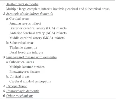
血管性認知症は,脳血管障害にともなって認知機能低下を 示す症候群であり,異質な病態を包含している.研究用の診断 基準としてもっとも広くもちいられている NINDS-AIREN (National Institute of Neurological Disorders and Stroke and Association Internationale pour la Recherche et l Enseigne-ment en Neurosciences)診断基準(1990),およびその分類表 を示した(Table 1)5).本基準では,大血管の閉塞によって大小の脳梗塞が多発し認知機能障害を呈する多発梗塞性認知 症,小血管病変によって認知症を発症する小血管性認知症 (Small vessel disease with dementia),記憶に重要な部位の障 害による Strategic single-infarct dementia の 3 型が基本形で ある.これらの病型分類では病変部位は皮質,皮質下いずれを もふくむが,多発梗塞性認知症の梗塞巣は,大脳皮質領域が主 体である.これに対して,小血管性認知症の血管病変は皮質下 領域が主体であり,Strategic single-infarct dementia では皮
*
Corresponding author: 三重大学大学院医学系研究科神経病態内科学講座(神経内科)〔〒514―8507 三重県津市江戸橋 2―174〕 三重大学大学院医学系研究科・神経病態内科学
Table 1 Classification ofVaD in the reportofNINDS-AIREN inter
-nationalworkshop (1993;summarized by the author)[Ref.4].
1.Multi-infarctdementia
Multiple large complete infarctsinvolving coritcaland subcorticalareas. 2.Strategic single-infarctdementia
a;Corticalareas Angulargyrusinfarct
Posteriorcerebralartery (PCA)infarcts Anteriorcerebralartery (ACA)infarcts Middle cerebralartery (MCA)infarcts b;Subcorticalareas
Thalamicdementia Basalforebrain infarcts 3.Small-vesseldisease with dementia
a.Subcorticalareas Multiple lacunarstrokes Binswanger’ sdisease b.Corticalareas
Cerebralamyloid angiopathy 4.Hypoperfusion
5.Hemorrhagic dementia 6.Othermechanisms
Table 2 Diagnosticcriteria forSVD (2000 by Erkinjuntti;summarized by
the author)[Ref.7].
I.The criteria include allofthe following A.Cognitive syndrome including both
Dysexcutive syndrome Memory deficit(may be mild) B.Cerebrovasculardisease including
Evidence ofcerebrovasculardisease by brain imaging
Presence ora history ofneurologicsignsasevidence forcerebrovasculardisease II.Clinicalfeaturessupporting the diagnosis
a.Episodesofmild uppermotorneuron involvement b.Early presence ofa gaitdisturbance
c.History ofunsteadinessand frequent,unprovoked falls d.Early urinary frequency,urgency,and otherurinary symptoms e.Dysarthria,dysphagia,extrapyramidalsigns
f.Behavioraland psychologicalsymptoms
III.Featuresthatmake the diagnosisuncertain orunlikely
a.Early onsetorprogressive worsening ofmemory and othercognitive functions b.Absence ofcerebrovascularlesionson CT orMRI
質,皮質下いずれの病巣でも発症する. 小血管性認知症はわが国の血管性認知症の約半数を占める もっとも重要な病型であり6),ラクナ梗塞が多発する多発ラク ナ 梗 塞 性 認 知 症,お よ び 広 汎 白 質 病 変 を 特 徴 と す る Binswanger 型脳梗塞に分類される.ラクナ梗塞,白質病変い ずれも穿通枝の小血管病変に起因するため,その病変は皮質 下領域に分布している.2000 年,本基準が策定された NINDS-AIREN ワークショップ(1990 年)のメンバーであった Erkin-juntti は,より均一な臨床病理像を呈する血管性認知症の亜 型として「皮質下血管性認知症」の用語を提唱した7)8).SVD はその大部分が小血管性認知症と重複しているが,皮質に分 布する小血管病変をふくまないことに注意が必要である.ア ミロイド血管症は大脳皮質を主座とする小血管性認知症の代 表であり,白質病変をともなうもののアミロイド沈着は皮 質・皮質内血管に限局している.基本 3 病型のほか,心不全な どによる低灌流性血管性認知症,脳出血後遺症による脳出血 性認知症,遺伝性血管性認知症などがある.さらに,遺伝性 血管性認知症として遺伝性 ア ミ ロ イ ド 血 管 症 や Cerebral autosomal dominant arteriopathy with subcortical infarcts and leukoencephalopathy(CADASIL),Cerebral autosomal recessive arteriopathy with subcortical infarcts and leukoen-cephalopathy(CARASIL),Retinal vasculopathy with cere-bral leukodystrophy(RVCL),Fabry 病などが挙げられる.
Fig. 1 A schema ofmouse chroniccerebralhypoperfusion.The bilateralcommon carotid arteries
ofmice were narrowed (bilateralcommon carotid artery stenosis;BCAS)using microcoilswith a
varying internaldiameter.Depending on the diameterofthe microcoils,the survivalrate (A,left)
decreasesand cerebralblood flow increases(B,right).Compared to the sham-operated mice (Cont),
the BCAS mice showed white matterlesionsconsisted ofredundantnerve fibersand vacuole for
-mation (BCAS).Magnification ×400 (2004.by Shibata etal.)[Ref.23,24]
Cont Control 100 80 60 40 Surv ival Rate (%) CBF (%) 20 0 100 110 80 90 60 70 40 50 30 0 1 2 3 7 14 30days Pre 2hr 1d 3d 7d 14d 30d 0.22mm 0.20mm 0.18mm 0.16mm BCAS A A B B
3.皮質下血管性認知症の概念
SVD の診断基準を Table 2 に示した7).SVD の中核をなす ビンスワンガー型脳梗塞は,1894 年に報告され,その記録は ドイツの神経科医である Otto Binswanger の講演録にみる ことができる9).彼は,脳血管の動脈硬化,後頭葉・側頭葉白 質の高度萎縮,側脳室後角・下角の開大など皮質下血管病変 を主座とするが,大脳皮質はほぼ正常であった症例を呈示し, 動 脈 硬 化 に 起 因 す る 病 態 と し て encephalitis subcorticalis chronica progressiva の名称のもとに記載した.1964 年には, Jellinger らが病理学的な所見に基づき,progressive subcorti-cal vascular encephalopathy of Binswanger type として疾 患 概念を記載している10).さらに,1990 年 Bennett らは画像所 見を考慮してビンスワンガー型脳梗塞の臨床診断基準を提唱 した11).彼らは剖検例を検討し,本診断基準を満たした患者の 大部分がビンスワンガー型脳梗塞であり,アルツハイマー病 で本基準を満たしたものは 184 名中 3 名のみであったと報告 している. 穿通枝領域の細動脈硬化は,血管壊死(angionecrosis),リ ポヒアリノーシスなどと呼ばれ,ラクナ梗塞や出血の原因と なる.いっぽう,白質の髄質動脈では中膜平滑筋細胞の変性と 中外膜の膠原線維の増生がみられ,フィブロヒアリノーシス (fibrohyalinosis)と呼ぶ.Fibrohyalinosis では血管内腔の閉塞 はまれであるが,血管反応性が低下するため白質組織の慢性 低灌流の原因となる12).これらの細動脈硬化は,高血圧を共通 の基盤として発症するため,白質病変とラクナ梗塞は通常は 混在する.以上の事実を背景に,ビンスワンガー型脳梗塞と多 発ラクナ梗塞性認知症を総称し,SVD の名称が汎用されるよ うになっている.4.皮質下血管性認知症の臨床症状
ビンスワンガー型脳梗塞を特徴づける白質病変に対し,そ の主要な危険因子として加齢,高血圧,糖尿病,喫煙,低教育 歴などが指摘されてきた13)∼15).臨床症状は大別すると,1)認 知症,2)歩行障害,3)アパシー,抑うつなどの精神症状,4) めまいなどの非特異的神経症状がある.白質病変は軽度であFig. 2 A schema ofthe pathogeneticmechanism ofSVD.
Smallvesselchangesmay cause itsocclusion and lacunar
infarction,whereaslossofvasomotorreactivity due to fi
brohyalinosisin the smallvesselsmay cause chroniccere
bralhypoperfusion in the white matter.Subsequentglial
activation and BBB damage are likely to induce neuroin
flammation,and thence white matterlesions.
Chronic cerebral hypoperfusion
Neuroinflammation Oxidative stress Apoptosis of oligodendroglia Demyelination Axonal damage MMP activation BBB damage
Lacunar infarction White matter lesions Small vessel changes
Angionecrosis / lipohyalinosis Fibrohyalinosis
Occlusion
Subcortical vascular dementia Host factors
Vascular risk factors, Age, Genetic factors, Education
Atherosclerosis
Cortico-subcortical disconnection Synaptic & neuronal loss, Brain atrophy
Activated microglia Reactive astroglia / clasmatodendrosis
れば無症状で経過するが,広汎になると症状の原因となる.白 質病変に関連する認知機能障害は実行機能障害が主体である が,前向き縦断研究では白質病変の進行がいちじるしい群ほ ど認知症の発症が有意に多かった16).歩行障害についても,白 質病変が高度なほど歩行障害の増悪傾向が強いことが示され ている17). ビンスワンガー型脳梗塞患者の多くは 50∼70 歳台に発症 し,緩徐進行性または階段状の経過をとる.神経症状として は,ラクナ梗塞・白質病変の程度や分布に応じて,固縮・姿勢 反射障害をともなうパーキンソニズム,偽性球麻痺,錐体路障 害,失禁などがある.皮質下病変が主体であっても,けいれん をともなうばあいがある.健忘は比較的軽度であり,むしろ前 頭葉機能を反映する実行機能障害や判断力の低下がめだつ. 精神症状としては無為・抑うつ,自発性の低下などがある. 高血圧はビンスワンガー型脳梗塞の大半の患者でみとめる が,縦断的にみると白質病変の進行期に血圧は低下し,受診時 には血圧は正常のことがしばしばである.血圧の日内変動で は,夜間の血圧低下が 10mmHg 以下に留まる non dipper が 大部分を占める18).血小板凝集能や凝固線溶能は亢進し,とく に脱水・感染症などにともなって凝固能が亢進するときに神 経症状が増悪しやすい19)20).典型例では末期に寝たきり状態 となりやすく,その予後規定因子には脳卒中の合併,誤嚥性肺 炎が挙げられる21).歩行障害や嚥下障害が早期から発症する ため寝たきりになりやすく,医療経済的にも問題となりやす い.
5.皮質下血管性認知症の発症機序
Fazekas 分類は白質病変の代表的な重症度分類であるが, 脳室周囲病変と深部白質病変を分けて評価する.Fazekas 分 類の grade 1∼2 に相当する非融合性の白質病変が神経症状 の責任病変となることはないが,その広がりと障害の強さが 一定の閾値を超えると,認知症,パーキンソニズム,アパシー, 抑うつなどの原因となる.Roman は小血管の閉塞がおきれば ラクナ梗塞となるが,髄質血管の fibrohyalinosis では血管反 応性が低下する結果,白質血流がびまん性に低下し白質病変 が形成されるとの仮説を呈示している4). われわれは,げっ歯類で脳血流を慢性的に低下させる実験 モデルを開発してきたが,長期間にわたって脳血流を低下さ せると,脳梗塞を生じることなく白質病変を安定的に作成可 能である22)∼24)(Fig. 1).ヒト白質病変の脳病理では,病変部位 に血液脳関門の障害,ミクログリア活性化,アストログリアの 増生と変性(clasmatodendrosis),オリゴドログリアのアポ トーシスが観察される25)∼28).いっぽう,実験的白質病変でも 同様の現象が観察され,慢性脳低灌流 1∼3 日で血液脳関門の 障害,ミクログリアの活性化,3∼7 日でアストログリアの反 応性変化,これらのグリア細胞に TNFα,IL-1β などの炎症性 サイトカインや MMP2 の発現がみられる22)∼24)29)30).治療介入 として FK506 や cyclosporin A などの免疫抑制剤を投与す ると,白質病変の形成が抑制される31).また,基底膜を構成す る細胞外マトリックス,ミエリンを構成するミエリン塩基性 蛋白(MBP)は MMP で分解され,その活性は慢性脳低灌流 で亢進する.反対に,MMP 活性を低下させる MMP 阻害薬の 投与,MMP2 遺伝子ノックアウトをおこなうと白質病変は抑 制される32).以上の結果から,慢性脳虚血によってグリア細胞 が活性化されるとともに,MMP2 活性化,血液脳関門の障害 をきたす.さらにグリア細胞から炎症性サイトカインやフ リーラジカルが放出され,オリゴデンドログリアのアポトー シスを誘導する機序が推定されている(Fig. 2).SVD の患者 脳や脳脊髄液で MMP2!3 が増加しているとの報告がある が33),この仮説を支持するものである. 白質病変は高度であっても無症候に経過するばあいがあ り,認知症などの責任病変にならないとの考えが従来は支配 的であった.しかし,縦断的前向き観察研究の結果から,白質 病変は進行すると神経症候の原因となることが明らかになっ た.実際,LADIS 研究では,ラクナ梗塞と白質病変は認知機 能低下の独立した危険因子であり,とくに白質病変の関与が 大きいとされている34). それでは,どのような白質病変が神経症候の原因となるの であろうか.この点を明らかにするため,われわれはび慢性の 白質病変を呈する 12 名の患者を,認知症あるいはパーキンソ ニズムの有無によって各々 2 群に分け,ベンゾジアゼピン! GABAA 受容体のリガンドであるフルマゼニルをもちいたFig. 3 Statisticalparametricmapping (SPM)analysison the relationship between clinicalfeatures
and decreased neuronalviability by [11C]flumazenil-positron emission tomography (FMZ-PET).
Among patientswith severe white matterlesions,those with dementia showed a decreased bi
nd-ing in the the bilateralfrontopolarand frontal/insularareas,the lefttemporo-occipitalborderar
-eas,and the leftmarginalcorticalareas.On the contrary,those with parkinsonism showed a
de-crease in the lateralstriatum (2004,2007 by Ihara etal.)[Ref.35,36].
A. Dementia
R
R
B. Parkinsonism
Fig. 4 A schema ofneuralnetwork dysfunction in white
matterlesions. Axonsare damaged and neuronalsoma
become atrophicand degenerated by retrograde degen
eration,when the degree ofwhite matterlesionsprogress
to some extent.Dysfunction ofneuralnetworksare partly
accelerated by cholinergicdenervation.
Axonopathy BBB dysfunction Glial activation Neuroinflammation Apoptosis (Oligodendroglia) Neuronal Dysfunction Brain atrophy M y el inopath y Chol inerg ic defic it Cerebral cortex White mater PET で神経細胞への結合量をしらべた35)36).その結果,認知症 群では前頭領域をはじめとする広範な領域において,パーキ ンソニズム群では外側線条体において,各々結合量の低下が みとめられた(Fig. 3).この事実は,同じ程度のび慢性白質病 変であっても有症候群では灰白質の機能障害をきたしている ことを意味する.さらに,われわれは大脳皮質に投射するコリ ン神経投射路が白質病変の好発部位である外包を経由する点 に着目し,コリン神経が戦略的に障害される可能性を検討し た.その結果,SVD では対照群と比較してコリン神経投射路 の著明な脱落がみとめられた37).以上の結果から,白質障害は 通過線維を障害してニューロンの機能障害を惹起するものと 考えられる(Fig. 4). 白質病変は必ずしも純粋な脱髄ではなく,病変の進行にと もなって軸索障害を呈する.MR スペクトロスコピーで,白質 病変部位の NAA!Cre 比が認知症発症と相関して低下すると の報告があり,軸索障害が進行するにつれ有症候となってい くものと考えられる38).軸索障害に進展した病変は,MRI 拡 散テンソル画像で高感度に検出される可能性があり,実際 Nave らは T2強調画像に比較して,拡散テンソル画像の異常 が運動機能,認知機能障害とより良く相関したと述べてい る39).
6.皮質下血管性認知症とアルツハイマー病の鑑別診断
血管性認知症とアルツハイマー病が合併する頻度は偶然の 合併より高率であり,その機序として脳血管へのアミロイド 沈着による二次的循環障害などが指摘されている.アルツハ イマー病患者脳ではアミロイド血管症が 70∼100% と高率に みとめられる.その沈着は皮質血管や皮質内髄質血管に限局 し,白質血管にはほとんど分布しない.アミロイド血管症は, それ自体が皮質小動脈の BBB を障害して認知機能低下の原 因になるほか,破綻,閉塞を生じやすく,脳葉型出血や微小出 血(Microbleeds;MBs)40),クモ膜下出血,CAA-relatedin-flammation41),皮質微小梗塞(Cortical microinfarction)を惹
起する.また,皮質領域で血管狭窄を生じるため深部白質の血 流低下をきたし,白質病変の原因となる可能性がある.
Table 3 Differentialdiagnosisbetween SVD and Alzheimer’ sdisease based on pathologicaland radiological
findings.
Alzheimer’ sdisease Subcorticalvasculardementia
Cerebralcortex CerebralWM
Site oflesions
Senile plaque & Neurofibrillary tangle Smallvesseldisease
Core pathology
Pathological& radiologicalfindings
Lessthan moderate May be severe
・WM lesions(degree)
Rare in deep WM Periventricular& deep WM
(location) Rare Frequent ・Lacunarinfarction Occasional Frequent
・Microbleeds(number)
Dominantin the occipitallobe; Centrencephalic;
(location)
Subcorticalarea Basalganglia,thalamus
Frequent None
・Corticalmicroinfarctions
May be severe Lessthan moderate
・Hippocampalatrophy WM;white matter SVD はその約半数が緩徐進行性の経過をとるため6),アル ツハイマー病との鑑別がとくに問題になる.剖検所見からみ た鑑別点を Table 3 にまとめた.アミロイド血管症にともな う MBs は皮質・皮髄境界に分布する特徴があり,通常の高 血圧性でみられる基底核・視床などの皮質下領域のものと分 布がことなっている.皮質微小梗塞はアルツハイマー病に特 徴的で,SVD ではほとんどみとめない42). アルツハイマー病の白質病変は,上記のように皮質アミロ イド血管症による白質低灌流が原因と考えられるが,ワラー 変性の関与も推定されている.SVD にくらべると,深部白質 病変が少なく程度も軽い傾向がある.海馬萎縮はアルツハイ マー病で高度で,SVD では中等度以下である.血管性認知症 で海馬萎縮がみられるとの剖検報告があるが43),海馬萎縮は 数カ月の慢性脳低灌流負荷で実験的にも再現可能である44).
7.皮質下血管性認知症の治療
SVD では,再発予防の目的で高血圧の管理と抗血小板薬の 投与が一般的である.カルシウム拮抗薬ニトレンジピンをも ちいた Syst-Eur 試験や,ACE 阻害薬ぺリンドプリルによる PROGRESS-dementia などでは,これらの降圧療法が認知症 の発症抑制に一定程度有効である.いっぽう,白質病変は微小 出血をともなうことが多いため,抗血小板薬の使用は血圧を 厳重に管理したうえで必要に応じて考慮する.このばあい,血 管内皮保護作用をあわせもち,出血性合併症が少ないシロス タゾールが望ましい.近年,三環系抗うつ薬による血圧変動が 白質病変の増悪と関連するとの報告があり45),抑うつには SSRI,自発性の低下には少量の塩酸アマンタジン,焦燥や幻 覚妄想に対しては抑肝散,テグレトールなどの抗てんかん薬, 少量のミアンセリン,非定型抗精神病薬などが推奨される. 血管性認知症では白質病変によるコリン神経系の障害があ り,アセチルコリンエステラーゼ阻害薬が有効であったとの 報告があるが46),保険適応はわが国ではみとめられていない. また,遺伝性血管性認知症である CADASIL でアセチルコリ ンエステラーゼ阻害薬の有効性が証明されなかった点も問題 である47).この他,臨床試験で有用性が報告されている薬剤と して,NMDA 受容体拮抗薬である塩酸メマンチンがあげられ るが48),わが国では未承認である.8.結
語
血管性認知症とアルツハイマー病の境界はますます不明瞭 となりつつある.近年では脳虚血によるアルツハイマー病理 の加速や49),アルツハイマー病の危険因子としての生活習慣 病が注目されており,血管病変の立場からも改めて認知症を 見直す必要性に迫られている.SVD は,時に緩徐進行性の経 過を示すことがあることから,臨床症状,病理,機能画像に基 づき総合的にアルツハイマー病との鑑別をおこなうことが重 要である.最後に,VCI の概念は以上を背景に提唱されたが, Hachinski らは VCI を VCI-ND(VCI-no dementia),すなわち 血管性の原因による MCI の意味で狭義にもちいており,いず れの意味でもちいているかに注意が必要である.文 献
1)Tomlinson BE, Blessed G, Roth M. Observations on the brains of demented old people. J Neurol Sci 1970;11:205-242.
2)Petrovitch H, Ross GW, Steinhorn SC, et al. AD lesions and infarcts in demented and non-demented Japanese-American men. Ann Neurol 2005;57:98-103.
3)Bancher C, Jellinger K, Lassmann H, et al. Correlations between mental state and quantitative neuropathology in the Vienna Longitudinal Study on Dementia. Eur Arch Psychiatry Clin Neurosci 1996;246:137-146.
4)Román GC, Erkinjuntti T, Wallin A, et al. Subcortical is-chaemic vascular dementia. Lancet Neurology 2002;1:426-436.
dementia: diagnostic criteria for research studies. Report of the NINDS-AIREN international workshop. Neurology 1993;43:250-260.
6)Yoshitake T, Kiyohara Y, Kato I, et al. Incidence and risk factors of vascular dementia and Alzheimer s disease in a defined elderly Japanese population: the Hisayama Study. Neurology 1995;45:1161-1168.
7)Erkinjuntti T, Inzitari D, Pantoni L, et al. Research crite-ria for subcortical vascular dementia in clinical tcrite-rials. J Neural Transm Suppl 2000;59:23-30.
8)Erkinjuntti T, Inzitari D, Pantoni L, et al. Limitations of clinical criteria for the diagnosis of vascular dementia in clinical trials. Is a focus on subcortical vascular dementia a solution? Ann N Y Acad Sci 2000;903:262-272.
9)Blass JP, Hoyer S, Nitsch R. A translation of Otto Binswanger s article, The delineation of the generalized progressive paralyses . 1894. Arch Neurol 1991;48:961-972. 10)Jellinger K, Neumayer E. Binswanger s Progressive subcortical vascular encephalopathy. A clinico-neuropathological study. Arch Psychiatr Nervenkr 1964; 205:523-554.
11)Bennett DA, Gilley DW, Wilson RS, et al. Clinical diagno-sis of Binswanger s disease. J Neurol Neurosurg Psychia-try 1990;53:961-965.
12)Brun A. Pathology and pathophysiology of cerebrovascu-lar dementia: pure subgroups of obstructive and hypop-erfusive etiology. Dementia 1994;5:145-147.
13)Lechner H, Schmidt R, Bertha G, et al. Nuclear magnetic resonance image white matter lesions and risk factors for stroke in normal individuals. Stroke 1988;19:263-265. 14)Liao D, Cooper L, Cai J, et al. The prevalence and severity
of white matter lesions, their relationship with age, eth-nicity, gender, and cardiovascular disease risk factors : the ARIC Study. Neuroepidemiology 1997;16:149-162. 15)Gouw AA, van der Flier WM, Fazekas F, et al.
Progres-sion of white matter hyperintensities and incidence of new lacunes over a 3-year period: the Leukoaraiosis and Disability study. Stroke 2008;39:2921-2922.
16)Longstreth WT Jr, Arnold AM, Beauchamp NJ Jr, et al. Incidence, manifestations, and predictors of worsening white matter on serial cranial magnetic resonance imag-ing in the elderly : the Cardiovascular Health Study. Stroke 2005;36:56-61.
17)Baloh RW, Ying SH, Jacobson KM. A longitudinal study of gait and balance dysfunction in normal older people. Arch Neurol 2003;60:835-839.
18)Yamamoto Y, Akiguchi I, Oiwa K, et al. Twenty-four-hour blood pressure and MRI as predictive factors for dif-ferent outcomes in patients with lacunar infarct. Stroke 2002;33:297-305.
19)Iwamoto T, Kubo H, Takasaki M. Platelet activation in the cerebral circulation in different subtypes of ischemic stroke and Binswanger s disease. Stroke 1995;26:52-56. 20)Tomimoto H, Akiguchi I, Ohtani R, et al. The
coagulation-fibrinolysis system in patients with leukoaraiosis and Binswager disease. Arch Neurol 2001;58:1620-1625. 21)Frisoni GB, Galluzzi S, Bresciani L, et al. Mild cognitive
impairment with subcortical vascular features. Clinical characteristics and outcome. J Neurol 2002;249:1423-1432. 22)Wakita H, Tomimoto H, Akiguchi I, et al. Glial activation
and white matter changes in the rat brain induced by chronic cerebral hypoperfusion: An immunohistochemi-cal study. Acta Neuropathol 1994;87:484-492.
23)Shibata M, Ohtani R, Ihara M, et al. White matter lesions and glial activation in a novel mouse model of chronic cerebral hypoperfusion. Stroke 2004;35:2598-2603. 24)Shibata M, Yamasaki N, Miyakawa T, et al. Selective
im-pairment of working memory in a mouse model of chronic cerebral hypoperfusion. Stroke 2007;38:2826-2832. 25)Tomimoto H, Akiguchi I, Akiyama H, et al. T cell infiltra-tion and expression of MHC class II antigen by macro-phage!microglia in a heterogeneous group of leukoen-cephalopathy. Am J Pathol 1993;143:579-586.
26)Tomimoto H, Akiguchi I, Suenaga T, et al. Alterations of the blood-brain barrier and glial cells in white matter le-sions in cerebrovascular and Alzheimer s disease pa-tients. Stroke 1996;27:2069-2074.
27)Tomimoto H, Akiguchi I, Suenaga T, et al. Regressive change of astroglia in white matter lesions in cerebrovas-cular and Alzheimer s disease patients. Acta Neuropathol 1997;94:146-152.
28)Tomimoto H, Ihara M, Wakita H, et al. Chronic cerebral hypoperfusion induces white matter lesions and loss of oligodendroglia with DNA fragmentation in the rat. Acta Neuropathol 2003;106:527-534.
29)Tomimoto H, Akiguchi I, Kinoshita A, et al. Glial expres-sion of cytokines in the brains with cerebrovascular dis-ease patients. Acta Neuropathol 1996;92:281-287. 30)Ihara M, Tomimoto H, Kinoshita M, et al. Chronic
cere-bral hypoperfusion induces MMP-2 but not MMP-9 ex-pression in the microglia and vascular endothelium of the white matter. J Cereb Blood Flow Metab 2001;21:828-834. 31)Wakita H, Tomimoto H, Akiguchi I, et al.
Dose-dependent, protective effect of FK506 against white mat-ter changes in the rat brain afmat-ter chronic cerebral hypop-erfusion. Brain Res 1998;792:105-113.
32)Nakaji K, Ihara M, Takahashi C, et al. Matrix metalloproteinase-2 plays a critical role in the pathogene-sis of white matter lesions after chronic cerebral hypop-erfusion in rodents. Stroke 2006;37:2816-2823.
33)Rosenberg GA. Inflammation and white matter damage in vascular cognitive impairment. Stroke 2009;40:S20-S23. 34)van der Flier WM, van Straaten EC, Barkhof F, et al.
Small vessel disease and general cognitive function in nondisabled elderly: the LADIS study. Stroke 2005 ; 36 : 2116-2120.
35)Ihara M, Tomimoto H, Ishizu K, et al. Decrease in cortical benzodiazepine receptors in symptomatic patients with leukoaraiosis : a positron emission tomography study. Stroke 2004;35:942-947.
36)Ihara M, Tomimoto H, Ishizu K, et al. Association of vas-cular parkinsonism with impaired neuronal integrity in the striatum. J Neural Transm 2007;114:577-584.
37)Tomimoto H, Ohtani R, Shibata M, et al. Loss of choliner-gic pathway in vascular dementia of the Binswanger type. Dement Geriatr Cogn 2005;19:282-288.
38)Brooks WM, Wesley MH, Kodituwakku PW, et al. 1 H-MRS differentiates white matter hyperintensities in sub-cortical arteriosclerotic encephalopathy from those in normal elderly. Stroke 1997;28:1940-1943.
39)Della Nave R, Foresti S, Pratesi A, et al. Whole-brain his-togram and voxel-based analyses of diffusion tensor im-aging in patients with leukoaraiosis: correlation with mo-tor and cognitive impairment. AJNR 2007;28:1313-1319. 40)Pettersen JA, Sathiyamoorthy G, Gao F-Q, et al.
Mi-crobleed topography, leukoaraiosis,and cognition in prob-able Alzheimer disease from the Sunnybrook Dementia study. Arch Neurol 2008;65:790-795.
41)Kinnecom C, Lev MH, Wendell L, et al. Course of cerebral
amyloid angiopathy-related inflammation. Neurology 2007;68:1411-1416.
42)Okamoto Y, Ihara M, Fujita Y, et al. Cortical microin-farcts in Alzheimer s disease and subcortical vascular de-mentia. Neuroreport 2009;20:990-996.
43)Du AT, Schuff N, Laakso MP, et al. Effects of subcortical ischemic vascular dementia and AD on entorhinal cortex and hippocampus. Neurology 2002;58:1635-1641.
44)Nishio K, Ihara M, Yamasaki N, et al. A mouse model characterizing features of vascular dementia with hip-pocampal atrophy. Stroke 2010;41:1278-1284.
45)Steffens DC, Chung H, Krishnan KR, et al. Antidepres-sant treatment and worsening white matter on serial cra-nial magnetic resonance imaging in the elderly: the Car-diovascular Health Study. Stroke 2008;39:857-862. 46)Wilkinson D, Doody R, Helme R, et al. Donepezil in
vascu-lar dementia : a randomized, placebo-controlled study. Neurology 2003;61:479-486.
47)Dichgans M, Markus HS, Salloway S, et al. Donepezil in patients with subcortical vascular cognitive impairment: a randomised double-blind trial in CADASIL. Lancet Neurol 2008;7:310-318.
48)Orgogozo JM, Rigaud AS, Stoffler A, et al. Efficacy and safety of memantine in patients with mild to moderate vascular dementia: a randomized, placebo-controlled trial (MMM 300). Stroke 2002;33:1834-1839.
49)Kitaguchi H, Tomimoto H, Ihara M, et al. Chronic cere-bral hypoperfusion accelerates amyloidβ deposition in APPSwInd transgenic mice. Brain Res 2009;1294:202-210.
Abstract
Diagnosis and treatment of subcortical vascular dementia
Hidekazu Tomimoto, M.D.
Department of Neurology, Mie University Graduate School of Medicine
Subcortical vascular dementia(SVD) exhibits relatively uniform clinical and pathological features among
vari-ous subtypes of vascular dementia, and constitutes approximately half of vascular dementia in Japan. This
sub-type is further classified into Binswanger s sub-type infarction and multiple lacunar infarctions. The former is
charac-terized by diffuse white matter lesions, and the latter by lacunar infarctions. However, the both have common
pa-thologic mechanism related to hypertensive small vessel changes. This paper overview the notion, clinical
fea-tures, pathogenesis and treatment for SVD, and also refer to the recent topics related to Alzheimer s disease.
(Clin Neurol 2010;50:539-546)
Key words: Vascular dementia, white matter lesions, Binswanger s type infarction, chronic cerebral hypoperfusion, Al-zheimer s disease