異時性肝転移に対し肝切除を行った 非浸潤性乳管癌の 1 例
昭和大学医学部外科学講座(消化器一般外科学部門)
箱崎 智樹* 村上 雅彦 松田 和広 草野 智一 古泉 友丈 藤 森 聰
青木 武士
抄録:症例は 43 歳,女性.前医で右乳癌に対し右乳房部分切除術を施行.病理診断で非浸潤 性乳管癌(Ductal Carcinoma In Situ:DCIS)と診断され,術後補助放射線療法後に外来経過 観察されていた.術後 2 年 8 か月に健診で施行した腹部超音波検査で,肝臓 S3, 4 に 3.6 cm 大 の腫瘤を指摘された.前医で同腫瘤を針生検し,病理診断で乳癌異時性肝転移疑いと診断さ れ,当科へ手術目的に紹介された.過去に DCIS の異時性肝転移切除例で,長期生存を得た報 告がある事を考慮し,肝部分切除術を施行した.病理診断では乳癌異時性肝転移に矛盾しない と診断された.DCIS は間質浸潤を示さない乳癌であり,現在本邦において増加傾向にあるが,
外科的切除後に再発・遠隔転移を認める症例は稀である.今回われわれは DCIS の術後 2 年 8 か月で異時性単発肝転移を認め,肝切除を施行した 1 例を経験したので文献的考察を加えて報 告する.
キーワード:非浸潤性乳管癌(DCIS),異時性肝転移,肝切除
緒 言
非浸潤性乳管癌(DCIS)は間質浸潤を示さない 乳癌であり,検診の普及に伴い DCIS の段階で発見 される割合が増加傾向にある.Jorgensen ら1)の報 告によれば DCIS の発見率は 1980 年代中ごろと比 較し,50 歳未満の女性では 4 倍に増加し,50 歳以 上の女性では 8 倍になっているとされるが,外科的 切除後に再発・遠隔転移を認める症例は稀である.
今回 DCIS に対し乳房温存術を施行し,術後 2 年 8 か月に異時性単発肝転移が出現したため,肝切除 を施行した症例を経験したので文献的考察を加え報 告する.
症 例 患者:43 歳 女性.
家族歴・既往歴:橋本病 内服薬なし.
現病歴:健診で発見された右乳癌に対し,前医で 右乳房温存術を施行.病理学的診断で非浸潤性乳管 癌(DCIS)の診断となり,術後補助放射線療法を
施行した後に経過観察していた.術後 2 年 8 か月に 健 診 の 腹 部 超 音 波 検 査 で, 肝 臓 の 外 内 側 区 域
(S3, 4)にかけて 3.6 cm 大の腫瘤を指摘された.画 像検査では質的診断が困難であったため,前医で肝 腫瘤に対し針生検を施行した.病理学的診断から乳 癌の異時性肝転移として矛盾しない所見を得て,当 科へ手術目的に紹介となった.
身体所見:右乳房に乳房部分切除の瘢痕を認め た.腹部に有意な所見は認めなかった.
血液検査所見:WBC;3,400
×
104/µl,Hb;12.6 g/dl,CRP;0.24 mg/dl,ICGR15;8%.
腫瘍マーカー:AFP;5 ng/ml,PIVKA-Ⅱ;
21 mAU/m,CEA;1.0 ng/ml,CA19-9;16.2 U/ml.
HBs 抗原,HCV 抗体共に陰性で,上記の如く著明 な炎症反応や腫瘍マーカーの上昇は認めなかった.
病理学的診断(乳房温存術で採取した標本:前医 提供):
検体は 5 mm 幅全割.右乳房円状部分切除検体で 内下部(B)領域に腫瘍を認める.
腫瘍径は 1.0
×
1.0×
0.7 cm (Fig. 1).症例報告
*
責任著者
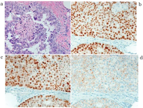
核異型や免疫染色の結果は下記の如くである.
核異型 score;3,核分裂 score;3(11/10 HPF),
核 grade 3.
ER;Allred PS5+IS3 TS8,PgR:Allred PS4+
IS2 TS6(Fig. 2a, b, c, d).
HER2 score1,強陽性;0%,中等度陽性;0%,
弱陽性;20%,ly0,v0.
切除断端は皮膚側,深部側,側方,乳頭側の何れ もが陰性であった.
腫瘍胞巣には Comedo 様の壊死と,間質へのリ ンパ球浸潤がそれぞれ中等度にみられ,一部には石 灰化も認めた.検体中に明らかな浸潤巣は認めな かった.上記より乳癌取り扱い規約第 17 版におい て solid,comedo type の DCIS,pTis pNx pM0 Stage 0 と診断された.手術標本は免疫染色のプレ パラートを含め全て当院で改めて病理診断の見直し を行い,浸潤巣は認めなかった.
腹部超音波検査(B mode):肝 S3, 4 の門脈臍部 直上に類円形を呈す 3.6 cm 大の腫瘤を認めた.腫 瘤 は 高 echo を 呈 し, 厚 い 辺 縁 低 echo 帯(bull's
Fig. 2 Pathological findings of the primary tumor
a:Microscopic photo showed comedo-DCIS with central necrosis (H&E stain, × 20).
b:Immunohistochemistry( × 20.) show ER positive.
c:Immunohistochemistry( × 20.) show PgR positive.
Fig. 1 Gross appearance of the specimen from a breast pre- serving surgery. The resected specimen of the lower inner quadrant of the right breast measured 6.0 cm
× 4.5 cm × 2.3 cm (B region).
The primary tumor measured 1.0 cm × 1.0 cm ×
0.7 cm, and surgical margin was confirmed with
negative by pathological findings. Ductal carcinoma
in situ was shown by the red line.
eye pattern)を認めた(Fig. 3).
腹部造影 CT:肝 S3, 4 に類円形で平衡相にて周 囲肝実質より低濃度を呈し,門脈相で弱い造影効果 を示す 3.6 cm 大の腫瘤を認めた(Fig. 4a, b).肺な
ど他臓器への転移は認めなかった.画像所見からは 転移性肝癌や原発性肝癌が鑑別に挙がった.
EOB-MRI:肝細胞相にて,肝 S3/4 に 41 mm 大の 境界明瞭な八つ頭状の腫瘤を認めた(Fig. 5).門脈 臍部に近接していたが,圧排所見は認めなかった.
PET-CT:肝腫瘍に一致して高集積を認め,同部 位は SUV max 9.1 を呈した.右乳房には局所再発 を疑う異常集積は無く,単発の肝腫瘍と診断された
(Fig. 6).
Fig. 3
abdominal ultrasound(B mode)
Ultrasonography showed bull's eye pattern of a well-defined mass 36 mm in size of the liver.
Fig. 5 EOB-MRI findings
EOB-MRI showed that hypointense of the liver tumor in the hepatocyte phase.
Fig. 6 PET-CT findings
FDG-PET showed strong uptake in the liver.
No other abnormal uptake was observed.
Fig. 4 Contrast Enhanced CT findings a: CECT showed that heterogenous hypo-enhance-
ment of the liver tumor in the arterial phase.
b: CECT showed that heterogenous hypo-enhance- ment of the liver tumor in the portal phase.
a
b
上下部消化管内視鏡検査:腫瘍性病変を含め異常 所見を認めなかった.
治療経過:前医で肝腫瘍を経皮的針生検してお り,病理学的所見は異型細胞が充実性,索状に増殖 浸潤しており carcinoma の所見を呈した.免疫染 色では ER(Allred PS5+IS3=TS8),PgR(allred PS5+IS3=TS8),mammaglobin 陽 性,GCDFP-15 少数陽性,CK7 陽性,CK20 陰性,AE1/AE 強陽性,
Heppar 1 陰性,AFP 陰性で肝原発腫瘍か転移性肝 腫瘍の可能性も示唆された.Heppar 1 陰性であっ
た事から肝細胞癌の可能性は低いと考えられ,バイ オマーカーが既往の乳癌とほぼ一致しており,
mammoglobin や GCDFP-15 と言った乳癌関連のバ イオマーカーも陽性であった.上記より既往の乳癌 は非浸潤癌であったものの,乳癌の肝転移として矛 盾しない所見を得ていた.その後当科へ外科的切除 目的に紹介を受け,開腹肝部分切除術を施行した.
手術所見:開腹で肝部分切除術(S3/4)を施行 した.術中に腫瘍の露出は認めなかった.
肝腫瘍の病理組織学的診断:病理学的診断では肉 眼的に 55
×
48×37 mm 大の白色調を呈する結節性 病変を認め,組織学的には卵円形の核を有する異型 細胞が大小の巣状,癒合腺管状を呈して肝実質に浸 潤・増殖していた.切除断端の挫滅した組織内に腫 瘍細胞を認めたが,追加切除した腫瘍底部肝組織内 に腫瘍細胞は認めなかった(Fig. 7).免疫染色では HER2 score 0,ER 陽性,PgR 一部陽性,Mamma- globin 陽性,Ki-67 60〜70%で乳癌の肝転移として矛 盾しない所見と診断された(Fig. 8a, b, c).術後経過:本症例は術後合併症を有することなく 退院したが,術後 4 か月の時点で多発肝転移を生じ 前医にて治療中である.
Fig. 7 A cross section of the resected liver The tumor was well-circumscribed, solid, and white in color.
Fig. 8 Pathological findings of the liver tumor
a: microscopic examination of the liver tumor (H&E stain, × 20).
b: Immunohistochemistry( × 20)
show ER positive.
c: Immunohistochemistry( × 20)
show PgR positive.
考 察
近年本邦ではマンモグラフィ検診と穿刺吸引針生 検の普及により,非触知石灰化病変の診断が増加し ている.そのため原発乳癌において非浸潤性乳管癌 の割合が増加傾向にあるが,外科的切除を受けた非 浸潤性乳管癌の予後は非常に良く,明石らの報告で は DCIS 患者 336 名の 15 年生存率は 98.5%2)であっ た.術後の遠隔転移も稀であり,かつ初発遠隔 転移臓器が肝臓である頻度も約 1〜6%と低い3). 遠隔転移が 1 か所もしくは数か所の症例を「Oligo- metastases」というが,これは 1995 年に Hellman らによって提唱された概念4)で,遠隔転移が発見さ れた時点で腫瘍細胞が全身に広がっているという従 来の概念とは異なり,局所から全身へ広がっていく 中間段階5)と考えられるとしている.本症例におい て,初発遠隔転移臓器が頻度の高い肺ではなく,肝 臓となったのもその一過程である可能性も考慮され る.Meijnen らは DCIS 504 例を 8 年間にわたり追 跡調査しているが,それによると乳房温存術後と乳 房切除術後のそれぞれの遠隔転移は 4%,0.9%と温 存術後に高い6)と報告している.
DCIS に関しては病理検索状況に施設間で大きな 差異がある7)との報告があるが,本症例の手術検体 は 5 mm で全割され十分に検索されており,当院で 病理学的検索を再検討した結果,浸潤巣は認めな かった.また病理学的腫瘍径は断端陽性率や浸潤巣 の見落としなどと関連する事が示されており,広範 に広がった DCIS においては病変全体の病理検索は 実用的でなく,浸潤成分が見落とされる可能性も指 摘されている8).この点に関し本症例は,腫瘍径は 1.0
×
1.0×0.7 cm と小さく,病理標本でも十分な切 除断端が確保(Fig. 1 参照)されているものの,結 果的に切り出されていない箇所に微小浸潤部位が あった可能性は否定できない.DCIS の乳房温存術後における局所再発リスク因 子 と し て,Silverstein ら は Van Nuys 予 後 係 数
(VNPI)分類を提唱している9)が,本症例は VNPI リスクが 7 点で術後に計 50 Gy を照射しており,必 要十分な術後補助療法が施行されていた.しかしな がら本症例は核異型が強く,van Nuys 分類で High grade に分類される.DCIS を van Nuys 分類に従っ て grade 分類し,再発率について検討した Solin
ら10)の報告によると,切除と放射線照射を行った場 合でも High grade の再発率は 20.3%と高い.本症 例では適切な前治療が施行されているが,前述の報 告からも DCIS の遠隔転移を考慮する必要があり,
肝腫瘍の鑑別として転移性肝癌,胆管細胞癌,肝細 胞癌が挙げられた.画像検索からは特徴的な所見を 得られなかったが,免疫染色で Heppar 1 陰性であ る事から肝細胞癌の可能性は低いと考えられた.
Nash らは 92 症例の肝腫瘍に免疫染色を施行し,胆 管細胞癌の 31%が PgR 陽性となり,ER 陽性となっ た症例は乳癌の肝転移のみであったと報告してい る11).また胆管細胞癌において CK7 の染色率は 90〜
100%,CK20 の染色率は 5〜100%,乳癌肝転移で はそれぞれ 78%,8%であるという報告12–16)と本症 例 の ER 陽 性,PgR 陽 性,CK7 陽 性,CK20 陰 性,
mammoglobin や GCDFP-15 と言った乳癌関連のバイ オマーカーも陽性という免疫染色の結果から,肝腫瘍 は乳癌肝転移の可能性が高いと考えられた.
乳癌肝転移後の生存中央値は 1〜14 か月で,5 年 生存率は 3%と予後不良17)であり,本邦における乳 癌診療ガイドライン18)だと乳癌肝転移に対する肝 切除が推奨グレード C2 となっている. 医学中央雑 誌において「DCIS」「肝転移」「肝切除」を key word に,1980 年から 2019 年まで会議録を除外し た条件で検索すると,現在まで本邦における報告例 は 1 例のみ19)存在する.この症例は乳腺の原発巣を 針生検し,DCIS の疑いが示唆されたが,外科的切 除した後の病理検索で浸潤巣が発見されており,浸 潤性乳管癌(T1a)の肝転移であった事が判明して いる.実臨床において乳癌肝転移に対する肝切除は 選択されにくく,DCIS の肝転移は切除されていて も,報告されていない例が存在すると思われる.
しかし現在までに肝転移巣が単発で切除可能な場 合,長期生存例も報告20–24)されており,肝切除の 安全性の向上に伴い乳癌肝転移に対し切除術が適応 される事がある25, 26).
乳癌肝転移切除の良好な予後因子として disease free interval が 1 年以上,他臓器転移無し,転移個 数が 1 個,切除断端陰性,原発巣の estrogen re- ceptor が陽性,肝切除前の全身化学療法奏功例,
physical status が良好,背景肝が正常,転移巣の腫 瘍量が少ない,などが挙げられる27).また画像によ る鑑別が困難な場合は診断的治療を目的とした外科
的切除が選択されることがある28).本症例では前述 の内容を考慮し,診断的治療目的の意味も含め外科 的切除が選択肢となった.
残念ながら本症例は前述の通り肝切除後 4 か月の 時点でびまん性の肝転移を認め前医で治療を継続し ている.一般的に DCIS は予後良好であるが,日常 診療で普及している van Nuys 分類による grade 分 類をはじめ,Oncotype DCIS などの多重遺伝子診 断法による最新の DCIS の予後予測に関する解析29)
も活用され始めている.個々の症例に合わせつつ,
術後再発高危険群においては慎重な術後経過観察が 必要である.また乳癌術後の再発症例においては,
外科的切除が選択肢に挙がる可能性は現時点におい て低いが,症例を蓄積し予後因子の解析に努めたい.
利益相反
本論文投稿に際して,開示すべき利益相反はない.
文 献
1) Jorgensen KJ, Keen JD, Gotzsche PC. Is mam- mographic screening justifiable considering its substantial overdiagnosis rate and minor effect on mortality? Radiology. 2011;260:621︲627.
2) Akashi-Tanaka S, Fukutomi T, Nanasawa T, et al. Treatment of noninvasive carcinoma : fif- teen-year results at the National Cancer Center Hospital in Tokyo. Breast Cancer. 2000;7:341︲344.
3) Sakamoto Y, Yamamoto J, Yoshimoto M, et al.
Hepatic resection for metastatic breast cancer:
prognostic analysis of 34 patients. World J Surg. 2005;29:524︲527.
4) Hellman S, Weichselbaum RR. Oligometastases.
J Clin Oncol. 1995;13:8︲10.
5) Weichselbaum RR, Hellman S. Oligometastases revisited. Nat Rev Clin Oncol. 2011;8:378︲382.
6) Meijnen P, Oldenburg HS, Peterse JL, et al.
Clinical outcome after selective treatment of patients diagnosed with ductal carcinoma In situ of the breast. Ann Surg Oncol. 2008;15:235︲
243.
7) 坂元吾偉,浜 善久,秋山 太.非浸潤性乳管癌 の病理学的診断の現状.癌の臨.2000;46:71︲77.
8) Lester SC, Bose S, Chen YY, et al. Protocol for the examination of specimens from patients with invasive carcinoma of the beast. Arch Pathol Lab Med. 2009;133:1515︲1538.
9) Silverstein MJ. The University of Southern California/Van Nuys prognostic index for duc- tal carcinoma in situ of the breast. Am J Surg.
2003;186:337︲343.
10) Solin LJ, Yeh IT, Kurtz J, et al. Ductal carcinoma in situ (intraductal carcinoma) of the breast treated with breast-conserving surgery and de- finitive irradiation. Correlation of pathologic pa- rameters with outcome of treatment. Cancer.
1993;71:2532︲2542.
11) Nash JW, Morrison C, Frankel WL. The utility of estrogen receptor and progesterone receptor im- munohistochemistry in the distinction of meta- static breast carcinoma from other tumors in the liver. Arch Pathol Lab Med. 2003;127:1591︲1595.
12) Al-Muhannadi N, Ansari N, Brahmi U, et al.
Differential diagnosis of malignant epithelial tumours in the liver: an immunohistochemical study on liver biopsy material. Ann Hepatol.
2011;10:508︲515.
13) Maeno S, Kondo F, Sano K, et al. Morphometric and immunohistochemical study of cholangiolo- cellular carcinoma: comparison with non-neoplas- tic cholangiole, interlobular duct and septal duct.
J Hepatobiliary Pancreat Sci. 2012;19:289︲296.
14) Tot T. Cytokeratins 20 and 7 as biomarkers: use- fulness in discriminating primary from metastatic adenocarcinoma. Eur J Cancer. 2002;38:758︲763.
15) Rullier A, Le Bail, Fawaz R, et al. Cytokeratin 7 and 20 expression in cholangiocarcinomas varies along the biliary tract but still differs from that in colorectal carcinoma metastasis.
Am J Surg Pathol. 2000;24:870︲876.
16) Shimonishi T, Miyazaki K, Nakamura Y. Cyto- keratin profile relates to histological subtypes and intrahepatic location of intrahepatic cholan- giocarcinoma and primary sites of metastatic adenocarcinoma of liver. Histopathology.
2000;37:55︲63.
17) 神野浩光,池田 正,北島政樹.乳癌肝転移治 療方法の変遷.乳癌の臨.2005;20:45︲51.
18) 日本乳癌学会編.肺,骨,肝転移巣に対する外 科的切除は勧められるか.科学的根拠に基づく 乳癌診療ガイドライン 1.治療編.2015 年版.
東京: 金原出版; 2015.pp278︲280.
19) 木村弘子,小川朋子,山下雅子,ほか.7.5 cm の単発肝転移を伴った T1a 乳癌の 1 例.乳癌の 臨.2013;28:337︲343.
20) Caralt M, Bilbao I, Cortes J, et al. Hepatic re- section for liver metastases as part of the“on- cosurgical”treatment of metastatic breast cancer. Ann Surg Oncol. 2008;15:2804︲2810.
21)Selzer M, Morse MA, Vredenburgh JJ, et al.
Liver metastases from breast cancer : long- term survival after curative resection. Surgery.
2000;127:383︲389.
22) Maksan SM, Lehnert T, Bastert G, et al. Cura-
tive liver resection for metastatic breast can- cer. Eur J Surg Oncol. 2000;26:209︲212.
23) Vlastos G, Smith DL, Singletary SE, et al. Long- term survival after an aggressive survival ap- proach in patients with breast cancer hepatic metastases. Ann Surg Oncol. 2004;11:869︲874.
24) Lubrano J, Roman H, Terrab S, et al. Liver re- section for breast cancer metastasis: does in im- prove survival? Surg Today. 2008;38:293︲299.
25) 高見裕子,和田幸之,高橋宏樹,ほか.乳癌肝 転移の治療 マイクロ波凝固壊死療法を中心と した集学的治療.臨消内科.2011;26:469︲476.
26) 檜 垣 健 二. 転 移 の み ら れ る 乳 が ん へ の 対 応
肺・肝転移へのアプローチ その方法は? 臨 腫瘍プラクティス.2010;6:396︲402.
27) 小杉奈津子,明石定子.乳癌の肝転移における肝 切除の考え方.Web 医事新報.2015;(4743):59.
(2019 年 3 月 20 日アクセス) https://www.jmedj.
co.jp/journal/paper/detail.php?id=3197
28) 千鳥隆司,田中邦哉,遠藤 格.乳癌の浸潤・
転移 乳癌肝転移の臨床的特徴.日臨.2012;
70:124︲130.
29) Solin LJ, Gray R, Baehner FL, et al. A multi- gene expression assay to predict local recur- rence risk for ductal carcinoma in situ of the breast. J Natl Cancer Inst. 2013;105:701︲710.
HEPATECTOMY FOR METACHRONOUS SOLITARY LIVER METASTASIS OF DUCTAL CARCINOMA IN SITU AFTER BREAST PRESERVING SURGERY:
A CASE REPORT
Tomoki H
AKOZAKI
, Masahiko MURAKAMI
, Kazuhiro MATSUDA
, Tomokazu KUSANO
, Tomotake KOIZUMI
, Akira FUJIMORI
and Takeshi A
OKI
Department of Surgery, Division of General and Gastroenterological Surgery, Showa University School of Medicine
Abstract A 43 year-old-woman had an operation of breast preserving surgery for right breast cancer in a previous hospital. The diagnosis of right breast cancer was ductal carcinoma in situ(DCIS)by pathological examination. Two years and eight months after the surgery, she had a liver tumor re- vealed by abdominal ultrasound. The liver tumor was suspected as metachronous liver metastasis of breast cancer, based on examination by core needle biopsies. The patient underwent partial resection of the liver(S2, 3). We report a case of hepatectomy for metachronous solitary liver metastasis of DCIS subsequent to breast preserving surgery, with a review of the literature.
Key words: DCIS, metachronous liver metastasis, hepatectomy
〔特別掲載(査読修正後受理)〕