Plasma insulin-like peptide 3 concentrations
are acutely regulated by luteinizing hormone
in pubertal Japanese Black beef bulls
著者
Hannan Minhaj A., Fukami Yuri, Kawate
Noritoshi, Sakase Mitsuhiro, Fukushima
Moriyuki, Pathirana Indunil N., Bullesbach
Erika E, Inaba Toshio, Tamada Hiromichi
journal or
publication title
Theriogenology
volume
84
number
9
page range
1530-1535
year
2015-12
権利
(C) 2015. This manuscript version is made
available under the CC-BY-NC-ND 4.0 license
(http://creativecommons.org/licenses/by-nc-nd/
4.0/).
The full-text file will be made open to the
public on 1 January 2017 in accordance with
publisher's 'Terms and Conditions for
Self-Archiving'.
URL
http://hdl.handle.net/10466/15010
Acute regulation of insulin-like peptide 3 secretion in peripheral blood
1
by LH in pubertal Japanese Black beef bulls
2 3
Minhaj A. Hannana, Yuri Fukamia, Noritoshi Kawatea,*, Mitsuhiro Sakaseb,
4
Moriyuki Fukushimab, Indunil N. Pathiranac, Erika E. Büllesbachd,
5
Toshio Inabaa, and Hiromichi Tamadaa
6 7
a
Department of Advanced Pathobiology, Graduate School of Life and Environmental
8
Sciences, Osaka Prefecture University, Izumisano, Osaka, Japan
9 b
Northern Center of Agricultural Technology, General Technological Center of Hyogo
10
Prefecture for Agriculture, Forest and Fishery, Wadayama, Hyogo, Japan
11 c
Department of Animal Science, Faculty of Agriculture, University of Ruhuna,
12
Kamburupitiya, Sri Lanka
13 d
Department of Biochemistry and Molecular Biology, Medical University of South
14
Carolina, Charleston, South Carolina, USA
15 16
*Corresponding author: N. Kawate, Department of Advanced Pathobiology, Graduate
17
School of Life and Environmental Sciences, Osaka Prefecture University, Izumisano,
18
Osaka 598-8531, Japan. Tel.: +81-72-463-5354; Fax: +81-72-463-5354.
19
E-mail address: [email protected]
20
*Manuscript
1
Abstract
21
Insulin-like peptide 3 (INSL3) is a major secretory product of testicular Leydig cells.
22
The mechanism of acute regulation of INSL3 secretion is still unknown. The present
23
study was undertaken in pubertal beef bulls to: (1) determine the temporal relationship
24
of pulsatile secretion among LH, INSL3 and testosterone; and (2) monitor acute
25
regulation of INSL3 secretion by LH using GnRH analogue and hCG. Blood samples
26
were collected from Japanese Black beef bulls (n=6) at 15-min intervals for 8 h.
27
Moreover, blood samples were collected after GnRH (−0.5 h, 0 h, 1 h, 2 h, 3 h, 4 h, 5 h,
28
and 6 h)and after hCG (−0.5 h, 0 h, 2 h, 4 h, 8 h, day 1, day 2, day 4, day 8 and day 12)
29
treatments. Concentrations of LH, INSL3 and testosterone determined by enzyme-
30
immunoassays (EIA) indicated that secretion in the general circulation was pulsatile.
31
The frequency of LH, INSL3 and testosterone pulses was 4.7 ± 0.9, 3.8 ± 0.2 and 1.0 ±
32
0.0, respectively, during the 8 h period. Seventy percent of these INSL3 pulses peaked
33
within 1 h after a peak of an LH pulse had occurred. The mean increasing rate
34
(peak/basal concentration) of testosterone pulses was higher (P<0.001) than those of
35
INSL3 pulses. After GnRH treatment, LH concentrations increased (P<0.01)
36
dramatically 1 h post-treatment and remained high (P<0.01) until 5 h, while an elevated
37
(P<0.05) INSL3 concentration was observed at 1 h, 2 h and 6 h after treatment.
2
Testosterone concentrations increased (P<0.01) 1 h after the treatment and remained
39
high till the end of sampling. After hCG treatment, an increase of INSL3 concentration
40
was observed at 2 h, 4 h, day 2, day 4 and day 8 of treatment (P<0.05), whereas in case
41
of testosterone, concentrations remained significantly (P<0.01) high till 8 day after
42
treatment. The increasing rate (maximum/pre-treatment concentration) of testosterone
43
concentrations after injecting GnRH or hCG was much higher (P<0.001) than that of
44
INSL3. Our results demonstrate that the secretory pattern of INSL3 in the peripheral
45
blood is pulsatile in bull and that endogenous and exogenous LH can stimulate INSL3
46
secretion soon after the treatment. This suggests an acute regulation of INSL3 by LH in
47
beef bulls. Moreover, the increasing rate of INSL3 pulses are much smaller than those
48
of testosterone pulses, and therefore INSL3 can be used as a less-fluctuating marker
49
than testosterone to evaluate functions of testicular Leydig cells in the pubertal beef
50
bulls.
51
Keywords: INSL3; LH; Testosterone; GnRH; HCG; Beef bull
3
1. Introduction
53
Insulin-like peptide 3 (INSL3) is a major secretory product of testicular Leydig
54
cells in all mammalian species examined so far [1–2]. The two main known functions of
55
INSL3 in the male are the endocrine regulatory effect involved in completing the
56
trans-abdominal phase of testicular descent in mice [3, 4] and a paracrine function
57
exhibiting an anti-apoptotic effect to protect male germ cells in rats [5]. According to
58
studies on human, secretion of INSL3 is related to the differentiation status of testicular
59
Leydig cells and is stimulated by the long-term trophic effects of LH [1, 6–9]. However,
60
the process of acute regulation of INSL3 secretion is mostly unknown. Detection of
61
INSL3 in the peripheral blood of humans [7, 10, 11], dogs [12], and cattle [13] indicate
62
that INSL3 may have additional endocrine effects in mammalian male species.
63
According to recent studies in our laboratory, dynamics of secretory patterns of INSL3
64
and testosterone in peripheral plasma are different during sexual development in male
65
dogs [12] and beef bulls [13], although both hormones are secreted from the unique
66
source of testicular Leydig cells. It is well documented that in many species including
67
bull [14, 15] secretion of LH occurred in a pulsatile manner stimulating testicular
68
Leydig cells to produce pulsatile secretion of testosterone. However, under
69
physiological conditions the pulsatile secretory pattern of INSL3 and its relation with
4
LH has not been elucidated.
71
Endogenous LH increased by gonadotropin releasing hormone (GnRH) or human
72
chorionic gonadotropin (hCG), which possesses LH activity, caused a significant
73
increase of testosterone in the general circulation of bulls [16–19], male goats [20], and
74
rams [21]. In men testosterone concentrations in peripheral blood taken daily for 8 days
75
increased after hCG treatment while INSL3 concentrations did not change [22]. It
76
remains unknown whether endogenous and exogenous LH can acutely regulate the
77
secretion of INSL3 in domestic animals.
78
The objectives of this paper are: (1) to determine the temporal relationship of
79
pulsatile secretion among LH, INSL3 and testosterone; and (2) to monitor acute
80
regulation of INSL3 secretion by LH using GnRH analogue and hCG in pubertal beef
81
bulls.
82 83
2. Materials and methods
84
2.1. Animals
85
Japanese Black beef bulls (n=6, aged 10–19 mo) raised in an experimental beef
86
cattle station in the Northern Center of Agriculture Technology of Hyogo Prefecture in
87
Japan were used for the present study. The selected beef bulls had no apparent
5
abnormalities of the reproductive status and testicular presence was checked manually
89
to confirm the presence of both testes inside the scrotum. These bulls remained normal
90
in appearance and health during all experiments. Bulls were kept under natural light in
91
an open shelter covered by a roof and were maintained by ad libitum hey and
92
concentrate to meet or exceed Japanese Feeding Standard recommendations for the beef
93 bulls. 94 95 2.2. Experiment 1 96
Experiment 1 was done to determine the temporal relationship among INSL3, LH
97
and testosterone at 15-min intervals sampling for an 8 h session in beef bulls (aged,
98
10–11 mo; n=6). Blood sampling for all bulls was started at 10:00 AM and ended at
99
6:00 PM. An indwelling jugular venous catheter (Argyle™Covidien Ltd., Dublin,
100
Ireland) was inserted about 1 h before the beginning of sampling. No sedation was
101
performed before inserting the intravenous catheter and during sampling. Head restraint
102
by either a stanchion or a halter was not used, except during insertion of the intravenous
103
catheter. The bulls were given access to water and hay at every 2 to 3 h during
104
collection of the samples. Blood samples were collected into heparinized tubes and
105
immediately placed in ice before centrifuging (1700 × g for 15 min at 4°C). The plasma
6
was decanted and stored (−30°C) until assay.
107 108
2.3. Experiment 2
109
A single injection of GnRH analogue (fertirelin acetate; ConceralR, Intervet,
110
Tokyo) was given im at a dose of 0.5 μg/kg (n=6). The same beef bulls that were used in
111
experiment 1 were used for experiment 2, which took place at least 1 wk after
112
completion of experiment 1. The blood samples for assaying INSL3, LH and
113
testosterone were collected at –0.5 h, 0 h, 1 h, 2 h, 3 h, 4 h, 5 h, and 6 h after treatment.
114
The treatment was given immediate after the 0 h sample was drawn. Thus, blood
115
sampling taken at −0.5 h and 0 h are pre-treatment samples. Blood samples were
116
collected into heparinized vacutainers by jugular venipuncture and processed as above
117 mentioned in experiment 1. 118 119 2.4. Experiment 3 120
Six beef bulls that were used for experiments 1 and 2 were also used for
121
experiment 3. This experiment was conducted about 6 mo after completion of
122
experiment 2. A singledose ofhCG (5 IU/kg, im; Veterinary PuberogenR, Novartis
123
Animal Health, Tokyo) was administered. Two pre-treatment blood samples were taken
7
at –0.5 h and immediate before the hCG treatment (0 h). The sampling was then
125
continued at 2 h, 4 h, 8 h, day 1, day 2, day 4, day 8 and day 12 of the post-treatment.
126
Blood collection and processing of plasma were done as mentioned above in experiment
127 2. 128 129 2.5. Hormone assays 130
2.5.1. INSL3 and testosterone
131
Plasma concentrations of INSL3 were measured using an
132
enzyme-immunoassay (EIA). A homologous bovine plasma EIA developed and
133
validated in our laboratory [13] was used with a minor variation using a biotinylated
134
canine INSL3 instead of biotinylated bovine INSL3. An anti-bovine INSL3 mouse
135
monoclonal antibody (2-8F) and synthetic bovine INSL3 [23] were used. The minimum
136
detection limit of the INSL3 EIA was 0.31 ng/mL, and the detection was reliable in the
137
range from 0.31 to 20 ng/mL. The intra- and inter-assay CVs were 7.5% and 13.7%,
138
respectively. Plasma testosterone concentrations were determined by an EIA using the
139
procedure previously described by us [13]. An anti-testosterone rabbit polyclonal
140
antibody and horseradish peroxidase (HRP) -labeled testosterone (Cosmo Bio Co., Ltd.,
141
Tokyo) were used. The minimum detection limit was 0.07 ng/mL, and the reliable
8
detection range for testosterone EIA was 0.07 to 20 ng/mL. The intra- and inter-assay
143
CVs were 6.6% and 11.3%, respectively.
144 145
2.5.2. LH
146
An EIA procedure described below was used to measure LH concentrations in
147
the bovine plasma. Eight-well strips (Corning Inc. Life Sciences, Lowell, MA, USA)
148
were coated with 100 μL per well of anti-rabbit IgG mouse polyclonal antibody (MP
149
Biochemicals , Solon, OH, USA; 5 μg/mL in 0.05 M sodium bicarbonate, pH 9.7) for 2
150
h at room temperature. The wells were then drained and washed three times with 300
151
μL of 0.15 M sodium chloride. Next, 200 μL of assay buffer (0.01M PBS, pH 7.4)
152
supplemented with 2% bovine serum albumin (BSA; Cohn Fraction V, Sigma-Aldrich,
153
St. Louis, MO), and 0.02% ProClin 950 (Sigma-Aldrich) was added and kept overnight
154
at 4°C to block areas of the well that were not coated with antibody. Various
155
concentrations of bovine LH standards (AFP11743B, NIDDK, USA; 0.31 to 40 ng/mL)
156
were diluted with assay buffer. The plasma was centrifuged at 15,000 × g for 5 min at
157
4°C to sediment fibrin and other particles and then supernatant was collected and
158
diluted 2- times with assay buffer. Immediately before the assay, the wells were drained,
159
and 50 μL of standards or plasma samples followed by 50 μL of the anti-bovine LH
9
antibody (Immunodiagnostik AG, Bensheim, Germany; 1: 50,000 dilution in assay
161
buffer) were added and incubated for 2 h while shaking (180 rpm). Thereafter, 50 μL of
162
the biotinylated bovine LH was added (1: 50,000 dilution in assay buffer) and incubated
163
for 1 h. The LH (AFP11743B) was biotinylated with EZ-Link NHS-PEG4-Biotin 164
(Thermo Fisher Scientific, Waltham, MA USA). After the reaction, the wells were
165
drained and washed three times with 300 μL of washing buffer (0.15 M sodium chloride
166
containing 0.05% Tween 20). Then, 100 μL of the HRP-labeled streptavidin (KPL,
167
Gaithersburg, MD; 100 ng/mL in assay buffer) was added to the wells and incubated for
168
30 min. The wells were then again washed three times with saline containing 0.05%
169
Tween 20 and incubated for another 30 min at room temperature with 100 μL substrate
170
solution containing 3,3`,5,5`-tetramethylbenzidine (TMB; St. Louis, MO, USA). The
171
reaction was stopped by adding 100 μL of 2 M sulfuric acid, and the optical density was
172
measured at 450 nm using an xMark microplate absorbance spectrophotometer
173
(Bio-Rad Laboratories). The assay detection range was from 0.31 to 40 ng/mL. The
174
intra- and inter-assay coefficients of variation were 4.0% and 10.7%, respectively.
175 176
2.6. Data analyses
177
Pulses of LH, INSL3 and testosterone concentrations in plasma samples at
10
15-min intervals during 8 h were detected with Pulse XP software kindly provided by
179
Prof. Michael L. Johnson, University of Virginia [24]. Basal concentrations of INSL3
180
and testosterone pulses were also determined with the Pulse XP software. Pre-treatment
181
values at time 0 h were included in the data analysis while data of –0.5 h were excluded.
182
The increasing rate (peak/basal concentration) of INSL3 and testosterone pulses was
183
calculated from 15-min interval sampling. In addition, the increasing rate
184
(maximum/pre-treatment concentration) of INSL3 and testosterone concentrations after
185
administration of GnRH and hCG was calculated. Evaluation of LH, INSL3 and
186
testosterone data were performed by a two-way analysis of variance (ANOVA) using
187
the Generalized Estimating Equations (GEE) procedure of SPSS version 22 software
188
(IBM, Somers, NY) to assess the effects of GnRH and hCG treatments. Differences in
189
hormone concentrations were compared using pairwise comparisons of the GEE
190
procedure by the least significant difference (LSD) post hoc test. Data are expressed as
191
mean ± SEM. Differences were considered significant at P<0.05.
192 193
3. Results
194
3.1. Pulsatile interrelationships among plasma concentrations of INSL3, LH and
195
testosterone at 15-min interval for 8 hours
11
We studied the secretory pattern of INSL3 and compared its secretion with LH
197
and testosterone. Sampling was spaced in 15-min intervals over an 8 h session. Data
198
analysis using the Pulse XP software showed that apart from the known pulsatile
199
secretion of LH and testosterone, the secretion of INSL3 in the general circulation of
200
beef bulls was also pulsatile. Fig. 1 shows the hormone profiles and detected LH,
201
INSL3 and testosterone pulses for two representative beef bulls. In six beef bulls, during
202
the 8 h period, a total of 28, 23 and 6 pulses occurred for LH, INSL3 and testosterone,
203
respectively. Of the 23 INSL3 pulses, 16 (69.6%) pulses peaked within 1 h period after
204
a peak of an LH pulse. Five bulls showed that testosterone levels started increasing
205
within 30 min from a peak of an LH pulse. In case of the remaining bull (Fig. 1B), we
206
were unable to detect the beginning of the testosterone pulse. In this case the
207
testosterone pulse might have started before sampling and therefore we were unable to
208
detect the LH pulse that induced the testosterone pulse. The frequency of LH, INSL3
209
and testosterone pulses during an 8 h period was 4.7 ± 0.9, 3.8 ± 0.2 and 1.0 ± 0.0,
210
respectively. The mean increasing rate (peak/basal concentration) of testosterone pulses
211
(12.9 ± 2.0 fold, n=6) was significantly higher (P<0.001) than those of INSL3 pulses
212
(1.5 ± 0.1 fold, n=23).
213
12
3.2. Effect of GnRH treatment on LH, INSL3 and testosterone secretion
215
A single dose of a GnRH analogue was administered to stimulate LH secretion to
216
determine how increased plasma LH concentration facilitated the secretion of INSL3
217
from the testicular Leydig cells. Mean plasma LH concentrations increased (P<0.01)
218
dramatically 1 h after treatment and reached a maximum concentration at 2 h (Fig. 2A).
219
Thereafter, the concentration slowly decreased but remained significantly high (P<0.01)
220
up to 5 h post GnRH treatment but approached basal LH levels at 6 h.
221
Mean plasma INSL3 concentrations increased (P<0.01) 1 h after the treatment
222
and remained significantly high until 2 h (P<0.05) (Fig. 2B). From 3 h to 5 h INSL3
223
concentrations did not differ significantly when compared with the pre-treatment value.
224
However, a significant increase (P<0.05) of INSL3 concentrations was again observed
225
at 6 h.
226
Mean plasma testosterone concentrations were increased (P<0.01) at all time
227
points when compared with the pre-treatment value. Testosterone levels rose at 1 h post
228
treatment and remained significantly high until the end of sampling at 6 h (Fig. 2C). The
229
mean increasing rate (maximum/pre-treatment concentration) of testosterone
230
concentrations (7.6 ± 2.2-fold, n=6) after administration of GnRH analogue was higher
231
(P<0.01) than that of INSL3 (1.6 ± 0.2-fold, n=6).
13 233
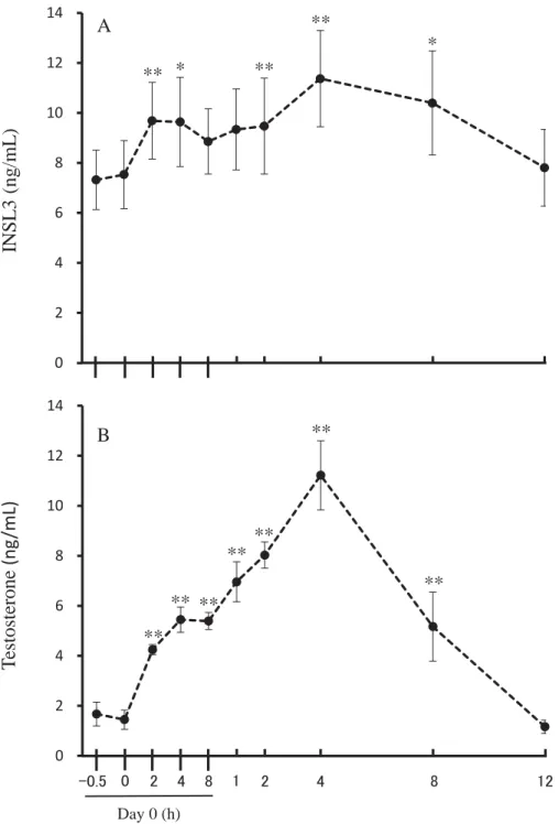
3.3. Effect of hCG treatment on INSL3 and testosterone secretion
234
A single injection of hCG was administered to determine the effect of sustained
235
levels of LH on INSL3 secretion. Mean plasma INSL3 and testosterone concentrations
236
after administration of hCG are presented in Fig. 3. Plasma INSL3 concentrations
237
increased (P<0.01) 2 h after treatment and remained significantly high (P<0.05) till the
238
next sampling at 4 h. When compared to control no significant changes were observed
239
at 8 h and 1 day after treatment. However, INSL3 concentrations again increased
240
significantly on days 2 through 8 (day 2, P<0.01; day 4, P<0.01; day 8, P<0.05),
241
approaching pre-treatment level on day 12 (Fig. 3A).
242
A dramatic increase (P<0.01) of mean plasma testosterone concentrations after
243
treatment was observed from 2 h and continued till day 4. Thereafter, concentrations
244
started to decrease but remained significantly elevated (P<0.01) until day 8, reaching
245
basal level on day 12 post-treatment (Fig. 3B). After administration of hCG, the mean
246
increasing rate (maximum/pre-treatment concentration) of testosterone concentrations
247
(10.4 ± 2.2-fold) was higher (P<0.001) than that of INSL3 (1.8 ± 0.2-fold).
248 249
4. Discussion
14
For many species including bulls [14, 15] the pulsatile release of LH from the
251
anterior pituitary stimulates immediate pulsatile secretion of testosterone from the
252
testicular Leydig cells. Conversely, it has been reported that secretion of INSL3 is not
253
acutely regulated by LH [22], but is stimulated by the long-term trophic effects of LH in
254
men [1, 6–9]. However, the short-term secretory pattern of INSL3 and its relationship
255
with LH with frequent blood sampling has not been reported. To the best of our
256
knowledge, this is the first study to evaluate circulating INSL3 levels at 15-min
257
intervals. We found that the nature of releasing INSL3 from the testicular Leydig cells
258
into the general circulation of beef bulls is pulsatile, and a temporal relationship
259
between LH and INSL3 secretion exists.
260
The secretion of INSL3 in the general circulation of beef bulls is pulsatile with an
261
average pulse frequency of about 4 in an 8 h sampling session. Themean increasing rate
262
of INSL3 pulses are much smaller than those of testosterone pulses, suggesting that
263
INSL3 can act as a less-fluctuating marker than testosterone to evaluate the testicular
264
Leydig cells status in bulls.
265
The frequency of testosterone pulses in the present study is in accordance with
266
the previous reports [14, 15] in bulls. It has been reported that LH pulses precede
267
testosterone pulses in bulls [14], which has been the case not only for testosterone
15
pulses but also for INSL3 pulses as shown in our present study. We noticed that 70% of
269
INSL3 pulses peak within 1 h period from the peak of an LH pulse, indicating that in
270
most cases INSL3 pulses are associated with LH pulses. The fewer number of
271
testosterone pulses compared with LH pulses in an 8 h sampling session demonstrate
272
that not all LH pulses are capable of generating a testosterone pulse, and therefore, there
273
might be a minimum threshold value for an LH pulse to initiate a testosterone pulse
274
whereas in case of INSL3 pulses, it seems that compared with testosterone pulses a
275
comparatively lower minimum threshold value of LH pulses is required.
276
Upon treatment with GnRH, we noticed that similar to LH and testosterone,
277
INSL3 concentrations also increased significantly within 1 h. The increasing rate
278
(maximum/pre-treatment concentration) by GnRH stimulation is much lower for INSL3
279
than for testosterone. A similar lower increasing rate of INSL3 pulses than testosterone
280
pulses was observed under physiological condition in experiment 1 with 15-min
281
intervals sampling. These results show that LH pulses precede INSL3 pulses in most
282
cases. The significant increase of INSL3 concentrations within 1 h period of GnRH
283
treatment in experiment 2 suggests that the INSL3 secretion is acutely regulated by LH.
284
Administration of hCG, which has LH-activity, provides additional evidence regarding
285
this issue. After hCG treatment, a significant increase of both testosterone and INSL3
16
levels was observed that sustained over a longer period of time. For both hormones, the
287
concentrations increased shortly after treatment, remained high till day 8, but again the
288
increasing rate is much smaller for INSL3 than for testosterone. Maintaining a
289
significant higher concentration of INSL3 and testosterone for a longer period of time
290
by hCG than GnRH treatment is probably due to the sustained longer activity of hCG
291
[25]. Previously, a significant increase of testosterone in the general circulation of bulls
292
has been shown after GnRH and hCG treatments [16–19]. In the present study, the
293
simultaneous increase of INSL3 and testosterone concentrations within 1 to 2 h after
294
those treatments, provide another new information that LH acutely regulates the
295
secretion of INSL3 in bull plasma. This acute regulation of INSL3 by LH in bulls is the
296
novel finding of our present study and is in difference to previous studies in men. One
297
study showed that when men were treated with hCG and peripheral blood was taken
298
daily for 8 days, testosterone concentrations increased after hCG treatment, but INSL3
299
did not change [22] whereas other studies showed that hCG can increase INSL3
300
concentrations in blood after 4 or 10 days of treatment when endogenous LH secretion
301
was inhibited by androgen analogues or GnRH antagonist [26, 27].
302
In conclusion, the secretion of INSL3 in the general circulation of beef bulls
303
occurs in a pulsatile manner. Endogenous and exogenous LH can stimulate INSL3
17
secretion soon after the treatment, suggesting the acute regulation of INSL3 by LH in
305
bulls.Moreover, the increasing rate of INSL3 pulses is much smaller than those of
306
testosterone pulses, and therefore we suggest that INSL3 can be used as a
307
less-fluctuating marker than testosterone when testing the functions of testicular Leydig
308 cells in bulls. 309 310 Acknowledgements 311
The authors thank NIDDK, NIH, USA and Dr. A. F. Parlow, National Hormone &
312
Pituitary Program, Harbor, UCLA Medical Center, for providing the bovine LH
313 (AFP11743B). 314 315 References 316
[1] Ivell R, Balvers M, Domagalski R, Ungefroren H, Hunt N, Schulze W. Relaxin-like
317
factor: a highly specific and constitutive new marker for Leydig cells in the human
318
testis. Mol Hum Reprod 1997;3:459–66.
319
[2] Adham IM, Burkhardt E, Benahmed M, Engel W. Cloning of a cDNA for a novel
320
insulin-like peptide of the testicular Leydig cells. J Biol Chem 1993;268:26668 –72.
321
[3] Nef S, Parada LF. Cryptorchidism in mice mutant for Insl3. Nat Genet 1999;22:295
18
9.
323
[4] Zimmermann S, Steding G, Emmen JM, Brinkmann AO, Nayernia K, Holstein AF,
324
et al. Targeted disruption of the Insl3 gene causes bilateral cryptorchidism. Mol
325
Endocrinol 1999;13:681–91.
326
[5] Kawamura K, Kumagai J, Sudo S, Chun SY, Pisarska M, Morita H, et al. Paracrine
327
regulation of mammalian oocyte maturation and male germ cell survival. Proc Natl
328
Acad Sci U S A 2004;101:7323–8.
329
[6] Foresta C, Bettella A, Vinanzi C, Dabrilli P, Meriggiola MC, Garolla A, et al.
330
Insulin-like factor 3: a novel circulating hormone of testis origin in humans. J Clin
331
Endocrinol Metab 2004;89:5952–8.
332
[7] Bay K, Hartung S, Ivell R, Schumacher M, Jürgensen D, Jorgensen N, et al.
333
Insulin-like factor 3 serum levels in 135 normal men and 85 men with testicular
334
disorders: relationship to the luteinizing hormone-testosterone axis. J Clin
335
Endocrinol Metab 2005;90:3410–8.
336
[8] Ferlin A, Garolla A, Rigon F, Rasi CL, Lenzi A, Foresta C. Changes in serum insulin
337
like factor 3 during normal male puberty. J Clin Endocrinol Metab
338
2006;91:3426–31.
339
[9] Wikstrom AM, Bay K, Hero M, Andersson A-M, Dunkel L. Serum insulin-like
19
factor 3 levels during puberty in healthy boys and boys with Klinefelter syndrome. J
341
Clin Endocrinol Metab 2006;91:4705–8.
342
[10] Anand-Ivell R, Wohlgemuth J, Haren MT, Hope PJ, Hatzinikolas G, Wittert G, et al.
343
Peripheral INSL3 concentrations decline with age in a large population of
344
Australian men. Int J Androl 2006;29:618–26.
345
[11] Büllesbach EE, Rhodes R, Rembiesa B, Schwabe C. The relaxin-like factor is a
346
hormone. Endocrine 1999;10:167–9.
347
[12] Pathirana IN, Yamasaki H, Kawate N, Tsuji M, Büllesbach EE, Takahashi M, et al.
348
Plasma insulin-like peptide 3 and testosterone concentrations in male dogs:
349
changes with age and effects of cryptorchidism. Theriogenology 2012;77:550–7.
350
[13] Kawate N, Ohnari A, Pathirana IN, Sakase M, Büllesbach EE, Takahashi M, et al.
351
Changes of plasma concentrations of insulin-like peptide 3 and testosterone from
352
birth to pubertal age in beef bulls. Theriogenology 2011;76:1632–8.
353
[14] Stumpf TT, Wolfe MW, Roberson MS, Kittok RJ, Kinder JE. Season of the year
354
influences concentration and pattern of gonadotropins and testosterone in
355
circulation of the bovine male. Biol Reprod 1993;49:1089–95.
356
[15] Finnerty M, Enright WJ, Roche JF. Testosterone, LH and FSH episodic secretory
357
patterns in GnRH-immunized bulls. J Reprod Fertil 1998;114:85–94.
20
[16] D’Occhio MJ, Aspden WJ. Characteristics of luteinizing hormone LH and
359
testosterone secretion, pituitary responses to LH-releasing hormone LHRH and
360
reproductive function in young bulls receiving the LHRH agonist deslorelin: effect
361
of castration on LH responses to LHRH. Biol Reprod 1996;54:45–52.
362
[17] Mongkonpunya K, Hafs HD, Convey EM, Tucker HA, Oxender WD. Serum
363
luteinizing hormone, testosterone and androstenedione in pubertal and prepubertal
364
bulls after gonadotropin releasing hormone. J Anim Sci 1975;40:682–6.
365
[18] Sundby A, Tollman R, Velle W. Long-term effect of hCG on plasma testosterone in
366
bulls. J Reprod Fert 1975;45:249–54.
367
[19] Murase T, Okuda K, Sato K. Assessment of bull fertility using a mucus penetration
368
test and a human chorionic gonadotrophin stimulation test. Theriogenology
369
1990;34:801–12.
370
[20] Saito H, Sawada T, Yaegashi T, Goto Y, Jin J, Sawai K, et al. Kisspeptin-10
371
stimulates the release of luteinizing hormone and testosterone in pre- and
372
post-pubertal male goats. Anim Sci J 2012;83:487–92.
373
[21] Falvo RE, Buhl AE, Reimers TJ, Foxcroft GR, Dunn MH, Dziuk PJ. Diurnal
374
fluctuations of testosterone and LH in the ram: effect of hCG and
375
gonadotrophin-releasing hormone. J Reprod Fert 1975;42:503–10.
21
[22] Bay K, Andersson AM. Human testicular insulin-like factor 3: in relation to
377
development, reproductive hormones and andrological disorders. Int J Androl
378
2011;34:97–109.
379
[23] Büllesbach EE, Schwabe C. The primary structure and the disulfide links of the
380
bovine relaxin-like factor (RLF). Biochemistry 2002;41:274–81.
381
[24] Johnson ML, Pipes L, Veldhuis PP, Farhy LS, Boyd DG, Evans WS. AutoDecon, a
382
deconvolution algorithm for identification and characterization of luteinizing
383
hormone secretory bursts: description and validation using synthetic data. Anal
384
Biochem 2008;381:8–17.
385
[25] Boime I, Ben-Menahem D. Glycoprotein hormone structure-function and analog
386
design. Recent Prog Horm Res 1999;54:271–88.
387
[26] Bay K, Matthiesson KL, McLachlan RI, Andersson A-M. The effects of
388
gonadotropin suppression and selective replacement on insulin-like factor 3
389
secretion in normal adult men. J Clin Endocrinol Metab 2006;91:1108–11.
390
[27] Roth MY, Lin K, Bay K, Amory JK, Anawalt BD, Matsumoto AM, et al. Serum
391
INSL3 is highly correlated with intratesticular testosterone in normal men with
392
acute, experimental gonadotropin deficiency stimulated with low-dose hCG: a
393
randomized-controlled trial. Fertil Steril 2013;99:132–9.
22 395
Figure legends
396 397
Fig. 1. Changes of plasma LH, INSL3 and testosterone concentrations in blood samples
398
taken from two individual representative beef bulls (A: #1, B: #2). Blood samples were
399
taken at 15-min intervals for 8 h. The peaks for INSL3 ( ), LH ( ), and testosterone
400
( ) pulses were determined by the Pulse XP software.
401 402
Fig. 2. Plasma LH (A), INSL3 (B), and testosterone (C) concentrations in response to
403
GnRH treatment (0.5 μg/kg) in beef bulls. Data are expressed as mean ± SEM (n=6). *P
404
<0.05, **P <0.01 compared with the pre-treatment value of the corresponding hormone
405
at 0 h.
406 407
Fig. 3. Plasma INSL3 (A), and testosterone (B) concentrations in response to hCG
408
treatment (5 IU/kg) in beef bulls. Data are expressed as mean ± SEM (n=6). *P <0.05,
409
**P <0.01 compared with the pre-treatment value of the corresponding hormone at 0 h.
410 411
0 1 2 3 4 5 6 7 8 0 1 2 3 4 5 6 7 8 9 10 0 2 4 6 8 10 12 0 1 2 3 4 5 6 Testosterone INSL3 LH B (#2) INSL3 LH Testosterone A (#1) Hours of sampling Fig. 1 IN SL 3 (ng /mL ) L H or T estosterone (ng /mL ) 㻜 㻝 㻞 㻟 㻠 㻡 㻢 㻣 㻤 L H or T estosterone (ng /mL ) IN SL 3 (ng /mL ) Figure
Fig. 2
LH ( n g /m L) 0 5 10 15 20 25 30**
**
**
**
**
0 1 2 3 4 5 6 7 8**
*
*
0 2 4 6 8 10 12**
**
**
**
**
**
Hours after GnRH treatment A B C 㻜 㻝 㻞 㻟 㻠 㻡 㻢 㻙㻜㻚㻡 T estosterone (ng /mL ) INSL 3 (ng /mL )
Fig. 3
T estoster one (n g /mL) 0 2 4 6 8 10 12 14 ** ** ** ** ** ** ** 㻞 㻠 0 2 4 6 8 10 12 14 ** * ** ** * A BDays after hCG treatment
㻙㻜㻚㻡 㻜 㻞 㻠 㻤 㻝 㻤 㻝㻞 Day 0 (h) IN SL 3 (ng /mL )