Effects of hypoxia exposure and eccentric contraction on skeletal muscle
1
3
5
6
7
8
10
14
22
22
25
26
30
37
37
39
40
43
48
50
51
64
66
1 Dumont et al. 2015
Murach wt al. 2017; Christov et al. 2007
1 Hypoxia Inducible Factor-1 : HIF-1 Favier et al. 2015; Semenza, 2012 HIF-1 HIF-
HIF-HIF-
HIF-Prolyl-hydroxylases PHD
HIF-2- Fe2
HIF- von Hippel-Lindau
PHD
HIF-HIF-
HIF-HIF- HeLa
HIF-1 % O2 3 Bagnall et al. 2014 1%O2 4
Hep 3B 15 Wang et al. 1995 HIF-1
in vitro 1%O2
HIF-6 O2 5000m Favier et al. 2015
HIF-Reactive Oxygen Species ROS
nitric-oxide : NO Chaudhary et al. 2012; Guzy et al. 2005; Balligand et al. 2009
ROS PHD Fe2 Fe3+ HIF- NO
PHD S-
HIF-HIF-1 Vascular Endothelial Growth Factor VEGF
Hoier and Hellsten, 2014 VEGF VEGF
Hoier and Hellsten, 2014 VEGF mRNA
Ohno et al 2012; de Theije et al. 2015; Flann et al. 2014 VEGF HIF-1 AMP-activated protein kinase AMPK
2
AMPK VEGF mRNA
VEGF Zwetsloot et al. 2008
AMPK VEGF
Peroxisome proliferator-activated receptor Gamma Coactivator 1 alpha PGC1
-estrogen-related receptor- ERR- AMPK
PGC1 mRNA Lee et al. 2006 VEGF ERR
PGC1 ERR- ERR- VEGF
Chinsomboon et al. 2008 NO 1
NO NOS endothelial NOS eNOS
NOS neuronal NOS : nNOS Clanton, 2007
-VEGF eNOS Williams et al. 2006 nNOS
Wild VEGF-A mRNA
Ward et al. 2005 1/2 Fibroblast growth factors :
FGF1/2 1
Yun et al. 2010; Brindle et al. 2006
Dumont et al. 2015 in vitro
Jash and Adhya, 2015; Li et al.
2007; Liu et al. 2012 HIF-1 HIF-1
in vivo Jash and Adhya, 2015
in vivo Christov et al. 2007 myogenin VEGF-A FGF2 HGF in vivo Latroche Latroche et al. 2017 Rhoads et al 2013
Snijders et al. 2017 Type II
3 Adult Murach et al. 2017 10 20 15 12 O2 15 5 PGC1 VEGF-A
mRNA Suzuki, 2016 VEGF-A mRNA PGC1
1
3 C/F 1
ROS Lavie, 2015
ROS
10 O2 ROS
Domínguez-Álvarez et al. 2017 COPD
Mateika et al. 2015 VEGF Mateika et al. 2015 Serebrovskaya ROS Serebrovskaya et al. 2008 Living
High-Training High LHTH Living High-Training Low LHTL
Living Low-Training High LLTH Vogt and Hoppeler, 2010
LHTH LHTL LLTH
LLTH
4
Vogt and Hoppeler, 2010 LLTH 16%O2 Okabe et al. 2017 VEGF mRNA AMPK Morales-Alamo AMPK Thr172 Morales-Alamo et al. 2012 AMPK AMP/ATP AMPK LKB1 Sirt1
Sirt1 NAD+/NADH.H+ ROS
ROS
Morales-Alamo et al. 2017 ROS NAD+/NADH.H+
AMP/ATP AMPK
Christiansen et al. 2018
ROS AMPK
ROS NADPH
ROS Bretón-Romero et al 2012 AMPK
ROS AMP/ATP NAD+/NADH.H+
25%O2
VEGF-A mRNA ROS
ROS ROS Clanton,
2007 ROS
AMPK- -VEGF-A
ROS
Pax7 MyoD Myogenin
5
Chen et al. 2012; Lavender
et al. 2008; Maeo et al. 2017 2 1
Lavender et al. 2008; Maeo et al. 2017
Takagi et al. 2010
Ikezaki et al. 2017 3
MyoD mRNA
Roig et al. 2009; Hedayatpour et al. 2015 1~2
DOMS Nosaka et al. 2002; Chen et al. 2012 2
6
Semenza, 2012 Radak et al. 2013 (ROS)
ROS Zuo et al.
2013 ROS (RNS)
Nagahisa et al. 2016; Merry and Ristow, 2016 Nagahisa et al. 2016
de Theije et al. 2015; Carberry et al. 2014 ROS RNS ROS
ROS Semenza, 2012; Lavie, 2015
Suzuki, 2016
ROS ROS
Gliemann et al. 2016 ROS Merry and
Ristow, 2016 VEGF-A Chinsomboon et al. 2008
VEGF-A mRNA
Croley et al. 2005 ROS
RT-PCR 10
20
1
Kyudo company Tosu, Japan 16 10 week-old, body weights: 30.9±0.51g 17 20 month-old, body weights: 49.3±1.75g ICR-JCL strain
N: FIO2 = 0.21 H: FIO2 = 0.16
7
gamagori, Japan carbon dioxide monitor COZY-1; JIKCO, Tokyo, Japan : 28 x 35 x 24 cm 16%O2 0.1%CO2 5 5 12 1 16%O2 1 12 12 23±2 55±7% (70mg/kg) -80 Fig 1A 2
(%), cross-sectional area (CSA: m2),
Nagahisa et al. 2016 20
CM510,Leica,Nussloch,Germany
0.1M phosphate buffered saline PBS;pH7.6 1 normal goat
serum Millipore Chemicon, Billerica, Massachusetts, USA 10 10
PBS 3
: 1 MHC -II Fast myosin
1:2000, Sigma,St Louis, Missouri, USA 2 MHC-IIa SC-71 1:1000,
Developmental Studies Hybridoma Bank, Iowa, USA PBS horseradish
peroxidase HRP goat anti-mouse IgG, Bio-Rad, Hercules, California, USA 3 diaminobenzine tetrahydrochloride Sigma HRP
BZ-X710, KEYENCE, Osaka, Japan TypeI IIa IIxb
200
0.1MPBS 2% paraformaldehyde10
PBS PBS 10% normal goat serum 2% bovine serum albumin
30 PBS 2%bovine serum albumin/PBS
mouse anti-paired box protein-7 Pax7, Developmental Studies Hybridoma Bank; 1: 1,000 rabbit anti-laminin Sigma Aldrich, 1: 1,000 2
PBS :
Cy3-conjugated AffiniPure goat anti-mouse IgG 1:1000,Jackson ImmunoResearch, West Grove, Pennsylvania, USA AlexaFlour488 goat anti-rabbit IgG 1:1000,Molecular Proves, Breda,
Netherlands 2 PBS PBS
4,6-diamino-2-phenylindole DAPI, Molecular Probes 5
Pax7 laminin DAPI BZ-X710, KEYENCE
Fig1 B laminin
8
the number of capillaries per 1 mm2
/ CD31 10 m
anti-laminin anti-CD311 1: 1000; Sigma Aldrich
3 RNA RT-PCR
mRNA RT-PCR
Nagahisa et al. 2016 TRIZOL reagent Molecular Probes, Breda,
Netherlands total RNA Total RNA 260nm 280nm
total RNA TURBOTM DNase Ambion, Austin,
USA 30 37 DNA DNase
Exscript RT reagent kit Takara, Tokyo, Japan cDNA
cDNA StepOneTM Real Time PCR System Applied Biosystems Japan, Tokyo, Japan
SYBR Green PCR Master Mix
95 10 +95 30 58 1
/ 40 Glyceraldehyde-3-phosphate dehydrogenase GAPDH
mRNA mRNA cycle
threshold CT GAPDH Ct target
PRE cDNA
Table 1 PCR pax7
MyoD myogenin Fujimaki et al. 2014 Atrogin1 ATG5 de Theije et al. 2015 BDNF
Naumenko et al. 2015 Primer Express
software v3.0, Applied Biosystems Japan FASMAC
Kanagawa, Japan
±SE RT-PCR
two-way ANOVA hypoxic method and age differences t-test
Bonferroni mRNA coefficients
p 0.05
3
N SOL: 16.0±1.1g, GA-S:193.0±7.3g H SOL: 13.3±0.3g, GA-S:173.6±17.4g IH SOL: 13.4±0.6g, GA-S:173.7±5.2g
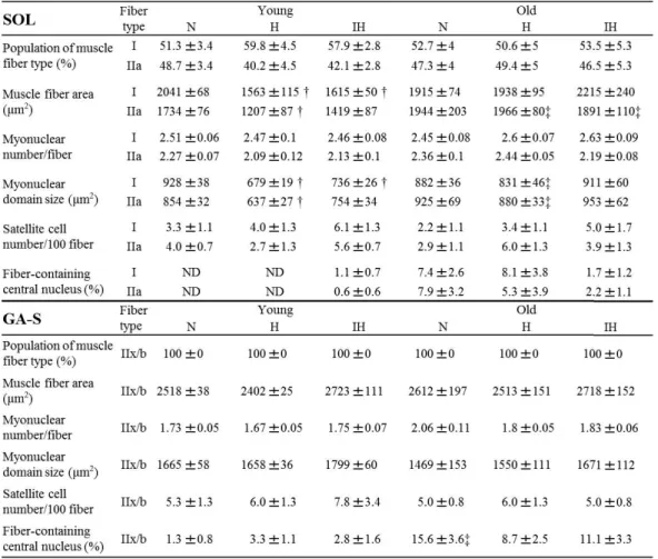
9 15.9±1.1g, GA-S:197.6±16.1g 2 Table 2 TypeI IIa TypeI Fig 2 / / Fig 3A, B 15 / Fig 3C, D / 3 mRNA Soleus muscle mRNA N 1 Fig 4 H
MyoD BDNF MHCe mRNA Atrogin1 mRNA
Fig 4A VEGF-A
IH Myostatin mRNA
H
H VEGF-A mRNA Fig
4B nNOS H IH Myostatin
mRNA
VEGF-A nNOS mRNA H mRNA
H BDNF IH FGF2 mRNA Gastrocnemius muscle H VEGF-A mRNA Fig 4C IH FGF-2 mRNA IH MyoD VEGF-A FGF-2 PGC-mRNA
10 mRNA
Fig 4D H Myogenin ATG5 mRNA IH
Correlations between factors
NO De Palma and
Clementi, 2012 SC VEGF-A FGF2 Rhoads et al.
2009 nNOS eNOS mRNA
VEGF-A FGF2 MyoD mRNA Table 3
VEGF-A nNOS mRNA
eNOS VEGF-A MyoD
1
Soleus muscle. 5 16%O2
TypeI TypeIIa
Atrogin1 Razeghi et al. 2006 mRNA IH H
Myostatin Rodriguez et al. 2014
mRNA H:P=0.3 IH:P 0.05 Myostatin mRNA
Myostatin mRNA H2O2 ROS NF-Sriram et al. 2011 IH 8%O2 3 de Theije et al. 2015 14-15%O2 8 Chen et al. 2010 Chen et al. 2010
MyoD MHCe BDNF mRNA
in vitro MyoD
Kook et al. 2008 BDNF
Yu et al. 2017 BDNF in
vitro myogenin MHCe Clow and
11
8-week-old adult 16-week-old
Murach wt al. 2017 SCs
Chen et al. 2010
Gastrocnemius muscle.
RT-PCR Atrogin1 Atg5 Myostatin mRNA
8% O2 de
Theije et al. 2015 16%O2
MyoD BDNF mRNA
IH MyoD mRNA H MyoD mRNA
H BDNF MHCe mRNA
MyoD mRNA
ROS Gliemann et al. 2016
Rhoads et al. 2013 NOS Casey et al. 2011
2
Soleus muscle. de Theije et al. 2015 12 8%O2
20 16%O2
VEGF-A mRNA nNOS mRNA
nNOS Huber-Abel et al. 2012
nNOS VEGF-A mRNA Baum et al. 2013
nNOS mRNA Ward et al. 2005
16%O2 nNOS mRNA VEGF-A mRNA
nNOS VEGF-A mRNA
VEGF-A mRNA VEGF-A mRNA
12
6% O2/ 2 h Gavin et al. 2016 12% O2/ 8 weeks Olfert et al. 2001
VEGF-A mRNA
VEGF mRNA 12% O2/ 8 weeks Olfert et al. 2001
ROS Lavie, 2015 ROS
Merry and Ristow, 2016
Mateika et al. 2015 H VEGF-A nNOS mRNA H Gastrocnemius muscle. / Verdijk et al. 2016 nNOS mRNA VEGF-A mRNA de
Theije et al. 2015 (8%O2)
16%O2
NO constitutive NOS eNOS nNOS
Ho et al. 2012 NO eNOS nNOS
Ho et al. 2012 nNOS TypeIIb
Hoshino et al. 2002 nNOS
8%O2
de Theije et al. 2015
nNOS Ward et al. 2005 NO
eNOS mRNA Egginton et al. 2016
eNOS nNOS
Williams et al. 2006a nNOS eNOS
13
eNOS VEGF-A Williams et al. 2006b
IH H VEGF-A P<0.05 eNOS mRNA P=0.063
eNOS VEGF-A mRNA
NOS Casey et al. 2011
eNOS Ho et al. 2012
eNOS VEGF-A mRNA
(SIT)
eNOS SIT
Cocks et al. 2013 eNOS
VEGF-A mRNA
MyoD mRNA eNOS-VEGF-A
NO
De Palma and Clementi, 2012 VEGF-A
FGF2 Rhoads et al. 2009
FGF2 mRNA
eNOS mRNA MyoD VEGF-A
NO VEGF-A
Conclusion 10
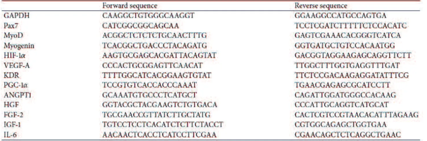
14 Table 1 Real-time RT-PCR primer sequences.
GAPDH; glyceraldehyde-3-phosphate dehydrogenase, Pax7; paired box transcription factor-7, MyoD; myogenic determination factor, VEGF-A; vascular endothelial growth factor-A, FGF2; fibroblast
growth factor 2, BDNF; brain-derived -activated receptor
gamma coactivator 1-alpha, NOS; nitric oxide synthase, MHCe; myosin heavy chain embryonic, -related gene 5
15
Table 2 Muscle fiber properties in each experimental group.
Data are shown for properties of muscle fiber types and CSA in the soleus muscle (SOL) and
superficial portion of the gastrocnemius (GA-S) in normoxic control (N), continuous hypoxia (H), and
16
Table 3 Pearson's correlation coefficients between each factor.
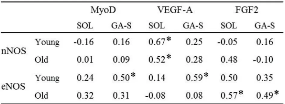
Pearson's correlation coefficients (R) between the ratio of the increase in nNOS, eNOS, and MyoD mRNA expression, and the ratio of the increase in MyoD, VEGF-A and FGF2 mRNA expression. Pearson's R was calculated based on total data for three experimental groups in each muscle from young and old mice. : Significant correlation between each factor (P < 0.05).
17
Figure 1 Images for MHC-IIa (A), satellite cells (B), central myonucleus (B), and capillaries (C) in the gastrocnemius. (A): Stained fibers represent MHC-IIa, and the area surrounded by the dashed line is the superficial portion of the gastrocnemius. (B) Image representing the basal lamina (green), myonucleus (blue), and satellite cells (red). White arrows indicate satellite cells (SCs) or central myonucleus (CMN). (C) Image representing co-localization of capillaries detected by laminin (green) and CD31 (red).
18
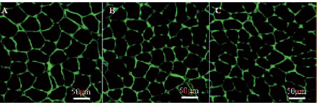
Figure 2 Images of muscle fibers in the young soleus muscle stained by laminin (green) from the N group (A), H group (B), and IH group (C).
19
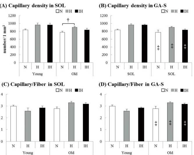
Figure 3 The capillary density (number/ 1 mm2) and capillary-to-fiber ratio of the soleus (A, C) and superficial portion of the gastrocnemius (B, D) muscles in each experimental group of young and old mice (N: white bar, H: gray bar, IH: black bar).
Values are the
difference from young mice in each group (P < 0.05). The capillary density (A) was higher in the old soleus muscle of H groups than of N groups (P=0.022). In old gastrocnemius muscles, capillary densities (B) and capillary-to-fiber ratios (D) in all groups were lower than those in young mice [capillary densities: N (P=0.049), H (P=0.001), IH (P=0.001), capillary-to-fiber ratios: N (P=0.001), H (P=0.001), IH (P=0.001)].
21
Figure 4 mRNA expression in both hypoxic groups compared with each N group in the soleus (A, B) and superficial portion of the gastrocnemius (C, D) muscles from young and old mice.
Gray and black bars represent continuous hypoxia (H) and intermittent hypoxia (IH) groups, 0.05). : Significant difference between H and IH groups for e
difference from young mice in each group (P < 0.05).
(A) In the young soleus muscle, a significant increase was observed in the MyoD (P=0.013 in H vs. N), BDNF (P=0.002 in H vs. N), MHCe (P=0.035 in H vs. N), Atrogin1 (P=0.011 in H vs. IH), and
myostatin (P=0.020 in IH vs. N) mRNA expression. On the other hand, a significant decrease was observed in the
VEGF-vs. N) mRNA expression. (B) In the old soleus muscle, VEGF-A (P=0.009) and nNOS (P=0.035) mRNA expression was significantly increased. In the IH groups, myostatin mRNA expression was significantly higher than that in the N groups (P=0.011). The mRNA expression in old mice was higher in VEGF-A (H:
P=0.001) than in the young mice, and was lower in BDNF (H: P=0.039) and FGF2 (IH: P=0.031) than in the young mice. (C) In the young gastrocnemius muscle, VEGF-A mRNA was significantly
increased (P=0.016 in H vs. N). FGF-2 mRNA expression was significantly higher than that in the N groups only in the gastrocnemius muscle of the IH groups (P=0.028). In the IH groups, the
expression of MyoD (P=0.049), VEGF-A (P=0.001), FGF-2 (P=0.005), and PGC
was significantly higher than that in H groups. (D) The expression of myogenin (P=0.024) and ATG5 (P=0.028) mRNA was higher in the H groups than in the IH groups. The expression of pax7 (P=0.022) and myogenin (P=0.048) in the old IH groups was lower than that in the young IH groups.
22
Vogt and Hoppeler, 2010; Wang et al. 2007; Stray-Gundersen et al. 4-8
4000-5000 m Vogt and
Hoppeler, 2010; Wang et al. 2007; de Theije et al.
- - Vogt and Hoppeler, 2010
VO2 max
vascular endothelial growth factor-A (VEGF-A) Vogt and Hoppeler, 2010
VEGF-A hypoxia inducible factor-1 (HIF-1 )
Favier et al. 2015; Semenza 2012 SCs
Christov et al. 2007; McClung et al. 2015; Rhoads et al. 2009 SCs
Christov et al. 2007 SCs SCs
Jash and Adhya, 2015; Li et al. 2007; Liu et al. 2012 SCs
SC Imaoka et al. 2014 SCs SCs SCs 1 8 5 3 6.5 ± 1.7 502 ± 14 kg Pascoe et al. 1999
23
Sato I, Sato AB, Uppsala, Sweden 4
6 2
16
Figure 1 = 4, FIO2 = 0.21
= 4, FIO2 = 0.15 4 6%
3 4 2 6
1.7 m/s for 1 min and 4m/s for 2 min
7 m/s for 1min 100% VO2max 11.7 ± 0.2 m/s 2
1.7m/s for 3 min 2
pretest normoxic training group pretest: Nor Con; hypoxic training group
pretest: Hypo Con 2 posttest
normoxic training group posttest: Nor Tr; hypoxic training group posttest: Hypo Tr 6%
S810, Polar, Kempele, Finland) 1.7m/s 3.5m/s
2 6m/s 2 2m/s O2 90 30 VO2 VO2 3 50 mg (lidocaine, Fujisawa
Pharmaceutical Co., Osaka, Japan) 2 cm
5cm pre post 4 4h 24
24h 3 3d 7 7d Lindholm
and Piehl, 1974 80
Kawai et al. 2013 7
m 20 CM510, Leica,Nussloch, Germany
0.1M phosphate buffered saline PBS, pH7.6 1% Millipore-Chemicon,
Billerica, MA, USA 10 (1) Myosin
heavy chain-IIa (MHC-IIa) and MHC-IIx fast myosin (Sigma,St. Louis, USA; 1 : 4,000) (2) MHC-IIa SC-71 (Developmental Studies Hybridoma Bank, Iowa,
USA;1 : 1,000) horseradish peroxidase (HRP, Bio-Rad,
24 tetrahydrochloride (Bio-Rad) HRP
(E600, Nikon, Tokyo, Japan) image-processing system (DS-U1, Nikon) Type I IIa IIx
300 4 Okabe et al. 2017 7 m 50 m 20 7 m 0.1M PBS 4% 10 10% 0.1M PBS 2% bovine serum
albumin 30 2% bovine serum albumin/PBS
mouse anti-paired box protein-7 Pax7, Developmental StudiesHybridoma Bank; 1 : 1,000 rabbit anti-laminin, Sigma, 1 : 1,000) 1
Pax7 Cy3-conjugated AffiniPure goat antimouse IgG (Jackson ImmunoResearch, West Grove, USA; 1 : 1,000) Alexa Fluor 488 goat anti-rabbit IgG (Molecular Probes, Breda, Netherlands; 1 : 1,000) PBS
4,6-diamidino-2-phenylindole DAPI 5 anti-Pax7
anti-laminin DAPI image-processing software Adobe Photoshop Elements 12, Adobe,
San Jose, CA, USA SCs Figure 2(a) SCs
DAPI Pax7 SCs/fibers 3
50 m (anti-laminin) 2 (Alexa Fluor 488 goat
antirabbit IgG) laser-scanning confocal system C1, Nikon
4 15 2 m 2
Figure 2(b)
the number of capillaries per 1mm2
5 RNA RT-PCR
Total RNA TRIzol Molecular Probes, Breda, Netherlands
RNA 260nm / 280nm Total RNA
DNA 37 30 TURBO DNase (Ambion, Austin, USA)
DNase RNA 0.5 g ExscriptTM RT reagent kit (Takara, Tokyo, Japan)
1 cDNA cDNA SYBR Green PCR Master Mix
protocol in StepOneTM Real-Time PCR System (Applied Biosystems Japan, Tokyo, Japan)
RT-PCR
10 95 40 30
1 glyceraldehyde-3-phosphate dehydrogenase GAPDH mRNA
25
Pre cDNA
Table 1 PCR Primer Express
software Applied Biosystems Japan FASMAC FASMAC,
Kanagawa, Japan 6
mean ± SEM t-test
mRNA
< 0.05
1
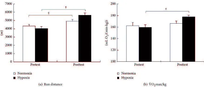
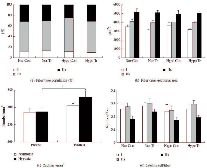
Nor: from 498 ± 6 to 489 ± 8 kg; = 0.034; Hypo: from 499 ± 6 to 483 ± 7 kg; = 0.006 Figure 3(a)
40.1% 14.0% (from 4317
± 217 to 4921 ± 232 m; = 0.0006 from 4036 ± 225 to 5653 ± 266 m;
= 0.0001 Figure 3 (b) VO2max
from 162.5 ± 5.3 to 166.2 ± 4.6 mL/min/kg from 159.5 ± 4.9 to 177.7 ± 2.8 mL/min/kg; = 0.011
2
Pre Figure 4(a)
type I: 11.0 13.1%; type IIa: 57.0 63.8%; type IIx: 24.7 31.4%
Figure 4(b) type I: 3145 3621 m2;type IIa: 3956 4072
m2; type IIx: 4967 5176 m2
Figure 4(c) Nor: from 286 ± 11 to 304 ± 6/mm2; Hypo: from287 ± 11 to 329 ± 18/mm2;
= 0.045 Figure 4(d) SC number/fiber Nor
Tr, type I: from 0.26 ±0.04 to 0.28±0.03; type IIa: from0.28±0.03 to 0.30±0.04; type IIx: from0.18±0.02 to 0.24±0.02 Hypo Tr, type I: from 0.24 ± 0.06 to 0.26 ± 0.03/fiber; type IIa: from 0.25 ± 0.03 to 0.30±0.03/fiber; type IIx: from0.17±0.02 to 0.2±0.01/fiber
3 mRNA
Pre mRNA Figure 5 Pax7 mRNA
Pre 7d
MyoD mRNA Hypo Tr Pre 2.3 MyoD mRNA
26
Tr myogenin mRNA Hypo Tr Pre = 0.023 Post = 0.002
myogenin mRNA Pax7 MyoD mRNA
24h 7d VEGF-A mRNA Hypo Tr Hypo Con
Pre = 0.029 Post e = 0.020 VEGF-A mRNA
4h 24h 7d
VEGF receptor-2 KDR mRNA Hypo Tr 4h = 0.031
Nor Tr 7d = 0.016 Peroxisome proliferator-activated receptor
coactivator-1 PGC-1 mRNA VEGF-A 4h
24h 3d Angiopoietin-1 ANGPT1 mRNA
Hypo Tr Pre Post Hypo Tr HIF-1
mRNA Pre Hypo Con = 0.042 Nor Tr = 0.041
Hypo Con 3d = 0.007 HIF-1 mRNA Nor Tr
Nor Con 7d = 0.040 HIF-1
mRNA 24h 7d
Hepatocyte growth factor HGF mRNA HIF-1 mRNA
HGF mRNA Pre Hypo Tr = 0.037 7d
Nor Tr = 0.023 HGF mRNA MyoD mRNA 4h
Fibroblast growth factor-2 FGF-2 and insulin-like growth factor-1
IGF-1 mRNA HGF HIF-1 Hypo Tr FGF-2 mRNA
3d = 0.013 Nor Tr FGF-2 mRNA
Pre = 0.007 7d
= 0.033 IGF-1 mRNA Hypo Tr 3d = 0.026 Nor Tr
Pre = 0.010 SC HGF, FGF-2, IGF-1, HIF-1
3d IL-6 mRNA 4h Hypo Tr 4h = 0.006 3d = 0.040 Nor Tr 1 VEGF-A ANGPT-1
VEGF-A mRNA Lloyd et al. 2003
capillary contacts 12 VEGF-A mRNA
8
5 mRNA VEGF-A
Gustafsson et al. 2007 VEGF-A mRNA
27
VEGF-A PGC-1
Okabe et al. 2017 VEGF-A PGC-1
mRNA Hypo Tr PGC-1 mRNA 4h
VEGF-A mRNA VEGF-A mRNA
Malek et al. 2010 VEGF-A
nitrite/nitrate NO nitric oxide NO NOS
Ren et al. 2010; Wang et al. 2014 NO
NO IL-6 mRNA Liu et al. 2015
NOS 4 7 IL-6 mRNA
Liu et al. 2015 IL-6 mRNA 4h 3d
NO
HIF-1 VEGF-A HIF-1 prolyl hydroxylase PHD
Semenza,
2012; Balligand et al. 2009 NO PHD HIF-1
Balligand et al. 2009 NO HIF-1 NO
PHD Balligand et al.
2009 HIF-1 NO
HIF-1 HGF IGF-1 FGF-2 mRNA HGF HIF-1 mRNA
Rhoads et al. 2009 IGFs Flann et al. 2014 FGF-2 Conte et al. 2008
HIF-1 HIF-1 mRNA
VEGF-A mRNA VEGF-A
KDR HIF-1 2
SCs SC HGF, VEGF, IGF-1, FGF-2
Christov et al. 2007
SCs VEGF ANGPT1 FGF-2 HGF Christov
et al. 2007; McClung et al. 2015; Rhoads et al. 2009 myogenin
SC VEGF ANGPT Christov et al.
2007; McClung et al. 2015 SCs
SC Rhoads et al.
2009 SC HIF-1 Jash and
Adhya, 2015; Li et al. 2007; Liu et al. 2012 in vivo
HIF-1 SCs
28
Jash and Adhya, 2015 NO HIF-1
NO SCs HIF- 1 mRNA Flann et al.
2014 HGF Rhoads et al. 2009 NOS nitrite/nitrate
posttest Hypo Tr
SC SC number/fiber
FGF IGF HGF IL-6
Filippin et al. 2009 IL-6
Liu et al. 2015 IL-6 mRNA 4h 3d
HIF-1 HGF FGF-2 IGF-1 KDR 3d
Yamaguchi et al. 2004
HGF FGF-2 mRNA SC
IGF-1 mRNA 3 SC IFG-1
McKay et al. 2008 IGF-1 mRNA SCs
NOS NO
Filippin et al. 2009 Pax7 HGF HIF-1
3d NO 3d
IGF-1 FGF-2 IL-6 mRNA NO
SCs Jash and
Adhya, 2015 posttest SCs
NO myogenin mRNA Pre
SCs myogenin mRNA 7 mRNA 3-7 myogenin Srikuea et al. 2010 SCs VEGF IGF-1 Borselli et al. 2010 SCs NO HIF-1
SCs myogenin mRNA Pre
SCs VEGF-A
ANGPT1 FGF-2 HGF 5
29
myogenin
VEGF-A HGF NO
HIF-1
30
Table 1 Real-time reverse transcriptional-PCR (RT-PCR) primer sequences.
GAPDH: glyceraldehyde-3-phosphate dehydrogenase; Pax7: paired box transcription factor-7; MyoD: myogenic determination factor; HIF-1 : hypoxia inducible factor-1 ; VEGF-A: vascular endothelial growth factor-A; KDR: vascular endothelial growth factor receptor-2; PGC-1 : peroxisome proliferator activated receptor coactivator 1 ; ANGPT1: angiopoietin-1; HGF: hepatocyte growth factor; FGF-2: fibroblast growth factor-2; IGF-1: insulin-like growth factor-1; IL-6: interleukin-6.
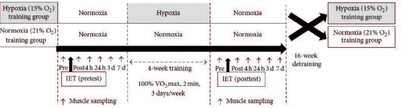
31
Figure 1 Schematic figure of the experimental schedule. The training protocol adopted a randomized crossover design, which was separated by a 16-week detraining period. Eight horses were assigned randomly into normoxic training ( = 4, FIO2 = 21%) and hypoxic training ( = 4, FIO2 = 15% O2)
groups. Incremental exercise tests (IET) were carried out before (pretest) and after (posttest) training. In each IET, horses were subjected to biopsy sampling of the gluteus medius six times, immediately (post) and 4 hours (4 h), 24 hours (24 h), 3 days (3 d), and 7 days (7 d) after IET).
32
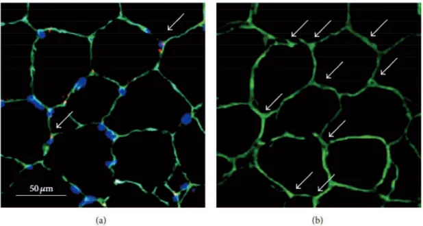
Figure 2 Typical photomicrographs of serial transverse sections of the gluteus medius muscle. Thicknesses of sections are 7 and 50 m in panels (a) and (b), respectively. (a) Triple-immunofluorescent stained for laminin (green), Pax7 (red), and nuclei (blue). The white arrow in (a) indicates a satellite cell (Pax7+ nuclei). (b) Single-immunofluorescent stained for laminin. White arrows in (b) indicate capillaries.
33
Figure 3 Changes in run distance (a) and maximal oxygen consumption (VO2max) (b) in the incremental exercise test under normoxia for the normoxic training group (white bar) and the Significant difference versus pretest ( < 0.05). Values are mean ± SEM.
34
Figure 4 Changes in fiber type population (a), fiber cross-sectional area (b), capillary density (c), and number of satellite cells (d) in pretest of control (Nor Con: normoxic control; Hypo Con: hypoxic control) and posttest (Nor Tr: normoxic training; Hypo Tr: hypoxic training) in normoxic or hypoxic training group. Measurements were performed on muscle samples obtained before the incremental exercise test (pre). White, grey, and black bars in (a), (b), and (d) represent fiber types I, IIa, and IIx, respectively.White and black bars in (c) represent normoxia and hypoxia, respectively.
36
Figure 5 (a f) Time-course changes in mRNA expression of Pax7 (a), MyoD (b), myogenin (c), VEGF-A (d), KDR (e), and PGC-1 (f) in pretest of control (Nor Con: normoxic control, light red; Hypo Con: hypoxic control, light blue) and posttest (Nor Tr: normoxic training, red; Hypo Tr: hypoxic training, blue) after normoxic or hypoxic training.Measurements were performed before (pre) and immediately (post) and 4 hours (4 h), 24 hours (24 h), 3 days (3 d), and 7 days (7 d) after the incremental exercise test. Values of mRNA expression were calculated as x-fold change from pretest < 0.05). Values are mean ± SEM. Significant difference versus normoxia training group ( < 0.05). (g l) Time-course changes in mRNA expression of ANGPT1 (g), HIF-1 (h), HGF (i), FGF-2 (j), IGF-1 (k), and IL-6 (l) in pretest of control (Nor Con: normoxic control, light red; Hypo Con: hypoxic control, light blue) and posttest (Nor Tr: normoxic training, red; Hypo Tr: hypoxic training, blue) after normoxic or hypoxic training.Measurements were performed before (pre) and immediately (post) and 4 hours (4 h), 24 hours (24 h), 3 days (3 d), and 7 days (7 d) after the incremental exercise test. Values of mRNA expression were calculated as
x- < 0.05). Values
37 (ECC)
delayed onset muscle soreness DOMS Nelson, 2013; Chen et al. 2012; Hyldahl and Hubal, 2014 ECC
Roig et al. 2009; Hedayatpour and Falla, 2015
ECC ECC DOMS
DOMS
Chen et al. 2012; Lavender and Nosaka, 2008; Maeo et al. 2017
Repeated-bout effect 2 1 Lavender and Nosaka,
2008; Maeo et al. 2017; Urai et al. 2013 6 Nosaka et al. 2013
Bradykinin BK B2 receptor mRNA Meotti et al. 2012
BKB2 receptor BKB2 receptor
Urai et al. 2013; Meotti et al. 2012 Prostaglandin
PG E2 PGE2 Cyclooxygenase COX -2 mRNA
Murase et al. 2013
Carrageenan COX-2 COX-2
PGE2 Murase et al. 2013; Zhang et al. 1997 PGE2
PGE PGE
-1 mPGES-1 COX-2 Samuelsson et al. 2007 PGE2
COX-2 mPGES-1 PGE2
BKB2 receptor COX-2 mPGES-1 PGE2
ECC Preconditioning; Precon ECC
mRNA
38
No. 290 No. 16-077
Wistar (9 n=36) Sankyo Labo Service Sapporo, Japan
12 (24±2 )
ECC Non-Precon ECC 2 ECC Precon
ECC CTL
Precon 100 ECC 2 Precon 10 ECC
ECC
Hesselink et al. 1996; Willems and Stauber, Precon
ECC (0 )
2 4
2
0°
90° S-14154, Takei Scientific Instruments, Tokyo, Japan
ECC ES 150°/ 0°
40° 4 1 45 V
1 ms 50 Hz
3
H E Evans blue
dye EBD 24 1% wt/vol EBD
solution 1 mg/10 g body wt H E EBD BIOREVO
BZ-9000 KEYENCE, Osaka, Japan
4 RT-PCR
RT-PCR Nagahisa et al.
2016 Total RNA TRIZOL reagent Invitrogen, Carlsbad, CA Total RNA
260 nm 280 nm Total RNA
TURBO DNase Ambion-Life Technologies Austin TX 37 30
DNA DNase-treated RNA 0.5 µg Exscript RT reagent kit TaKaRa Bio Otsu
Japan cDNA cDNA StepOne Real-Time PCR System
Applied Biosystems Japan, Tokyo, Japan SYBR-Green PCR Master Mix
95 10 40 95 ; 30 + 58 ;
1 / Glyseraldehyde-3-phosphate dehydrogenase
39
cycle threshold Ct GAPDH Ct Ct(target)
CTL cDNA
Primer Express software v3.0, Applied Biosystems Japan FASMAC Kanagawa, Japan
5
± SE mRNA One way-ANOVA
Bonferroni adjust t-test
Pearson < 0.05
1 ECC
0 day ECC non-Precon Precon H E
Figures 1(a) and 1(b) ECC2 4 H E
non-Precon
Figures 1(c) and 1(e) Precon Figures 1(d) and 1(f)
H E EBD 0 day
non-Precon ECC2 4 Figures 1(i) and 1(k)
Precon Figures 1(j) and 1(l)
2 mRNA
0d CTL 1 MHC-embryonic
MHC-neonatal MHC-embryonic mRNA ECC2
Precon CTL Figure 2(a) ECC4
CTL Non-Precon Precon
MHC-neonatal mRNA ECC 2 4 Non-Precon Precon
CTL Figure 2(b) Non-Precon Precon
HGF mRNA ECC2 4
Precon Figure 3(a) ECC2 4 HGF mRNA
non-Precon CTL Precon
/ Pax7 mRNA ECC2
4 Non-Precon CTL ECC4 Precon
Non-Precon Figure 3(b)
40
Non-Precon ECC2 Non-Precon CTL
Figure 3(c)
Myogenin mRNA 2 Precon
CTL 2 4 Non-Precon CTL
Figure 3(b) ECC2 4 Precon Myogenin mRNA
Non-Precon
3 mRNA
Precon BKB2 receptor mRNA ECC 2 4 CTL
Figure 4(a) Non-Precon BKB2 receptor mRNA ECC2
4 CTL ECC2 4
Precon Non-Precon
COX-2 mRNA Precon Non-Precon ECC 2 CTL
Figure 4(b) 4 Non-Precon CTL
Precon Non-Precon
mPGES-1 mRNA Non-Precon ECC2 4 CTL
Figure 4(c) Precon mPGES-1 mRNA ECC4
Non-Precon
ECC ECC
Precon ECC
Yamada et al. 2018 Precon MHC-embryonic MHC-neonatal mRNA
Pax7 MyoD Myogenin mRNA Precon
Precon
mRNA ECC
ECC mRNA
Precon mRNA non-Precon
0 day CTL Precon 2 days after
Precon Precon 10 ECC non-Precon 100
ECC Precon mRNA
Precon 2
1
MHC a x b MHC
41
MHC Schiaffino et al. 2015; Ciciliot and Schiaffino, 2010
MHC-embryonic noenatal mRNA ECC ECC4
MHC-embryonic Precon
Pax7 Dumont et al. 2015
Dumont et al. 2015 HGF
Yamada et al. 2010 MyoD myogenin
Dumont et al. 2015 ECC
Pax7 MyoD Myogenin mRNA ECC
Precon Precon
ECC Nelson et al, 2013; Chen et al. 2012 Kano et al. 2008; Sudo and Kano, 2009; Hyldahl et al. 2014
ECC Precon mRNA
2
PGE2 Karamouzis et al. 2005 PGE2
COX-2 COX-2
Lengthening Murase et al. 2013; Zhang et al. 1997
PGE2 mPGES-1 COX-2 PGE2
Samuelsson et al. 2007 PGE2 Dallaporta et al.
2010 COX-2 mPGES-1 PGE2
COX-2 /
Murase et al. 2013; Bachawaty et al. 2010; Novak et al. 2009 mPGES-1
Korotkova et al. 2008; Korotkova and Jakobsson,
2008 Precon COX-2 mPGES-1 mRNA PGE2
Precon Precon PGE2
BK Blais Jr et al. 1999
BK Langberg et al. 2002 BK BKB1 receptor BKB2 receptor
BKB1 receptor BKB2 receptor
Hamza et al. 2010; Su, 2014; Couture et al.
2001 BKB2 receptor mRNA ECC 2 4 Precon
1 4
42
BKB1 receptor Murase
et al. 2010 Formalin BKB1 B2 receptor
Formalin 7 B2 receptor
IL-6 mRNA Meotti et al. 2012 IL-6
Dina et al. 2008; Manjavachi et al. 2010 BKB2 receptor B1 receptor
30 BKB2 receptor BKB2 receptor
Murase et al. 2010
BKB2 receptor mRNA Precon 0 day ECC
non-Precon BKB2 receptor mRNA Precon
BKB2 receptor mRNA Precon BKB2 receptor mRNA BKB2 receptor mRNA Precon mRNA 3 ECC Precon
ECC ECC mRNA
Pax7
MyoD Myogenin mRNA Precon Dumont et al. 2015
COX-2 COX-2
Novak et al. 2009 COX-2
43 Table 1 Real-time RT PCR primer sequences.
GAPDH, glyceraldehydes-3-phosphate dehydrogenase; MHC, myosin heavy chain; HGF, hepatocyte growth factor; Pax7, paired box transcription factor-7; MyoD, myogenic determination factor; BKB2, bradykinin B2 receptor; COX-2, cyclooxygenase 2; mPGES-1, microsomal prostaglandin E
44
Figure 1 Photomicrograph of hematoxylin and eosin (a f) and Evans blue dye (g l) staining on sections of the left medial gastrocnemius muscles after damaging eccentric contractions.
45
Figure 2 Time course changes in relative expression of MHC-embryonic (a) and MHC-neonatal (b) mRNA. Thee mRNA expression of each time point was calculated as x-fold change from each CTL value at 0 d. CTL indicates intact right muscle of each experimental group. Values are means ± SE.
significant differences ( < 0.05) as compared with each CTL value.#significant differences ( <
46
Figure 3 Time course changes in relative expression of HGF (a), Pax7 (b), MyoD (c), and myogenin (d) mRNA. The mRNA expression of each time point was calculated as x-fold change from each CTL value at 0 d. CTL indicates intact right muscle of each experimental group. Values are means ± SE.
significant differences ( < 0.05) as compared with each CTL value. # significant differences <
47
Figure 4: Time course changes in relative expression of BKB2 receptor (a), COX-2 (b), and mPGES-1
(c) mRNA. The mRNA expression of each time point was calculated as x-fold change from each CTL value at 0 d. CTL indicates intact right muscle of each experimental group. Values are means ± SE.
significant differences ( < 0.05) as compared with each CTL value. # significant differences ( <
48
VEGF HIF1
AMPK-PGC1 NO ROS in vitro
in vivo in vivo 10 20 MyoD mRNA BDNF MyoD mRNA 3.2 1.3 BDNF mRNA VEGF-A mRNA
RT-PCR VEGF-A mRNA nNOS mRNA
R=0.52 n=17
nNOS NO
VEGF-A mRNA
VEGF-A eNOS mRNA R=0.59 n=16
eNOS-VEGF
FGF2 mRNA FGF2
MyoD mRNA VEGF-A mRNA
VEGF-A
VEGF-A mRNA
myogenin
mRNA mRNA posttest pre
49
NO NO
IL-6 NOS posttest IL-6
FGF2 mRNA
NOS eNOS Posttest 7 Pax7 mRNA
Posttest 4 IL-6
posttest 4 mRNA
IL-6 IL-6
6
BKB2 COX-2 mPGES-1 mRNA
mRNA
50 3
JRA 4
51
1. Arany Z, Foo SY, Ma Y, Ruas JL, Bommi-Reddy A, Girnun G, Cooper M, Laznik D,
Chinsomboon J, Rangwala SM, Baek KH, Rosenzweig A, Spiegelman BM (2008)
HIF-independent regulation of VEGF and angiogenesis by the transcriptional coactivator
PGC-1alpha. Nature 451, 1008-12.
2. Bachawaty T, Washington SL, Walsh SW (2010) Neutrophil expression of cyclooxygenase
2 in preeclampsia, Reprod Sci 17, 465-70.
3. Bagnall J, Leedale J, Taylor SE, Spiller DG, White MR, Sharkey KJ, Bearon RN, Sée V
(2014) Tight control of hypoxia-inducible factor-a transient dynamics is essential for cell
survival in hypoxia. J Biol Chem 289, 5549 5564.
4. Balligand JL, Feron O, Dessy C (2009) eNOS activation by physical forces: from
short-term regulation of contraction to chronic remodeling of cardiovascular tissues, Physiol
Rev 89, 481-534.
5. Baum O, Vieregge M, Koch P, Gu¨ l S, Hahn S, Huber-Abel FA, Pries AR, Hoppeler H
(2013) Phenotype of capillaries in skeletal muscle of nNOS-knockout mice. Am J Physiol
Regul Integr Comp Physiol 304, R1175-82.
6. Blais C Jr, Adam A, Massicotte D, Péronnet F (1999) Increase in blood bradykinin
concentration after eccentric weight-training exercise in men, J Appl Physiol (1985) 87,
1197-201.
7. Borselli C, Storrie H, Benesch-Lee F, Shvartsman D, Cezar C, Lichtman JW, Vandenburgh
HH, Mooney DJ (2010) Functional muscle regeneration with combined delivery of
angiogenesis and myogenesis factors, Proc Natl Acad Sci U S A 107, 3287-92.
8. Bretón-Romero R, González de Orduña C, Romero N, Sánchez-Gómez FJ, de Álvaro C,
Porras A, Rodríguez-Pascual F, Laranjinha J, Radi R, Lamas S (2012) Critical role of
hydrogen peroxide signaling in the sequential activation of p38 MAPK and eNOS in laminar
shear stress. Free Radic Biol Med 52, 1093-100.
9. Brindle NP, Saharinen P, Alitalo K (2006) Signaling and functions of angiopoietin-1 in
vascular protection. Circ Res 98, 1014-23.
52
10. Bruusgaard JC, Liestøl K, Gundersen K (2006) Distribution of myonuclei and
microtubules in live muscle fibers of young, middle-aged, and old mice. J Appl Physiol 100,
2024-2030.
11. Carberry JC
sustained hypoxia on sternohyoid and diaphragm muscle during development. Eur Respir J
43, 1149-58.
12. Casey DP, Walker BG, Curry TB, Joyner MJ (2011) Ageing reduces the compensatory
vasodilatation during hypoxic exercise: the role of nitric oxide. J Physiol 589, 1477-88.
13. Chaudhary P, Suryakumar G, Prasad R, Singh SN, Ali S, Ilavazhagan G. (2012) Chronic
hypobaric hypoxia mediated skeletal muscle atrophy: role of ubiquitin-proteasome pathway
and calpains. Mol Cell Biochem 364, 101-13.
14. Chen CY, Tsai YL, Kao CL, Lee SD, Wu MC, Mallikarjuna K, Liao YH, Ivy JL, Kuo CH
(2010) Effect of mild intermittent hypoxia on glucose tolerance, muscle morphology and
AMPK-PGC-1alpha signaling. Chin J Physiol 53, 62-71.
15. Chen TC, Chen HL, Pearce AJ, Nosaka K (2012) Attenuation of eccentric exercise-induced
muscle damage by preconditioning exercises. Med Sci Sports Exerc 44, 2090-2098.
16. Christiansen D, Murphy RM, Bangsbo J, Stathis CG, Bishop DJ (2018) Increased FXYD1
and PGC-
-restricted running is related to fibre type-specific
AMPK signalling and oxidative stress in human muscle. Acta Physiol (Oxf) 223, e13045.
17. Christov C, Chrétien F, Abou-Khalil R, Bassez G, Vallet G, Authier FJ, Bassaglia Y, Shinin
V, Tajbakhsh S, Chazaud B, Gherardi RK (2007) Muscle satellite cells and endothelial cells:
close neighbors and privileged partners, Mol Biol Cell 18, 1397-409.
18. Ciciliot S, Schiaffino S (2010) Regeneration of mammalian skeletal muscle. Basic
mechanisms and clinical implications, Curr Pharm Des 16, 906-14.
19. Clanton TL (2007) Hypoxia-induced reactive oxygen species formation in skeletal muscle.
J Appl Physiol (1985) 102, 2379-88.
20. Clow C, Jasmin BJ (2010) Brain-derived neurotrophic factor regulates satellite cell
differentiation and skeltal muscle regeneration. Mol Biol Cell 21, 2182-90.
53
21. Cocks M, Shaw CS, Shepherd SO, Fisher JP, Ranasinghe AM, Barker TA, Tipton KD,
Wagenmakers AJ (2013) Sprint interval and endurance training are equally effective in
increasing muscle microvascular density and eNOS content in sedentary males. J Physiol
591, 641-56.
22. Conte C, Riant E, Toutain C, Pujol F, Arnal JF, Lenfant F, Prats AC (2008) FGF2
translationally induced by hypoxia is involved in negative and positive feedback loops with
HIF-1 , PLoS One 3, Article ID e3078.
23. Couture R, Harrisson M, Vianna RM, Cloutier F (2001) Kinin receptors in pain and
inflammation, Eur J Pharmacol 429, 161-76.
24. Croley AN, Zwetsloot KA, Westerkamp LM, Ryan NA, Pendergast AM, Hickner RC,
Pofahl WE, Gavin TP (2005) Lower capillarization, VEGF protein, and VEGF mRNA
response to acute exercise in the vastus lateralis muscle of aged vs. young women. J Appl
Physiol (1985) 99, 1872-9.
25. Dallaporta M, Pecchi E, Thirion S, Jean A, Troadec JD (2010) Toward the management of
inflammation: recent developments of mPGES-1 inhibitors, Recent Pat CNS Drug Discov 5,
70-80.
26. De Palma C, Clementi E (2012) Nitric oxide in myogenesis and therapeutic muscle repair.
Mol Neurobiol 46, 682-92.
27. de Theije CC, Langen RC, Lamers WH, Gosker HR, Schols AM, Köhler SE (2015)
Differential sensitivity of oxidative and glycolytic muscles to hypoxia-induced muscle
atrophy, J Appl Physiol (1985) 118, 200-11.
28. Dina OA, Green PG, Levine JD (2008) Role of interleukin- 6 in chronic muscle
hyperalgesic priming, Neuroscience 152, 521-5.
29. Domínguez-Álvarez M, Gea J, Barreiro E (2017) Inflammatory Events and Oxidant
Production in the Diaphragm, Gastrocnemius, and Blood of Rats Exposed to Chronic
Intermittent Hypoxia: Therapeutic Strategies. J Cell Physiol 232, 1165-1175.
30. Dumont NA, Wang YX, Rudnicki MA (2015) Intrinsic and extrinsic mechanisms regulating
satellite cell function, Development 142, 1572-81.
54
31. Egginton S, Hussain A, Hall-Jones J, Chaudhry B, Syeda F, Glen KE (2016) Shear
stress-induced angiogenesis in mouse muscle is independent of the vasodilator mechanism
and quickly reversible, Acta Physiol (Oxf) 218, 153-166.
32. Favier FB, Britto FA, Freyssenet DG, Bigard XA, Benoit H (2015) HIF-1-driven
skeletalmuscle adaptations to chronic hypoxia: molecular insights into muscle physiology,
Cell Mol Life Sci 72, 4681-96.
33. Filippin LI, Moreira AJ, Marroni NP, Xavier RM (2009) Nitric oxide and repair of skeletal
muscle injury, Nitric Oxide 21, 157-63.
34. Flann KL, Rathbone CR, Cole LC, Liu X, Allen RE, Rhoads RP (2014) Hypoxia
simultaneously alters satellite cell-mediated angiogenesis and hepatocyte growth factor
expression, J Cell Physiol 229, 572-9.
35. Fujimaki S, Hidaka R, Asashima M, Takemasa T, Kuwabara T (2014) Wnt protein-mediated
satellite cell conversion in adult and aged mice following voluntary wheel running. J Biol
Chem 289, 7399-412.
36. Gavin TP, Westerkamp LM, Zwetsloot KA (2006) Soleus, plantaris and gastrocnemius
VEGF mRNA responses to hypoxia and exercise are preserved in aged compared with
young female C57BL/6 mice. Acta Physiol (Oxf) 188, 113 21.
37. Gliemann L, Nyberg M, Hellsten Y (2016) Effects of exercise training and resveratrol on
vascular health in aging. Free Radic Biol Med 98, 165-176.
38. Gustafsson T, Rundqvist H, Norrbom J, Rullman E, Jansson E, Sundberg CJ (2007) The
influence of physical training on the angiopoietin and VEGF-A systems in human skeletal
muscle, J Appl Physiol (1985) 103, 1012-20.
39. Guzy RD, Hoyos B, Robin E, Chen H, Liu L, Mansfield KD, Simon MC, Hammerling U,
Schumacker PT (2005) Mitochondrial complex III is required for hypoxia-induced ROS
production and cellular oxygen sensing. Cell Metab 1, 401-8.
40. Hamza M, Wang XM, Adam A, Brahim JS, Rowan JS, Carmona GN, Dionne RA (2010)
Kinin B1 receptors contributes to acute pain following minor surgery in humans, Mol Pain 6,
12.
55
41. Hedayatpour N, Falla D (2015) Physiological and neural adaptations to eccentric exercise:
mechanisms and considerations for training, Biomed Res Int 2015, Article ID 193741 7
pages,.
42. Hesselink MK, Kuipers H, Geurten P, Van Straaten H (1996) Structural muscle damage and
muscle strength after incremental number of isometric and forced lengthening contractions, J
Muscle Res Cell Motil 17, 335-41.
43. Hoier B, Hellsten Y (2014) Exercise-induced capillary growth in human skeletal muscle and
the dynamics of VEGF. Microcirculation 21, 301-14.
44. Ho JJ, Man HS, Marsden PA (2012) Nitric oxide signaling in hypoxia. J Mol Med (Berl) 90,
217-31.
45. Hoshino S, Ohkoshi N, Ishii A, Shoji S (2002) The expression of neuronal nitric oxide
synthase and dystrophin in rat regenerating muscles. J Muscle Res Cell Motil 23, 139-45.
46. Huber-Abel FA, Gerber M, Hoppeler H, Baum O (2012) Exercise-induced angiogenesis
correlates with the up-regulated expression of neuronal nitric oxide synthase (nNOS) in
human skeletal muscle. Eur J Appl Physiol 112, 155-62.
47. Hyldahl RD, Hubal MJ (2014) Lengthening our perspective: morphological, cellular, and
molecular responses to eccentric exercise, Muscle Nerve 49, 155-70.
48. Hyldahl RD, Olson T, Welling T, Groscost L, Parcell AC (2014) Satellite cell activity is
differentially affected by contraction mode in human muscle following a workmatched bout
of exercise, Front Physiol 5, 485.
49. Ikezaki K, Shibaguchi T, Sugiura T, Miyata H (2017) The effect of icing treatment on
recovery process of damaged muscle in the rat. Japanese Journal of Physical Fitness and
Sports Medicine 66, 345-354.
50. Imaoka Y, KawaiM, Mukai K, Ohmura H, Takahashi T, Hiraga A, Miyata H (2014)
Training and detraining effects on satellite cell response after exhaustive exercise in
thoroughbred horses, Japanese Journal of Physical Fitness and Sports Medicine 63,
177-187.
56
51. Jash S, Adhya S (2015) Effects of transient hypoxia versus prolonged hypoxia on satellite
cell proliferation and differentiation in vivo, Stem Cells Int 2015, 961307.
52. Kano Y, Masuda K, Furukawa H, Sudo M, Mito K, Sakamoto K. (2008) Histological
skeletal muscle damage and surface EMG relationships following eccentric contractions, J
Physiol Sci 58, 349-55.
53. Kawai M, Aida H, Hiraga A, Miyata H (2013) Muscle satellite cells are activated after
exercise to exhaustion inThoroughbred horses, Equine Vet J 45, 512-7.
54. Karamouzis M, Langberg H, Skovgaard D, Bülow J, Kjaer M, Saltin B (2001) In situ
microdialysis of intramuscular prostaglandin and thromboxane in contracting skeletal
muscle in humans, Acta Physiol Scand 171, 71-6.
55. Kook SH, Son YO, Lee KY, Lee HJ, Chung WT, Choi KC, Lee JC (2008) Hypoxia affects
positively the proliferation of bovine satellite cells and their myogenic differentiation
through up-regulation of MyoD. Cell Biol Int 32, 871-8.
56. Korotkova M, Helmers SB, Loell I, Alexanderson H, Grundtman C, Dorph C, Lundberg IE,
Jakobsson PJ (2008) Effects of immunosuppressive treatment on microsomal prostaglandin
E synthase 1 and cyclooxygenases expression in muscle tissue of patients with polymyositis
or dermatomyositis, Ann Rheum Dis 67, 1596-602.
57. Korotkova M, Jakobsson PJ (2011) Microsomal prostaglandin e synthase-1 in rheumatic
diseases, Front Pharmacol 1, 146.
58. Langberg H, Bjørn C, Boushel R, Hellsten Y, Kjaer M (2002) Exercise-induced increase in
interstitial bradykinin and adenosine concentrations in skeletal muscle and peritendinous
tissue in humans, J Physiol 542, 977-83.
59. Latroche C, Weiss-Gayet M, Muller L, Gitiaux C, Leblanc P, Liot S, Ben-Larbi S,
Abou-Khalil R, Verger N, Bardot P, Magnan M, Chrétien F, Mounier R, Germain S, Chazaud
B (2017) Coupling between Myogenesis and Angiogenesis during Skeletal Muscle
Regeneration Is Stimulated by Restorative Macrophages. Stem Cell Reports 9, 2018-2033.
60. Lavender AP, Nosaka K (2008) A light load eccentric exercise confers protection against a
57
61. Lavie L (2015) Oxidative stress in obstructive sleep apnea and intermittent
hypoxia-revisited-the bad ugly and good: implications to the heart and brain. Sleep Med Rev
20, 27-45.
62. Lee WJ, Kim M, Park HS, Kim HS, Jeon MJ, Oh KS, Koh EH, Won JC, Kim MS, Oh GT,
Yoon M, Lee KU, Park JY (2006) AMPK activation increases fatty acid oxidation in skeletal
muscle by activating PPARalpha and PGC-1. Biochem Biophys Res Commun 340, 291-5.
63. Lindholm A, Piehl K (1974) Fibre composition, enzyme activity and concentrations of
metabolites and electrolytes inmuscles of standardbred horses, Acta Vet Scand 15, 287-309.
64. Liu W, Wen Y, Bi P, Lai X, Liu XS, Liu X, Kuang S (2012) Hypoxia promotes satellite cell
selfrenewal and enhances the efficiency of myoblast transplantation, Development 139,
2857-2865.
65. Liu X, Wu G, Shi D, Zhu R, Zeng H, Cao B, Huang M, Liao H. (2015) Effects of nitric
oxide on notexin-induced muscle inflammatory responses, Int J Biol Sci 11, 156-67.
66. Li X, Zhu L, Chen X, Fan M (2007)Effects of hypoxia on proliferation and differentiation
ofmyoblasts, Med Hypotheses 69, 629-36.
67. Lloyd PG, Prior BM, Yang HT, Terjung RL (2003) Angiogenic growth factor expression in
rat skeletal muscle in response to exercise training, Am J Physiol Heart Circ Physiol 284,
H1668-78.
68. Maeo S, Yamamoto M, Kanehisa H, Nosaka K (2017) Prevention of downhill
walking-induced muscle damage by non-damaging downhill walking, PLoS One 12, Article
ID e0173909.
69. Malek MH, Olfert IM, Esposito F (2010) Detraining losses of skeletal muscle
capillarization are associated with vascular endothelial growth factor protein expression in
rats, Exp Physiol 95, 359-68.
70. Manjavachi MN, Motta EM, Marotta DM, Leite DF, Calixto JB (2010) Mechanisms
involved in IL-6-induced muscular mechanical hyperalgesia in mice, Pain 151, 345-55.
71. Mateika JH, El-Chami M, Shaheen D, Ivers B (2015) Intermittent hypoxia: a low-risk
58
72. McKay BR, O'Reilly CE, Phillips SM, Tarnopolsky MA, Parise G (2008) Co-expression of
IGF-1 family members with myogenic regulatory factors following acute damaging
musclelengthening contractions in humans, J Physiol 86, 5549-5560.
73. McClung JM, Reinardy JL, Mueller SB, McCord TJ, Kontos CD, Brown DA, Hussain SN,
Schmidt CA, Ryan TE, Green TD (2015) Muscle cell derived angiopoietin-1 contributes to
both myogenesis and angiogenesis in the ischemic environment, Front Physiol 6, 161.
74. Meotti FC, Campos R, da Silva K, Paszcuk AF, Costa R, Calixto JB (2012) Inflammatory
muscle pain is dependent on the activation of kinin B1 and B2 receptors and intracellular
kinase pathways, Br J Pharmacol 166, 1127-39.
75. Merry TL, RistowM (2016) Do antioxidant supplements interfere with skeletal muscle
adaptation to exercise training?. J Physiol 594, 5135-47.
76. Morales-Alamo D, Ponce-González JG, Guadalupe-Grau A, Rodríguez-García L, Santana A,
Cusso MR, Guerrero M, Guerra B, Dorado C, Calbet JA (2012) Increased oxidative stress
and anaerobic energy release, but blunted
Thr172-sprint exercise in severe acute hypoxia in humans. J Appl Physiol (1985) 113, 917-28.
77. Morales-Alamo D, Guerra B, Ponce-González JG, Guadalupe-Grau A, Santana A,
Martin-Rincon M, Gelabert-Rebato M, Cadefau JA, Cusso R, Dorado C, Calbet JAL (2017)
Skeletal muscle signaling, metabolism, and performance during sprint exercise in severe
acute hypoxia after the ingestion of antioxidants. J Appl Physiol (1985) 123, 1235-1245.
78. Murase S, Terazawa E, Hirate K, Yamanaka H, Kanda H, Noguchi K, Ota H, Queme F,
Taguchi T, Mizumura K. (2013) Upregulated glial cell line-derived neurotrophic factor
through cyclooxygenase-2 activation in the muscle is required for mechanical hyperalgesia
after exercise in rats, J Physiol 591, 3035-48.
79. Murach KA, White SH, Wen Y, Ho A, Dupont-Versteegden EE, McCarthy JJ, Peterson CA
(2017) Differential requirement for satellite cells during overload-induced muscle
hypertrophy in growing versus mature mice. Skelet Muscle 7, 14.
80. Murach KA, Confides AL, Ho A, Jackson JR, Ghazala LS, Peterson CA,
Dupont-Versteegden EE (2017) Depletion of Pax7+ satellite cells does not affect diaphragm
adaptations to running in young or aged mice. J Physiol 595, 6299-6311.
59
81. Murase S, Terazawa E, Queme F, Ota H, Matsuda T, Hirate K, Kozaki Y, Katanosaka K,
Taguchi T, Urai H, Mizumura K (2010) Bradykinin and nerve growth factor play pivotal
roles in muscular mechanical hyperalgesia after exercise (delayed-onset muscle soreness), J
Neurosci 30, 3752-61.
82. Nagahisa H, Okabe K, Iuchi Y, Fujii J, Miyata H (2016) Characteristics of skeletal muscle
fibers of SOD1 knockout mice, Oxid Med Cell Longev 2016, Article ID 9345970 8 pages.
83. Nagahisa H, Mukai K, Ohmura H, Takahashi T, Miyata H (2016) Effect of High-Intensity
Training in Normobaric Hypoxia on Thoroughbred Skeletal Muscle. Oxid Med Cell Longev
Article ID 1535367, 10 pages.
84. Naumenko VS, Kulikov AV, Kondaurova EM, Tsybko AS, Kulikova EA, Krasnov IB,
Shenkman BS, Sychev VN, Bazhenova EY, Sinyakova NA, Popova NK (2015) Effect of
actual long-term spaceflight on BDNF, TrkB, p75, BAX and BCL-XL genes expression in
mouse brain regions. Neuroscience 284, 730-6.
85. Nederveen JP, Joanisse S, Snijders T, Ivankovic V, Baker SK, Phillips SM, Parise G (2016)
Skeletal muscle satellite cells are located at a closer proximity to capillaries in healthy young
compared with older men. J Cachexia Sarcopenia Muscle 7, 547-554.
86. Nelson N (2013) Delayed onset muscle soreness: is massage effective?, J Bodyw Mov Ther
17, 475-82.
87. Nosaka K, Sakamoto K, Newton M, Sacco P. (2001) How long does the protective effect on
eccentric exercise-induced muscle damage last?, Med Sci Sports Exerc 33, 1490-5.
88. Nosaka K, Newton M, Sacco P (2002) Delayed-onset muscle soreness does not reflect the
magnitude of eccentric exercise-induced muscle damage. Scand J Med Sci Sports 12,
337-346.
89. Novak ML, Billich W, Smith SM, Sukhija KB, McLoughlin TJ, Hornberger TA, Koh TJ
(2009) COX-2 inhibitor reduces skeletal muscle hypertrophy in mice, Am J Physiol Regul
Integr Comp Physiol 296, R1132-9.
90. Okabe K, Mukai K, Ohmura H, Takahashi T, Miyata H (2017) Effect of acute high-intensity
exercise in normobaric hypoxia on Thoroughbred skeletal muscle. J Sports Med Phys
60
Fitness 57, 711-719.
91. Ohno H, Shirato K, Sakurai T, Ogasawara J, Sumitani Y, Sato S, Imaizumi K, Ishida H,
Kizaki T (2012) Effect of exercise on HIF-1 and VEGF signaling. Japanese Journal of
Physical Fitness and Sports Medicine 1, 5-16
92. Olfert IM, Breen EC, Mathieu-Costello O, Wagner PD (2001) Chronic hypoxia attenuates
resting and exercise-induced VEGF, flt-1, and flk-1 mRNA levels in skeletal muscle. J Appl
Physiol (1985) 90, 1532-8.
93. Pascoe JR, Hiraga A, Hobo S, Birks EK, Yarbrough TB, Takahashi T, Hada T, Aida H,
Steffey EP, Jones JH (1999)Cardiac output measurements using sonomicrometer crystals on
the left ventricle at rest and exercise, Equine Vet J Suppl 30, 148-52.
94. Radak Z, Zhao Z, Koltai E, Ohno H, Atalay M (2013) Oxygen consumption and usage
during physical exercise: the balance between oxidative stress and ROS-dependent adaptive
signaling. Antioxid Redox Signal 18, 1208-46.
95. Razeghi P, Baskin KK, Sharma S, Young ME, Stepkowski S, Essop MF, Taegtmeyer H
(2006) Atrophy, hypertrophy, and hypoxemia induce transcriptional regulators of the
ubiquitin proteasome system in the rat heart. Biochem Biophys Res Commun 342, 361-4.
96. Ren W, Yang X, Jiang X, Li Z, Zhang Z (2010) Chronichypoxia and exercise training affect
the NO content and NOS activity of rat skeletal muscle, International SportMed Journal 11,
244-257.
97. Rhoads RP, Johnson RM, Rathbone CR, Liu X, Temm-Grove C, Sheehan SM, Hoying JB,
Allen RE (2009) Satellite cellmediated angiogenesis in vitro coincides with a functional
hypoxia-inducible factor pathway. Am J Physiol Cell Physiol 296, C1321-C1328.
98. Rhoads RP, Flann KL, Cardinal TR, Rathbone CR, Liu X, Allen RE (2013) Satellite cells
isolated from aged or dystrophic muscle exhibit a reduced capacity to promote angiogenesis
in vitro. Biochem Biophys Res Commun 440, 399-404.
99. Rodriguez J, Vernus B, Chelh I, Cassar-Malek I, Gabillard JC, Hadj Sassi A, Seiliez I,
Picard B, Bonnieu A (2014) Myostatin and the skeletal muscle atrophy and hypertrophy
signaling pathways. Cell Mol Life Sci 71, 4361-71.
61
100.Roig M, O'Brien K, Kirk G, Murray R, McKinnon P, Shadgan B, Reid WD (2009) The
effects of eccentric versus concentric resistance training on muscle strength and mass in
healthy adults: a systematic review with meta-analysis. Br J Sports Med 43 556-568.
101.Samuelsson B, Morgenstern R, Jakobsson PJ (2007) Membrane prostaglandin E synthase-1:
a novel therapeutic target, Pharmacol Rev Sep 59, 207-24.
102.Schiaffino S, Rossi AC, Smerdu V, Leinwand LA, Reggiani C (2015) Developmental
myosins: expression patterns and functional significance, Skelet Muscle 5, 22.
103.Semenza GL (2012) Hypoxia-Inducible Factors in Physiology and Medicine. Cell 148,
399-408.
104.Serebrovskaya TV, Manukhina EB, Smith ML, Downey HF, Mallet RT (2008) Intermittent
hypoxia: cause of or therapy for systemic hypertension? Exp Biol Med (Maywood) 233,
627-50.
105.Snijders T, Nederveen JP, Joanisse S, Leenders M, Verdijk LB, van Loon LJ, Parise G
(2017) Muscle fibre capillarization is a critical factor in muscle fibre hypertrophy during
resistance exercise training in older men. J Cachexia Sarcopenia Muscle 8, 267-276.
106.Srikuea R, Pholpramool C, Kitiyanant Y, Yimlamai T (2010) Satellite cell activity in muscle
regeneration after contusion in rats, Clin Exp Pharmacol Physiol 37, 1078-86.
107.Sriram S, Subramanian S, Sathiakumar D, Venkatesh R, Salerno MS, McFarlane CD,
Kambadur R, Sharma M (2011) Modulation of reactive oxygen species in skeletal muscle by
myostatin is mediated through NF-
Aging Cell 10, 931-48.
108.Stray-Gundersen J, Chapman RF, Levine BD (2001) Living
high-training improves sea level performance in male and female elite runners, J Appl Physiol
(1985) 91, 1113-1120.
109.Sudo M, Kano Y (2009) Myofiber apoptosis occurs in the inflammation and regeneration
phase following eccentric contractions in rats, J Physiol Sci 59, 405-12.
110.Su JB (2014) Different cross-talk sites between the reninangiotensin and the kallikrein-kinin
systems, J Renin Angiotensin Aldosterone Syst 15, 319-28.
62
111.Suzuki J (2016) Short-duration intermittent hypoxia enhances endurance capacity by
improving muscle fatty acid metabolism in mice. Physiol Rep 4, e12744.
112.Takagi R, Fujita N, Arakawa T, Kawada S, Ishii N, Miki A (2011) Influence of icing on
muscle regeneration after crush injury to skeletal muscles in rats. J Appl Physiol 110, 382-8.
113.Urai H, Murase S, Mizumura K (2013) Decreased nerve growth factor upregulation is a
mechanism for reduced mechanical hyperalgesia after the second bout of exercise in rats,
Scand J Med Sci Sports 23, e96-101.
114.Verdijk LB, Snijders T, Holloway TM, VAN Kranenburg J, VAN Loon LJ (2016)
Resistance Training Increases Skeletal Muscle Capillarization in Healthy Older Men. Med
Sci Sports Exerc 48: 2157-2164.
115.Vogt M, Hoppeler H (2010) Is hypoxia training good formuscles and exercise performance?
Prog Cardiovasc Dis 52, 525-533.
116.Wang GL, Jiang BH, Rue EA, Semenza GL (1995) Hypoxia-inducible factor 1 is a
basic-helix-loop-helix-PAS heterodimer regulated by cellular O2 tension. Proc Natl Acad Sci
U S A 92, 5510-4.
117.Wang JS, Chen LY, Fu LL, Chen ML, Wong MK (2007) Effects of moderate and severe
intermittent hypoxia on vascular endothelial function and haemodynamic control in
sedentary men, Eur J Appl Physiol 100, 127-135,.
118.Wang JS, Lee MY, Lien HY, Weng TP (2014) Hypoxic exercise training improves
cardiac/muscular hemodynamics and is associated with modulated circulating progenitor
cells in sedentarymen, Int J Cardiol 170, 315-23.
119.Ward ME, Toporsian M, Scott JA, Teoh H, Govindaraju V, Quan A, Wener AD, Wang G,
Bevan SC, Newton DC, Marsden PA (2005) Hypoxia induces a functionally significant and
translationally efficient neuronal NO synthase mRNA variant. J Clin Invest 115, 3128-39.
120.Williams JL, Cartland D, Hussain A, Egginton S (2006) A differential role for nitric oxide
in two forms of physiological angiogenesis in mouse. J Physiol 570, 445-54.
121.Williams JL, Cartland D, Rudge JS, Egginton S (2006) VEGF trap abolishes shear stress-
and overload- dependent angiogenesis in skeletal muscle. Microcirculation 13, 499-509.
63
122.Willems ME, Stauber WT (2009) the effect of number of lengthening contractions on rat
isometric force production at different frequencies of nerve stimulation, Acta Physiol (Oxf)
196, 351-6.
123.Yamada M, Tatsumi R, Yamanouchi K, Hosoyama T, Shiratsuchi S, Sato A, Mizunoya W,
Ikeuchi Y, Furuse M, Allen RE (2010) High concentrations of HGF inhibit skeletal muscle
satellite cell proliferation in vitro by inducing expression of myostatin: a possible
mechanism for reestablishing satellite cell quiescence in vivo, Am J Physiol Cell Physiol 298,
C465-76.
124.Yamada R, Himori K, Tatebayashi D, Ashida Y, Ikezaki K, Miyata H, Kanzaki K, Wada M,
Westerblad H, Yamada T (2018) Preconditioning contractions prevent the delayed onset of
myofibrillar dysfunction after damaging eccentric contractions, J Physiol 596, 4427-4442.
125.Yamaguchi A, Ishii H, Morita I, Oota I, Takeda H (2004) mRNA expression of fibroblast
growth factors and hepatocyte growth factor in rat plantarismuscle following denervation
and compensatory overload, Pflugers Arch 448, 539-46.
126.Yun YR, Won JE, Jeon E, Lee S, Kang W, Jo H, Jang JH, Shin US, Kim HW (2010)
Fibroblast growth factors: biology, function, and application for tissue regeneration. J Tissue
Eng 2010, 218142.
127.Yu T, Chang Y, Gao XL, Li H, Zhao P (2017) Dynamic Expression and the Role of BDNF
in Exerciseinduced Skeletal Muscle Regeneration. Int J Sports Med 38, 959-966.
128.Zhang Y, Shaffer A, Portanova J, Seibert K, Isakson PC (1997) Inhibition of
cyclooxygenase-2 rapidly reversesvinflammatory hyperalgesia and prostaglandin E2
production, J Pharmacol Exp Ther 283, 1069-75.
129.Zuo L, Shiah A, Roberts WJ, Chien MT, Wagner PD, Hogan MC (2013) Low Po2
conditions induce reactive oxygen species formation during contractions in single skeletal
muscle fibers. Am J Physiol Regul Integr Comp Physiol 304, R1009-16.
130.Zwetsloot KA, Westerkamp LM, Holmes BF, Gavin TP (2008) AMPK regulates basal
skeletal muscle capillarization and VEGF expression, but is not necessary for the angiogenic
response to exercise. J Physiol 586, 6021-35.
64 SCs SCs SCs in vivo SCs 10 20 MyoD mRNA SCs VEGF-A mRNA
VEGF-A mRNA RT-PCR VEGF-A
mRNA nNOS mRNA nNOS NO
FGF2 mRNA
eNOS VEGF-A mRNA
SCs SCs SCs SCs VEGF-A mRNA myogenin mRNA SCs SCs NO IL-6 NO NOS SCs IL-6 IL-6 SCs QOL mRNA
65
SCs
Pax7 MyoD Myogenin mRNA
SCs SCs
66
: Hiroshi Nagahisa, Kazutaka Mukai, Hajime Ohmura, Toshiyuki Takahashi, Hirofumi Miyata
: Effect of High-Intensity Training in Normobaric Hypoxia on Thoroughbred Skeletal Muscle
: Oxidative Medicine and Cellular Longevity Volume 2016, Article ID 1535367, 10 pages
: 2016 9
: Hiroshi Nagahisa, Kazumi Ikezaki, Ryotaro Yamada, Takashi Yamada, Hirofumi Miyata
: Preconditioning Contractions Suppress Muscle Pain Markers after Damaging Eccentric Contractions
: Pain Research and Management Volume 2018, Article ID 3080715, 8 pages
: 2018 10
: Hiroshi Nagahisa, Hirofumi Miyata
: Influence of hypoxic stimulation on angiogenesis and satellite cells in mouse skeletal muscle
: PLOS ONE Volume 13, Article ID e0207040, 15 pages : 2018 11
67
: Hiroshi Nagahisa, Kazuma Okabe, Yoshihito Iuchi, Junichi Fujii, Hirofumi Miyata : Characteristics of Skeletal Muscle Fibers of SOD1 Knockout Mice
:Oxidative Medicine and Cellular Longevity Volume 2016, Article ID 9345970, 8 pages
: 2015 12
: Hiroshi Ichikawa Taiki Matsuo Yasuo Higurashi Hiroshi Nagahisa Hirofumi Miyata Takao Sugiura Naomi Wada
: Characteristics of Muscle Fiber-Type Distribution in Moles
: The Anatomical Record : Advances In Integrative Anatomy And Evolutionary Biology https://doi.org/10.1002/ar.24008 : 2018 10 (on line published)