北里大学大学院理学研究科 平成27年度博士論文
Regulatory mechanism of FilGAP activity by phosphorylation
森 下 祐 至 ( DS- 12904 )
指導教授
細胞機能制御学
太 田 安 隆Index
Abstract P.1
Introduction P.2
Materials and Method s P.3
Results
Part.1 Phosphorylation of FilGAP regulates its cellular localization P.7
Part.2 Determination of the critical phosphorylation site of FilGAP P.15
Part.3 FilGAP phosphorylated at S402 is mainly localized in the cytoplasm P.19
Part.4 Cell spreading on fibronectin induces dephosphorylation of FilGAP at S402
P.27
Part.5 Arf6 may regulate FilGAP activity co-operatively with phosphorylation of
FilGAP P.31
Discussion P.36
References P.39
Acknowledgement P.42
1
ABSTRACT
FilGAP is a Rho GTPase-activating protein (GAP), which specifically regulates Rac. FilGAP is phosphorylated by ROCK, and this phosphorylation stimulates its RacGAP activity. However, it is unclear how phosphorylation regulates cellular functions and localization of FilGAP. We found that non-phosphorylatable FilGAP (ST/A) mutant is predominantly localized to cytoskeleton along actin filaments and partially co-localized with vinculin around cell periphery, whereas phosphomimetic FilGAP (ST/D) mutant is diffusely cytoplasmic. Moreover, phosphorylated FilGAP detected by Phos-tag is also mainly localized in the cytoplasm. Of the six potential phosphorylation sites in FilGAP tested, only mutation of serine 402 to alanine (S402) resulted in decreased cell spreading on fibronectin. FilGAP phosphorylated at S402 is localized to cytoplasm but not at the cytoskeleton. Although S402 is highly phosphorylated in serum-starved quiescent cells, dephosphorylation of S402 accompanied with the cell spreading on fibronectin. Treatment of the cells expressing wild-type FilGAP with Calyculin A, a Ser/Thr phosphatase inhibitor, suppressed cell spreading on fibronectin whereas cells transfected with FilGAP S402A mutant was not affected cell spreading by Calyculin A. Expression of constitutively activate Arf6 Q67L mutant stimulated membrane blebbing activity of both non-phosphorylatable (ST/A) and phosphomimetic (ST/D) FilGAP mutants. Conversely, depletion of endogenous Arf6 suppressed membrane blebbing induced by FilGAP (ST/A) and (ST/D) mutants.
Our study suggests that Arf6 and phosphorylation of FilGAP may regulate FilGAP and
phosphorylation of S402 may play a role in the regulation of cell spreading on
fibronectin
.2
INTRODUCTION
Rho family small GTPases (Rho GTPases) are involved in the control of actin cytoskeleton and membrane dynamics and play essential roles in many cellular functions such as cell adhesion, cell migration, and vesicle traffickings (1-6). Rho GTPases function as molecular switches in cells. They exist in either an inactive GDP-bound state or an active GTP-bound state; in the active state, they stimulate downstream effectors. This cycle is mainly regulated by two classes of proteins.
Guanine nucleotide exchange factors (GEFs) activate Rho GTPases by loading GTP whereas GTPase-activating proteins (GAPs) facilitate the inactivation of Rho GTPases by stimulating their intrinsic GTPase activity (7-11).
FilGAP is a Rac-specific GAP that suppresses Rac-dependent lamellipodia formation and cell spreading (12-20). Phosphorylation of FilGAP by Rho/Rho-associated protein kinase (ROCK) stimulates its Rac GAP activity (12).
Depletion of endogenous FilGAP by siRNA induces a Rac-driven elongated mesenchymal morphology. Conversely, over-expression of FilGAP induces membrane blebbing and a rounded amoeboid morphology contingent upon Rho/ROCK-dependent phosphorylation of FilGAP (18). Thus, FilGAP mediates antagonism of Rac by Rho, which suppresses the elongated mesenchymal morphology and promotes rounded amoeboid migration (18,19,21,22).
While Rho/ROCK-dependent phosphorylation of FilGAP stimulates its
RacGAP activity in vivo, such phosphorylation has no effect on the catalytic activity of
FilGAP in vitro (12). In this report, we present evidence that phosphorylation of FilGAP
may regulate its subcellular localization. We also show that Ser 402 is an important
phosphorylation site for the regulation of FilGAP activity.
3
MATERIALS AND METHODS Proteins and Plasmids
The HA-tagged FilGAP (wild-type, ST/D, ST/A, S391A, S402A, S413A, S415A, S437A, and T452A) constructs in pCMV5 vector were described previously (12,18). The HA-tagged Arf6 (Q67L) construct in the pcDNA vector was provided by Dr. Nakayama (Kyoto University, Kyoto, Japan). The FLAG-tagged FilGAP (wild-type, ST/D, and ST/A) constructs in pCMV5 vector were described previously (20).
Cell culture
HEK 293, Hela, Cos-7, and MDA-MB-231 cells were grown at 37°C in
DMEM (Sigma-Aldrich, St. Louis, MO) supplemented with 10% (v/v) fetal bovine
serum (FBS) and 50 U/ml penicillin/streptomycin at 37°C. Because Cos-7 and
MDA-MB-231 express endogenous FilGAP high, they were used to analyze
endogenous FilGAP.The human melanoma cell lines A7 were grown in MEM (Sigma)
supplemented with 2% FBS, 8% newborn calf serum, 50 U/ml penicillin/streptomycin
and 50g/ml geneticin at 37°C. Because A7 cells are composed of a lot of cytoskeleton
protein, they were used to analyze cell spreading. For transfection, cells were
transfected with plasmid DNA using Lipofectamine 2000 as described by the
manufactures (Invitrogen, Carlsbad, CA). Immunofluorescent staining was performed
as described (12). Briefly, cells plated on coverslips were fixed in 3.7% formaldehyde,
permeabilized in 0.5% Triton-X 100, and stained with anti-HA or other antibodies. For
cytoskeletal staining, cells were washed once by PHEM buffer (20 mM PIPES, 2 mM
MgCl2, 50 mM KCl, 5 mM EGTA, 5 mM DTT, and 1 mM ATP), permeabilized in
PHEM buffer containing 0.5% Triton X-100 for 2 min and then fixed in PHEM buffer
containing 3.7% formaldehyde at room temperature. For visualization of F-actin, cells
4
were stained with Alexa Fluor 568 conjugated-phalloidin in PBS for 1 h. Cells were observed under an Olympus IX81 fluorescence microscope (Olympus, Tokyo, Japan).
Images were acquired by a charge-coupled device camera (ORCA-ER; Hamamatsu photonics, Hamamatsu, Japan) with constant exposure time (300 ms for transfected cells and 1s for detecting endogenous protein) and analyzed by MetaMorph software (Molecular Devices, Sunnyvale, CA).
Antibodies
Mouse anti-HA (12CA5) antibody was purchased from Roche Applied Science (Indianapolis, IN). Mouse monoclonal anti--tubulin and anti-vinculin antibodies were purchased from Sigma. Mouse monoclonal anti-vimentin antibody was purchased from Dako Cytomation. Mouse monoclonal anti-Arf6 antibody was purchased from Santa-Curz Cruz BiotchnologyBiotechnology. Polyclonal antibodies against FilGAP were raised in rabbits and purified as described previously (20). Secondary antibodies conjugated to Alexa Fluor 488 or 568, and Alexa Fluor 568-phalloidin were also purchased from Invitrogen. Rabbit anti-pS402 FilGAP polyclonal antibody was directed against amino acid residues 397-407 (CGSKTNpSPKNSV) of human FilGAP protein.
The peptide was coupled through cysteine at the NH2-terminal residue to keyhole limpet hemocyanin (KLH) and was used to raise the antiserum. The antiserum specific to pS402 FilGAP was affinity-purified with the immobilized peptides. The 1st column contains phosphorylated peptides (CGSKTNpSPKNSV) and the 2nd column holds non-phosphorylated peptide (CGSKTNSPKNSV).
Cell spreading assay
Cell spreading assay was performed as described (12). Briefly, quiescent cells
5
were trypsinized and suspended in serum-free MEM containing 0.2% BSA (CALBIOCHM) and incubated as a suspension for 1h at 37°C. Cells were then plated on fibronectin-coated cover slips and incubated for indicated time periods at 37°C. The cells were fixed and processed for immunofluorescence staining. For immunoblotting, cells were washed twice with 2 ml of PBS, suspended with 200 l of lysis buffer (RIPA) containing 50 mM Tris-HCl (pH 7.4), 500 mM NaCl, 0.5% Triton-X100, 0.5% sodium deoxycholate, 0.1% SDS, 1 mM EDTA, 1mM sodium orthovanadate, 30 mM sodium pyrophosphate, and 50 mM sodium fluoride with protease inhibitors. The cell lysates were pre-cleared and the supernatants were collected and subjected to SDS-PAGE and proteins were detected by immunoblot using anti-HA or anti-pS402 antibody.
Subcellular fractionation
Cells transfected with HA-FilGAP were washed twice with 2 ml of PBS and lysed in 120 l of PHEM buffer containing 0.5% Triton X-100 with protease inhibitors.
Cell suspensions were collected with a rubber policeman and centrifuged at 2,1600 x g
for 3 min. Supernatant fluids were removed, and the l
of PBS containing 1% SDS. Fractions were subjected to SDS-PAGE and proteins were detected by immunoblot using anti-HA antibody. The relative amount of HA-FilGAP protein in the cytoskeleton and supernatant was quantitated from digitized images of immunoblots by using the Image J analysis program.
Dephosphorylation assay
HA-FilGAP protein was immunoprecipitated using anti-HA agarose beads
from HEK cells transfected with pCMV5-HA-FilGAP. After immunoprecipitation, the
anti-HA-beads were washed once with PBS and then three times with calf intestine
6
alkaline phosphatase (CIAP) reaction buffer (TaKaRa) containing 10 mM Tris-HCl (pH 9.0), and 1 mM MgCl2. The precipitates were re-suspended in 40
l of CIAP bufferwith or without 20 units of CIAP (TaKaRa). The beads were incubated for 30 min at 37 °C in the presence or absence of phosphatase inhibitors containing 10 mM sodium fluoride, 2 mM β-glycerophosphate, and 2 mM sodium pyrophosphate. The reaction was terminated by adding 10
l of 1% SDS, boiled for 5 min, and centrifuged. Thesupernatants were collected and subjected to SDS-PAGE. Bound-proteins were detected by immunoblot using anti-HA or anti-pS402 antibody. For phosphatase inhibitor (Calyculin A) treatment, A7 cells transfected with HA-FilGAP were serum-starved. The quiescent cells were incubated with 10 M Calyculin A (Cell signaling) for 30 min at 37°C. After treatment, the cells were washed three times with 2ml PBS, lysed by 120
l of lysis buffer (RIPA) containing 50 mM Tris-HCl (pH 7.4), 500 mM NaCl, 0.5%
Triton-X100, 0.5% sodium deoxycholate, 0.1% SDS, 1 mM EDTA, 1mM sodium orthovanadate, 30 mM sodium pyrophosphate, and 50 mM sodium fluoride with protease inhibitors, and centrifuged at 200,000 x g for 20min. The supernatants were collected and subjected to SDS-PAGE. Bound-proteins were detected by immunoblot using anti-HA or anti- pS402 antibody.
P hos-tag SDS-PAGE
Phos-tag SDS-PAGE using a 6% polyacrylamide gel containing 25 μM Phos-tag acrylamide (Wako chemicals) and 100 μM MnCl
2was also carried out according to the manufacturer's instructions.
Statistical analysis
The statistical significance was assessed by two-tailed unpaired Student’s t-test
7
or one-way analysis of variance (ANOVA).
RNA Interference
siRNA oligonucleotides were purchased from Invitrogen. The targeting sequence of Arf6 was 5`-GGCAAGACAACAAUCCUGUACAAGU-3`. The targeting sequences of FilGAP was 5`-CAGUGGUAAAUUACAACCUCCUCAA -3`. A7 cells were transfected with Arf6 siRNA using Lipofectamine 2000. CosCOS-7 cells were transfected with FilGAP si RNA using Lipofectamine 2000. Forty-eight hours after transfection, the levels of each protein were measured by Western blot analysis using anti-Arf6 or anti-FilGAP antibody.
Ethics statement
The animal experiments were carried out in strict accordance with the protocols
approved by committee of Kitasato University (No.SA1010). All efforts were made to
minimize animal suffering.
8
RESULTS
Part.1 Phosphorylation of FilGAP regulates its subcellular localization
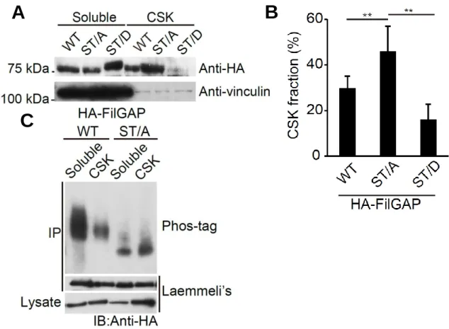
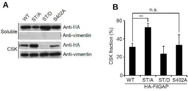
We have shown previously that FilGAP is phosphorylated at 5 serines and 1 threonine by ROCK (Fig.1)(12). a FilGAP mutant with all potential phosphorylation sites mutated to alanine (ST/A) failed to function as a RacGAP in cells, while a FilGAP mutant with all potential phosphorylation sites mutated to phosphomimetic aspartic acid (ST/D) suppressed Rac-driven lamellae formation in vivo(12). However, it is unclear how phosphorylation regulates FilGAP. To determine whether phosphorylation regulates subcellular localization of FilGAP, we compared the localization of non-phosphorylatable (ST/A) and phosphomimetic (ST/D) FilGAP mutants. We found more than 40% of the ST/A mutant in the Triton X-100 insoluble fraction (i.e., the cytoskeleton), whereas less than 20% of the ST/D mutant was found in this fracion (Fig.
2-1A and B). We next analyzed the localization of phosphorylated FilGAP using Phos-tag SDS-PAGE. Phosphorylated proteins can be detected by their delayed migration in Phos-tag SDS-PAGE gels (23,24). When FilGAP protein immunoprecipitated from transfected cells (HA-FilGAP) was resolved by Phos-tag SDS-PAGE, the migration of non-phosphorylatable FilGAP (ST/A) protein was much faster than that of wild-type FilGAP protein (Fig. 2-1C). This suggests that the serine and threonine residues mutated to alanine are major phosphorylation sites in vivo.
Moreover, while phosphorylated FilGAP was mainly recovered in the Triton X-100
soluble fraction, the mobility of non-phosphorylatable FilGAP (ST/A) did not differ
between Triton X-100 soluble and insoluble fractions (Fig. 2-1C). To further
demonstrate that phosphorylation is responsible for release of FilGAP from the
cytoskeleton, we determined the effect of Calyculin A, an inhibitor of protein
9
phosphatase 3A and protein phosphatase 1. As shown in Fig. 2-2 A and B, treatment of the cells with Calyculin A reduced the amount of HA-FilGAP localized to the cytoskeleton. Endogenous FilGAP was also released from the cytoskeleton following treatment with Calyculin A (Fig. 2-2C and D).
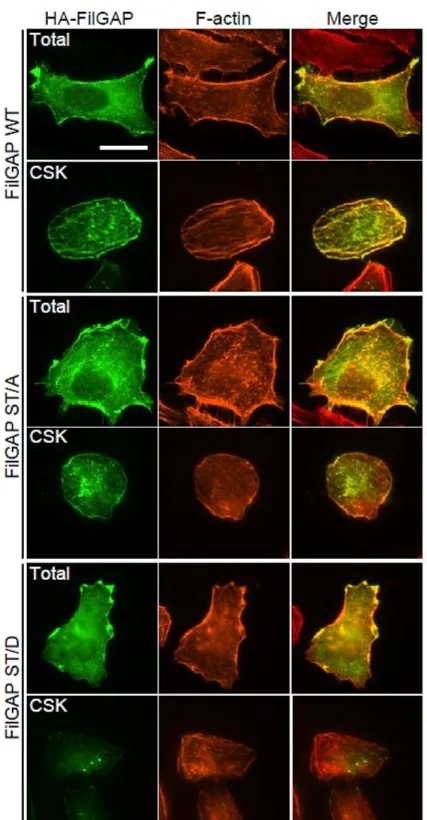
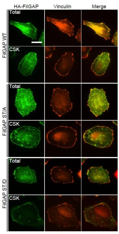
Following transient transfection of cells with HA-FilGAP constructs, wild-type
FilGAP localized with actin filaments (Fig. 3). Wild-type FilGAP is also localized to the
Triton X-100 insoluble cytoskeleton and co-localized with vinculin at the cell
peripheries (Fig. 4). Both the non-phosphorylatable FilGAP (ST/A) mutant and the
phosphomimetic FilGAP (ST/D) mutant localized with actin filaments and vinculin at
the cell peripheries. However, while the ST/A mutant localized to the cytoskeleton with
actin filaments and vinculin, little ST/D) mutant was detected at the cytoskeleton (Fig. 3
and 4). These results suggest that non-phosphorylated FilGAP may associate with the
cytoskeleton and that phosphorylation of FilGAP at critical residues may induce
translocation of FilGAP from the cytoskeleton to the cytoplasm, which may be required
for activation of FilGAP.
10
FIGURE 1. Schematic diagram of potential phosphorylation sites on FilGAP by
ROCK. Purified FilGAP protein was incubated with Myc-ROCK immune complex
beadfs and 1 mM ATP for 20 min at 30
oC. Phosphorylated FilGAP protein was
separated by SDS-PAGE, isolated from gels, and digested in-gel. The extracted peptides
were separated and processed for LC/MS/MS analysis at the Taplin Biological Mass
Spectrometry Facility at Harvard Medical School (12).
11
FIGURE 2-1. Phosphorylation of FilGAP regulates its cellular localization. A, HEK cells were transfected with HA-tagged wild-type FilGAP, non-phosphorylatable ST/A, or phosphomimetic ST/D FilGAP mutant. The cells were lysed and Triton-solubilized, and insoluble cytoskeletal (CSK) fractions were prepared. Samples of each fraction were immunoblotted with anti-HA antibody to detect HA-FilGAP.
Vinculin was used as a loading control. B, the relative amounts of HA-FilGAP in the cytoskeletons were quantitated from digitized images of autoradiograms of immunoblots by using Image J program. Each value represents the percentage of total and the mean ± SEM (n=7). **, p < 0.01. Statistical significance was determined by one-way ANOVA. C, Human melanoma A7 cells were transfected with HA-tagged wild-type FilGAP or non-phophorylatable ST/A FilGAP mutant. The cells were lysed and Triton-solubilized, and insoluble cytoskeletal fractions were prepared. HA-FilGAP proteins were immunoprecipitated from each fraction and subjected to Phos-tag SDS-PAGE (upper lane) or Laemmli’s SDS-PAGE (lower panel) and detected by immunoblot using anti-HA antibody.
A B
C
12
FIGURE 2-2. Calyculin A treatment change FilGAP localization by phosphorylation. A, A7 cells were transfected with HA-FilGAP (wild-type) and serum-starved. Quiescent cells were incubated with DMSO or 10 nM Calyculin A for 30 min at 37oC. Then, the cells were lysed and Triton-solubilized, and insoluble cytoskeletal (CSK) fractions were prepared. Samples of each fraction were immunoblotted with anti-HA antibody. Vimentin was used as a marker of cytoskeletal protein. B, the relative amounts of HA-FilGAP in the cytoskeleton were quantitated from digitized images of autoradiograms of immunoblots by using Image J program.
Each value represents the percentage of total and the mean ± SEM of triplicate determinations. *, p < 0.05. Statistical significance was determined by Student’s t test. C Human breast adenocarcinoma MDA-MB-231 cells were serum-starved. Quiescent cells were incubated with DMSO or 10 nM Calyculin A for 60 min at 37oC. Then, the cells were lysed and Triton-solubilized, and insoluble cytoskeletal (CSK) fractions were
A
C D
B
13
prepared. Samples of each fraction were immunoblotted with anti-FilGAP antibody.
Vimentin was used as a marker of cytoskeletal protein. D, the relative amounts of endogenous FilGAP in the cytoskeleton were quantitated from digitized images of autoradiograms of immunoblots by using Image J program. Each value represents the percentage of total and the mean ± SEM of triplicate determinations. **, p < 0.005.
Statistical significance was determined by Student’s t test.
14
FIGURE 3. Sub-cellular localization of FilGAP. A7 cells were transfected with
HA-tagged wild-type (WT) FilGAP, non-phosphorylatable (ST/A), or phosphomimetic
(ST/D) FilGAP mutant. The cells were fixed after treatment with (CSK) or without
(Total) 0.5% Triton X-100. HA-FilGAP (green) and F-actin (red) were localized by
staining the cells with anti-HA antibody and Alexa-Flour-phalloidin. Merged fluorescent
images are also shown. Scale bar, 20 μm.
15
FIGURE 4. Sub-cellular localization of FilGAP. A7 cells were transfected with
HA-tagged wild-type (WT) FilGAP, non-phosphorylatable (ST/A), or phosphomimetic
(ST/D) FilGAP mutant. The cells were fixed after treatment with (CSK) or without
(Total) 0.5% Triton X-100. HA-FilGAP (green) and vinculin (red) were localized by
staining the cells with anti-HA and anti-vinculin antibodies. Merged fluorescent images
are also shown. Scale bar, 20 μm.
16
Part.2 Determination of the critical phosphorylation site of FilGAP
Hela cells that were plated on fibronectin-coated coverslips adhered and then started to spread within 20 min (Fig. 5-1A) (12). Forced expression of wild-type FilGAP or the phosphomimetic FilGAP (ST/D) mutant abolished cell spreading, while over-expression of the non-phosphorylatable FilGAP (ST/A) mutant enhanced initial cell spreading on fibronectin (Fig. 5-1A). The spread area occupied by cells expressing the ST/A mutant expressing cells was larger than that occupied by cells expressing wild-type FilGAP or the ST/D mutant (Fig. 5-1B). Therefore, phosphorylation of FilGAP is required for efficient suppression of cell spreading on fibronectin.
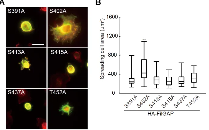
We used the cell-spreading assay to determine which phosphorylation site of FilGAP is critical for its activation. Among the FilGAP mutants with one potential phosphorylation site mutated to alanine, only the mutant with S402 mutated to alanine (S402A) stimulated initial cell spreading on fibronectin (Fig.5-2A and B). Thus, phosphorylation of Ser 402 (S402) is required for activation of FilGAP.
We next determined whether phosphorylation of FilGAP at S402 regulates
localization of FilGAP. Although the FilGAP S402A mutant did not suppress cell
spreading on fibronectin, localization of this mutant was not different from that of
wild-type FilGAP (Fig. 5-3 A and B). Thus, targeting of FilGAP to the cytoskeleton may
require de-phosphorylation of residues other than S402.
17
FIGURE 5-1. FilGAP inhibit cell spreading mediated adhesion to fibronecitn by phosphorylation. A, Hela cells were transfected without (control) or with HA-tagged wild-type FilGAP (WT), non-phosphorylatable (ST/A) or phosphomimetic (ST/D) FilGAP mutant and serum starved. The quiescent cells were trypsinized and cells in suspension were plated on coverslips coated with fibronectin and fixed 20 min after plating. Cells were stained with anti-HA antibody for HA-FilGAP (green) and Alexa-Flour-phalloidin for F-actin (red). Representative of merged images of cells are shown. Scale bar, 50 μm. B, the surface area of spreading cells 20 min after plating was calculated and plotted as the mean ± SEM (n=3). More than 50 cells were analyzed in each experiment. **p < 0.01. Statistical significance was determined by one-way ANOVA.
A B
18
FIGURE 5-2. Identification of Serine 402 of FilGAP as a critical phosphorylation site to regulate FilGAP activity. A, Hela cells were transfected with HA-tagged FilGAP mutants and serum starved. The quiescent cells were trypsinized and cells in suspension were plated on coverslips coated with fibronectin and fixed 20 min after plating. Cells were stained with anti-HA antibody for HA-FilGAP mutants (green) and Alexa-Flour-phalloidin for F-actin (red). Representative of merged images of cells are shown. Scale bar, 50 μm. B, the surface area of spreading cells 20 min after plating was calculated and plotted as the mean ± SEM (n=3). More than 50 cells were analyzed in each experiment. **p < 0.01. Statistical significance was determined by one way ANOVA.
A B
19
FIGURE 5-3. Multiple phosphorylation is necessary for translocation of FilGAP. A, A7 cells were transfected with HA-FilGAP constructs (wild-type, ST/A, ST/D, or S402A). The cells were lysed and Triton-solubilized, and insoluble cytoskeletal (CSK) fractions were prepared. Samples of each fraction were immunoblotted with anti-HA antibody to detect HA-FilGAP. Vimentin was used as a marker of cytoskeletal protein.
B, the relative amounts of HA-FilGAP in the cytoskeletons were quantitated from digitized images of autoradiograms of immunoblots by using Image J program. Each value represents the percentage of total and the mean ± SEM (n=3). **, p < 0.01.
Statistical significance was determined by Student’s t test.
A B
20
Part.3 FilGAP phosphorylated at S402 is mainly localized in the cytoplasm
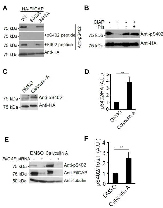
To study the functional significance of phosphorylation of FilGAP at S402, we prepared a rabbit polyclonal antibody that specifically recognizes FilGAP protein phosphorylated at S402 (anti-pS402 antibody). The anti-pS402 antibody recognized wild-type HA-FilGAP and the HA-FilGAP S413A mutant but not the HA-FilGAP S402A mutant (Fig. 6-1A). HA-FilGAP protein was detected when the anti-pS402 antibody was pre-adsorbed with non-phosphorylated S402 peptide but not when it was pre-adsorbed with phosphorylated peptide (Fig. 6-1A). Moreover, no signal for HA-FilGAP was detected when the protein was pre-incubated with calf intestine alkaline phosphatase (Fig. 6-1B). Consistent with these results, treatment of the cells with Calyculin A, a phosphatase inhibitor, increased the amount of FilGAP protein recognized by the anti-pS402 antibody (Fig. 6-1C and D). Calyculin A also increased the amount of endogenous FilGAP protein recognized by the anti-pS402 antibody (Fig.
6-1E and F). Moreover, depletion of endogenous FilGAP by siRNA abolished the signal (Fig. 6-1E and F). Thus, the anti-pS402 antibody specifically recognizes endogenous FilGAP protein phosphorylated at S402.
Treatment of the cells with LPA (lysophosphatidic acid) and FCS increased the
phosphorylation of FilGAP, which is consistent with our previous finding that
phosphorylation of FilGAP is Rho/ROCK-dependent (12,18). However, these
treatments did not increase phosphorylation of S402 (data not shown). Moreover,
treatment of the cells with the ROCK-specific inhibitor Y27632 did not abolish
phosphorylation of FilGAP at Ser 402 (Fig. 6-2A and B). These results may suggest that
protein kinases other than ROCK may be responsible for phosphorylation of S402.
21
We next determined the subcellular localization of FilGAP phosphorylated at
S402. A7 cells were transfected with wild-type HA-FilGAP and the Triton X-100
soluble and insoluble fractions were separated. As shown in Fig. 5-3A and B,
HA-FilGAP protein phosphorylated at S402 was mainly found in the Triton X-100
soluble fraction. Consistent with this observation, little HA-FilGAP protein
phosphorylated at S402 was detected at the cytoskeleton when HA-FilGAP was
transfected and its localization was determined (Fig. 6-3C and D). We also determined
the localization of endogenous FilGAP protein in COS-7 cells. We detected endogenous
FilGAP at the cytoskeleton but FilGAP protein phosphorylated at S402 mostly localized
in the cytoplasm (Fig. 7). The localization of endogenous FilGAP seems to be specific
because the fluorescent signals disappeared when the primary antibodies were
pre-adsorbed with antigens.
22
FIGURE 6-1. Characterization of anti FilGAP phosphorylated at Ser 402 antibody.A, A7 cells were transfected with HA-FilGAP constructs (wild-type, S402A, or S413A). Cell extracts were prepared and analyzed by Western blot using anti-HA or anti-pS402 FilGAP antibodies in the presence or absence of excess amounts of antigen-phosphopeptide or non-phosphopeptide. B, HA-FilGAP (wild-type) was
A B
C D
E F
23
isolated from transfected HEK cells using anti-HA agarose beads. The beads were washed and incubated in the presence or absence of alkaline phosphatase (CIAP) and phosphatase inhibitors (PIs) for 30 min at 37oC. The beads were washed and the bound FilGAP proteins were analyzed by immunoblot using anti-HA and anti-p402S FilGAP antibodies. C, A7 cells were transfected with HA-FilGAP (wild-type) and serum-starved.
Quiescent cells were incubated with DMSO or Calyculin A for 30 min at 37oC. Then, cell extracts were prepared and analyzed by immunoblot using anti-HA and anti-pS402 FilGAP antibodies. Asterisks indicate non-specific bands. D, the relative amounts of HA-FilGAP phosphorylated at S402 compared to total HA-FilGAP were quantitated from digitized images of autoradiograms of immunoblots by using Image J program.
Each value represents the mean ± SEM (n=3). **, p < 0.05. Statistical significance was determined by Student’s t test. E, CosCOS-7 cells treated with or without FilGAP siRNA were serum-starved. Quiescent cells were incubated with DMSO or Calyculin A for 60 min at 37oC. Cell extracts were prepared and analyzed by Western blot using anti-FilGAP and anti-pS402 FilGAP antibodies. Tubulin was used as a loading control.
F, the relative amounts of FilGAP phosphorylated at S402 compared to total FilGAP
were quantitated from digitized images of autoradiograms of immunoblots by using
Image J program. Each value represents the mean ± SEM (n=3). **, p < 0.05. Statistical
significance was determined by Student’s t test.
24
FIGURE 6-2 Y27632 inhibit phosphorylation at Ser402 slightly.A, A7 cells were transfected with HA-FilGAP (wild-type) and serum-starved. Quiescent cells were incubated with DMSO or 10 nM Y27632 for 60 min at 37oC. Cell extracts were prepared and analyzed by Western blot using anti-HA and anti-pS402 FilGAP antibodies. B, the relative amounts of HA-FilGAP phosphorylated at S402 compared to total HA-FilGAP were quantitated from digitized images of autoradiograms of immunoblots by using Image J program. Each value represents the mean ± SEM (n=3).
**, p < 0.05. Statistical significance was determined by Student’s t test.
A B
25
FIGURE 6-3. Phosphorylation of FilGAP at Serine 402 in cells. A, A7 cells transfected with HA-FilGAP (wild-type) were lysed and Triton-solubilized, and insoluble cytoskeletal (CSK) fractions were prepared. Sample of each fraction were immunoblotted with anti-HA and anti-pS402 antibodies. B, the relative amounts of HA-FilGAP (total protein and phosphorylated protein at S402) in the cytoskeletons wasere quantitated from digitized images of autoradiograms of immunoblots by using Image J program. Each value represents the percentage of total and the mean ± SEM (n=3). **, p < 0.05. Statistical significance was determined by Student’s t test. C, A7 cells transfected with HA-FilGAP (wild-type) were fixed after treatment with (CSK) or without (Total) 0.5% Triton X-100. HA-FilGAP (red) and HA-FilGAP phosphorylated at S402 (green) were localized by staining the cells with anti-HA and anti-pS402 antibodies. Scale bar, 20 μm. D, the relative intensities of HA-FilGAP phosphorylated at S402 compared to total HA-FilGAP were calculated and plotted as the means ±SEM (n=3). **, p < 0.01. Statistical significance was determined by Student’s t test.
A B
C D
26
Figure 7-1. Sub-cellular localization of endogenous FilGAP. CosCOS-7 cells were fixed after treatment with (CSK) or without (Total) 0.5% Triton X-100. Then the cells were stained with anti-FilGAP antibody for endogenous FilGAP (green) and Alexa-Flour-phalloidin for F-actin (red), which were nontreated or preabsorbed with antigen (GST-FilGAP). Merged fluorescent images are also shown. Scale bar, 2
40μm. Insets show magnification images of the boxed regions.
27
Figure 7-2 Sub-cellular localization of endogenous FilGAP. A, CosCOS-7 cells were fixed after treatment with (CSK) or without (Total) 0.5% Triton X-100. Then the cells were stained with anti-pS402S FilGAP antibody for phosphorylated FilGAP (green) and Alexa-Flour-phalloidin for F-actin (red), which were nontreated or preabsorbed with antigen peptide (pS402 peptide). Merged fluorescent images are also shown. Scale bar, 420 μm. Insets show magnification images of the boxed regions. B, the relative intensities of endogenous FilGAP (total protein and phosphorylated protein at S402p402S FilGAP) in the cytoskeletons was quantitated compared to total FilGAP was calculated and plotted as the means ± SEM (n=3). **, p < 0.01. Statistical significance was determined by Student’s t test.
A B
28
Part.4 Cell spreading on fibronectin induces dephosphorylation of FilGAP at S402
We next determined whether extracellular signals such as growth factors and cell adhesion molecules regulate phosphorylation of FilGAP at S402. We found that serum-starvation of A7 cells did not significantly decrease the phosphorylation of FilGAP at S402 and addition of growth factors such as EGF did not increase the phosphorylation of FilGAP at S402 (data not shown). Among the various conditions tested, spreading of cells on fibronectin induced dephosphorylation of FilGAP at S402 (Fig. 8-1A and B). In contrast, dephosphorylation of FilGAP at S402 did not occur when the cells were plated on poly-L-lysine (Fig. 8-1A and B). When cells overexpressing HA-FilGAP were plated on fibronectin and treated with Calyculin A, dephosphorylation of FilGAP at S402 was suppressed (Fig. 8-1C and D). A7 cells plated on fibronectin-coated dishes spread and achieved a maximal extent flattening by 1h (Fig.
8E). Treatment of control A7 cells with Calyculin A did not affect cell spreading or flattening on fibronectin (Fig. 8-2 A and B). However, Calyculin A treatment of A7 cells overexpressing wild-type FilGAP suppressed cell spreading on fibronectin, whereas spreading of A7 cells overexpressing FilGAP mutants (ST/A and S402A) was not affected by Calyculin A (Fig. 8-2 A and B). Thus, dephosphorylation of FilGAP at S402 may be necessary for efficient spreading on fibronectin.
We confirmed that dephosphorylation of endogenous FilGAP could be induced
by cell spreading on fibronectin. The amount of FilGAP phosphorylated at S402 in
COS-7 cells as detected by immunofluorescent staining was reduced after the cells were
plated on fibronectin (Fig. 9).
29
FIGURE 8-1. Cell adhesion to fibronectin induces dephosphorylation of FilGAP at Serine 402.A, A7 cells were transfected with HA-FilGAP (wild-type) and serum-starved. Quiescent cells were trypsinized and cells in suspension were plated on dishes coated with fibronectin or poly-L-lysine for indicated time. Cell extracts were prepared and analyzed by Western blot using anti-HA and anti-pS402 FilGAP antibodies. B, the relative amount of FilGAP protein phosphorylated at Ser 402 was calculated and plotted as the mean ± SEM (n=3). C, A7 cells were transfected with HA-FilGAP (wild-type) and serum-starved. Quiescent cells were incubated with DMSO or 10 nM Calyculin A for 30 min at 37oC. Then the cells were trypsinized and cells in suspension were plated on dishes coated with fibronectin or poly-L-lysine for indicated time. Cell extracts were prepared and analyzed by Western blot using anti-HA and anti-pS402 FilGAP antibodies. D, the relative amount of FilGAP protein phosphorylated at Ser 402 was calculated and plotted as the mean ± SEM (n=3).
C A B
D
30
FIGURE 8-2 Dephosphorylation of FIlGAP is necessary for cell spreading . A, A7 cells were transfected with HA-FilGAP constructs (wild-type, ST/A, or S402A) and serum-starved. Quiescent cells were incubated with DMSO or Calyculin A for 30 min at 37oC. Then the cells were trypsinized and cells in suspension were plated on coverslips coated with fibronectin and fixed 20 min after plating. Cells were stained with anti-HA antibody (green) and Alexa-Flour-phalloidin (red). Scale bar, 40 μm. B, The surface area of spreading cells 60 min after plating was calculated and plotted as the mean ± SEM (n=3) More than 50 cells were counted in each experiment. ***, p < 0.005.
Statistical significance was determined by Student’s t test.
A B
31
FIGURE 9. Cell adhesion to fibronectin induces dephosphorylation of endogenous FilGAP at Serine 402. COS-7 cells were serum-starved. Quiescent cells were trypsinized and cells in suspension were plated on coverslips coated with fibronectin for indicated time periods. Cells were fixed and stained with anti-FilGAP or anti-pS402 FilGAP antibodies. Dotted lines indicate cell peripheries Dotted lines are outlines determined by actin stain. Scale bar, 40m. B, the relative intensities of p402S FilGAP compared to total FilGAP was calculated and plotted as the means ± SEM (n=3).
A
B
32
Part.5 Regulation of plasma membrane blebbing by Arf6 and phosphorylation of FilGAP
We previously showed that Arf6 GTPase binds to FilGAP and stimulates its RacGAP activity to induce plasma membrane blebbing (20). We examined whether Arf6-mediated regulation has any role in the phosphorylation-dependent activation of FilGAP. Forced expression of FilGAP in A7 cells induced membrane blebbing around the cell periphery (Fig. 10A and C). The non-phosphorylatable FilGAP (ST/A) mutant failed to induce blebbing whereas the phosphomimetic FilGAP (ST/D) mutant induced blebbing as efficiently as wild-type FilGAP (Fig. 10A and
C).Forced expression of the constitutively activated mutant Arf6 Q67L stimulated
plasma membrane blebbing induced by wild-type FilGAP (Fig.9B and C). Arf6 Q67L
also stimulated both phosphomimetic (ST/D) FilGAP and non-phosphorylatable (ST/A)
FilGAP activity (Fig.10B and C). The smaller spreading area of cells expressing Arf6
Q67L also suggested that Arf6 stimulates FilGAP activity, because cell area is reduced
by the contraction of blebbing cells (Fig. 10D). We further examined the role of Arf6
using Arf6 siRNA. Depletion of endogenous Arf6 by siRNA suppressed bleb formation
induced by both wild-type and phosphomimetic (ST/D) FilGAP mutant (Fig. 11A and
C). Knockdown of endogenous Arf6 suppressed cell shrinkage induced by
over-expression of wild-type as well as the ST/A and ST/D mutants (Fig. 11D). We
generated a construct resistant to Arf6 siRNA (HA-Arf6 Q67LR) and examined whether
downregulation of FilGAP activity by Arf6 siRNA was prevented. At 48 h after
transfection with Arf6 siRNA, HA-Arf6 Q67LR, but not endogenous Arf6 protein, was
abundantly expressed in A7 cells (Fig. 11E) and HA-Arf6 Q67LR rescued the induction
of membrane blebbing by wild-type and phosphomimetic FilGAP (ST/D) (Fig. 11B and
33
C). Thus, Arf6-mediated regulation of FilGAP may be distinct from
phosphorylation-dependent activation of FilGAP.
34
.FIGURE 10. Arf6 and phosphorylation of FilGAP regulate bleb formation. A, A7 cells were transfected with FLAG-FilGAP constructs (WT, ST/A, and ST/D). After 24 h, the cells were fixed and stained with anti-FLAG antibody for FLAG-FilGAP (green) and Alexa-Fluor-phalloidin for F-actin (red). Scale bar, 40 μm. Arrowheads indicate the membrane blebbing cells. B, A7 cells were transfected with FLAG-FilGAP constructs (WT, ST/A, and ST/D) in the presence of constitutively-activated HA-Arf6 Q67L. After 24 h, the cells were fixed and stained with anti-FLAG antibody for FLAG-FilGAP (green) and anti-HA antibody for Arf6 Q67L (red). Scale bar, 40 μm. Arrowheads
A
C B
D
E
35
indicate the membrane blebbing cells. C, the percentage of blebbing cells was calculated, and the data are expressed as the mean ± S.E.M (n=3). **, p < 0.01. Statistical significance was determined by one-way ANOVA. D, the surface area of cells was calculated and plotted as the mean ± SEM (n=3) **, p < 0.01. Statistical significance was determined by one-way ANOVA. E, A7 cells were transfected with FLAG-FilGAP and HA-Arf6 Q67L. Cell extracts were prepared and analyzed by Western blot using anti-HA and anti-FLAG
antibodies.
36
FIGURE 11. Depression of Arf6 suppressed bleb formation induced by phosphorylation of FilGAP.A, A7 cells were treated with or without Arf6 siRNA in the
B A
C D
E
37
absence or presence of FLAG-FilGAP constructs. For co-transfection of plasmid DNA and
Arf6 siRNA, the cells were first transfected withsiRNA for 24 h and then cotransfected with plasmid DNA. After 24 h, cells were fixed and stained with anti-FLAG antibody for FLAG-FilGAP (green) and Alexa-Fluor-phalloidin for F-actin (red). Representative merged images are shown. Scale bar, 40 μm. Arrowheads indicate the membrane blebbing cells. B, A7 cells were transfected with Arf6 siRNA for 24 h and then co-transfected with FLAG-FilGAP constructs and HA-Arf6 Q67L resistant to
Arf6 siRNA (HA-Arf6 Q67LR). After 24 h, cells were fixed and stained with anti-FLAG antibody for FLAG-FilGAP (green) and anti-HA antibody for Arf6 Q67L
R(red). Representative merged images are shown. Scale bar, 40 μm. Arrowheads indicate
the membrane blebbing cells. C, the percentage of blebbing cells was calculated, and the
data are expressed as the mean ± S.E.M (n=3). Statistical significance was determined
by one-way ANOVA.**, p < 0.01. D, the surface area of cells was calculated and plotted
as the mean ± SEM (n=3) **, p < 0.01. Statistical significance was determined by
one-way ANOVA.E, A7 cells were transfected with FLAG-FilGAP with or without Arf6
siRNA. Cell extracts were prepared and analyzed by Western blot using anti-FLAG and
anti-Arf6 antibodies.
38
DISCUSSION
In this study, we demonstrated that FilGAP is localized to the cytoskeleton and cytoplasm and that phosphorylation of FilGAP may induce translocation of the protein from the cytoskeleton to the cytoplasm to activate its RacGAP activity. We identified S402 as a critical phosphorylation site for the activation of FilGAP to suppress cell spreading on fibronectin.
Several lines of evidence suggest that phosphorylation of FilGAP may regulate its subcellular localization. First, more than 40% of the non-phosphorylatable FilGAP (ST/A) mutant was found in the Triton X-100 insoluble fraction (the cytoskeleton), and this mutant localized with actin filaments and partially co-localized with the focal adhesion protein vinculin. In contrast, the phosphomimetic FilGAP (ST/D) mutant was found in the Triton X-100 soluble fraction and barely detectable at the cytoskeleton by immunofluorescent staining. Second, much of the phosphorylated FilGAP protein detected in Phos-tag SDS-PAGE was present in the Triton X-100 soluble fraction.
Moreover, treatment of the cells with Calyculin A reduced the amount of FilGAP protein localized to the cytoskeleton. We have shown previously that Rho/ROCK-dependent phosphorylation of FilGAP stimulates its RacGAP activity, but how phosphorylation regulates FilGAP remains unclear (12). It is possible that the phosphorylation-dependent release of FilGAP from the cytoskeleton induces translocation of FilGAP to the plasma membrane to inactivate Rac. This is consistent with our finding that bleb formation induced by FilGAP requires the pleckstrin homology domain, which binds to the plasma membrane (20).
Of the six potential phosphorylation sites in FilGAP, S402 seems to be
important for the protein’s activity. Among the FilGAP mutants with one potential
39
phosphorylation site mutated to alanine, only S402A failed to suppress cell spreading on fibronectin. This is consistent with our previous finding that S402A was the least effective in inducing membrane blebbing (12). However, our present study suggests that phosphorylation of S402 may not be responsible for release of FilGAP from the cytoskeleton. FilGAP protein phosphorylated at S402 is mostly localized in the cytoplasm as detected by anti-pS402 antibody. However, we found that localization of the non-phosphorylatable FilGAP S402A mutant was not different from that of wild-type FilGAP. Therefore, translocation of FilGAP from the cytoskeleton to the cytoplasm may be induced by phosphorylation of sites in FilGAP other than S402.
Phosphorylation of FilGAP at S402 may regulate FilGAP activity through an as yet unidentified mechanism other than release of FilGAP from the cytoskeleton.
Although FilGAP is phosphorylated downstream of Rho/ROCK signaling, our present study suggests that ROCK may not be the principal protein kinase responsible for phosphorylation of FilGAP at S402. We found that serum starvation of A7 cells did not significantly decrease the phosphorylation of FilGAP at S402, and stimulation with agonists such as LPA and EGF did not increase the phosphorylation of FilGAP at S402.
Moreover, treatment of cells with the ROCK-specific inhibitor Y27632 did not diminish
the phosphorylation of S402. Thus, Rho/ROCK-dependent phosphorylation of multiple
sites other than S402 may induce translocation of FilGAP from the cytoskeleton to
activate its RacGAP activity. FilGAP protein phosphorylated at S402 was mostly
detected in the cytoplasm; therefore, S402 may be phosphorylated in the cytoplasm after
FilGAP is translocated from the cytoskeleton. S402 matches the consensus
phosphorylation sequence defined for various protein kinases, and therefore multiple
kinases may be responsible for phosphorylation of S402 (25-27).
40
Cell spreading on extracellular matrix such as fibronectin initiates complex arrays of signaling through activation of integrin (28-32). We found that cell spreading on fibronectin induced dephosphorylation of S402, which may have a physiological significance. Forced expression of wild-type FilGAP suppressed initial cell spreading on fibronectin and maximal flattening was attained by 1h. Treatment of the cells with Calyculin A, a phosphatase inhibitor, further suppressed cell flattening on fibronectin, suggesting that dephosphorylation of S402 may occur downstream of integrin activation.
However, Calyculin A treatment of cells expressing the S402A mutant did not affect cell spreading on fibronectin. Therefore, dephosphorylation of S402 may be necessary and sufficient for inactivation of FilGAP to induce maximal flattening. Previous studies have suggested that protein serine/threonine phosphatases are involved in the control of cell spreading on fibronectin (33-36). Our present study suggests that dephosphorylation of endogenous FilGAP at S402 may occur during cell spreading on fibronectin. It is necessary to determine whether dephosphorylation of S402 of endogenous FilGAP has any role in the control of integrin-dependent cell spreading.
RhoGAPs are multidomain proteins that are regulated downstream of distinct
signaling cascades (7). Activated Arf6 recruits FilGAP to plasma membrane and
stimulates its RacGAP activity (20). Our present study suggests that Arf6 and
phosphorylation of FilGAP may independently regulate FilGAP to stimulate its
RacGAP activity. Forced expression of the constitutively activated mutant Arf6 Q67L
stimulated not only wild-type FilGAP and the phosphomimetic FilGAP (ST/D) mutant,
but also non-phosphorylatable FilGAP (ST/A) mutant. Conversely, depletion of
endogenous Arf6 by siRNA suppressed plasma membrane blebbing induced by the
phosphomimetic (ST/D) and non-phosphorylatable (ST/A) FilGAP mutants. This may
41
be consistent with our model in which activated Arf6 at the plasma membrane recruits FilGAP by binding its pleckstrin homology domain, allowing FilGAP to inactivate Rac at the membrane. Phosphorylation of FilGAP may induce translocation of FilGAP from the cytoskeleton to the cytoplasm increasing FilGAP’s access to the plasma membrane.
It remains to be determined how phosphorylation of S402 is involved in the activation of FilGAP. Phosphorylation of S402 may be required for release of an as yet unidentified inhibitor from FilGAP. Further study is necessary to understand the mechanism of regulation.
In conclusion, our study demonstrated that dephosphorylation of FilGAP
occurs during integrin-mediated cell adhesion on fibronectin. Both integrin-mediated
protein phosphorylation and integrin-mediated protein dephosphorylation may play a
role in the control of Rho GTPase signaling.
42
REFERENCES
1. Burridge, K., and Wennerberg, K. (2004) Rho and Rac take center stage. Cell 116, 167-179
2. Jaffe, A., and Hall, A. (2005) Rho GTPases: Biochemistry and biology. Annu.
Rev. Cell Dev. Biol. 21, 247-269
3. Heasman, S. J., and Ridley, A. J. (2008) Mammalian Rho GTPases: new insights into their functions from in vivo studies. Nat Rev Mol Cell Biol 9, 690-701 4. Parsons, J. T., Horwitz, A. R., and Schwartz, M. A. (2010) Cell adhesion:
integrating cytoskeletal dynamics and cellular tension. Nat Rev Mol Cell Biol 11, 633-643
5. Hall, A. (2012) Rho family GTPases. Biochem Soc Trans 40, 1378-1382
6. Sadok, A., and Marshall, C. J. (2014) Rho GTPases: Masters of cell migration.
Small GTPases 5
7. Bos, J. L., Rehmann, H., and Wittinghofer, A. (2007) GEFs and GAPs: critical elements in the control of small G proteins. Cell 129, 865-877
8. McCormack, J., Welsh, N. J., and Braga, V. M. (2013) Cycling around cell-cell adhesion with Rho GTPase regulators. J Cell Sci 126, 379-391
9. Miller, N. L., Kleinschmidt, E. G., and Schlaepfer, D. D. (2014) RhoGEFs in cell motility: novel links between Rgnef and focal adhesion kinase. Curr Mol Med 14, 221-234
10. Cook, D. R., Rossman, K. L., and Der, C. J. (2014) Rho guanine nucleotide exchange factors: regulators of Rho GTPase activity in development and disease.
Oncogene 33, 4021-4035
11. van Buul, J. D., Geerts, D., and Huveneers, S. (2014) Rho GAPs and GEFs:
Controling switches in endothelial cell adhesion. Cell Adh Migr 8
12. Ohta, Y., Hartwig, J. H., and Stossel, T. P. (2006) FilGAP, a Rho- and ROCK-regulated GAP for Rac binds filamin A to control actin remodelling. Nat Cell Biol 8, 803-814
13. Nakamura, F., Osborn, T. M., Hartemink, C. A., Hartwig, J. H., and Stossel, T.
P. (2007) Structural basis of filamin A functions. J Cell Biol 179, 1011-1025
14. Shifrin, Y., Arora, P., Ohta, Y., Calderwood, D., and McCulloch, C. (2009) The role of FilGAP-filamin A interactions in mechanoprotection. Mol. Biol. Cell 20, 1269-1279
15. Nakamura, F., Heikkinen, O., Pentikainen, O. T., Osborn, T. M., Kasza, K. E.,
Weitz, D. A., Kupiainen, O., Permi, P., Kilpelainen, I., Ylanne, J., Hartwig, J. H., and
Stossel, T. P. (2009) Molecular basis of filamin A-FilGAP interaction and its impairment
43
in congenital disorders associated with filamin A mutations. PLoS One 4, e4928
16. Nieves, B., Jones, C., Ward, R., Ohta, Y., Reverte, C., and LaFlamme, S. (2010) The NPIY motif in the integrin beta 1 tail dictates the requirement for talin-1 in outside-in signaling. J. Cell Sci. 123, 1216-1226
17. Ehrlicher, A. J., Nakamura, F., Hartwig, J. H., Weitz, D. A., and Stossel, T. P.
(2011) Mechanical strain in actin networks regulates FilGAP and integrin binding to filamin A. Nature 478, 260-263
18. Saito, K., Ozawa, Y., Hibino, K., and Ohta, Y. (2012) FilGAP, a Rho/Rho-associated protein kinase-regulated GTPase-activating protein for Rac, controls tumor cell migration. Mol Biol Cell 23, 4739-4750
19. Nakamura, F. (2013) FilGAP and its close relatives: a mediator of Rho-Rac antagonism that regulates cell morphology and migration. Biochem J 453, 17-25
20. Kawaguchi, K., Saito, K., Asami, H., and Ohta, Y. (2014) ADP Ribosylation Factor 6 (Arf6) Acts through FilGAP Protein to Down-regulate Rac Protein and Regulates Plasma Membrane Blebbing. J Biol Chem 289, 9675-9682
21. Guilluy, C., Garcia-Mata, R., and Burridge, K. (2011) Rho protein crosstalk:
another social network? Trends Cell Biol. 21, 718-726
22. Petrie, R. J., and Yamada, K. M. (2012) At the leading edge of three-dimensional cell migration. J Cell Sci 125, 5917-5926
23. Kinoshita, E., and Kinoshita-Kikuta, E. (2011) Improved Phos-tag SDS-PAGE under neutral pH conditions for advanced protein phosphorylation profiling. Proteomics 11, 319-323
24. Kinoshita-Kikuta, E., Aoki, Y., Kinoshita, E., and Koike, T. (2007) Label-free kinase profiling using phosphate affinity polyacrylamide gel electrophoresis. Mol Cell Proteomics 6, 356-366
25. Riento, K., and Ridley, A. (2003) ROCKS: multifunctional kinases in cell behaviour. Nat. Rev. Mol. Cell Biol. 4, 446-456
26. Huang, C., Jacobson, K., and Schaller, M. (2004) MAP kinases and cell migration. J. Cell Sci. 117, 4619-4628
27. Etienne-Manneville, S., and Hall, A. (2003) Cdc42 regulates GSK-3beta and adenomatous polyposis coli to control cell polarity. Nature 421, 753-756
28. Tomar, A., and Schlaepfer, D. D. (2009) Focal adhesion kinase: switching between GAPs and GEFs in the regulation of cell motility. Curr Opin Cell Biol 21, 676-683
29. Huveneers, S., and Danen, E. H. (2009) Adhesion signaling - crosstalk between
integrins, Src and Rho. J Cell Sci 122, 1059-1069
44
30. Wolfenson, H., Lavelin, I., and Geiger, B. (2013) Dynamic regulation of the structure and functions of integrin adhesions. Dev Cell 24, 447-458
31. Winograd-Katz, S. E., Fassler, R., Geiger, B., and Legate, K. R. (2014) The integrin adhesome: from genes and proteins to human disease. Nat Rev Mol Cell Biol 15, 273-288
32. Lawson, C. D., and Burridge, K. (2014) The on-off relationship of Rho and Rac during integrin-mediated adhesion and cell migration. Small GTPases 5, e27958
33. Bianchi, M., De Lucchini, S., Marin, O., Turner, D. L., Hanks, S. K., and Villa-Moruzzi, E. (2005) Regulation of FAK Ser-722 phosphorylation and kinase activity by GSK3 and PP1 during cell spreading and migration. Biochem J 391, 359-370 34. Kiely, P. A., O'Gorman, D., Luong, K., Ron, D., and O'Connor, R. (2006) Insulin-like growth factor I controls a mutually exclusive association of RACK1 with protein phosphatase 2A and beta1 integrin to promote cell migration. Mol Cell Biol 26, 4041-4051
35. Kong, M., Bui, T. V., Ditsworth, D., Gruber, J. J., Goncharov, D., Krymskaya, V. P., Lindsten, T., and Thompson, C. B. (2007) The PP2A-associated protein alpha4 plays a critical role in the regulation of cell spreading and migration. J Biol Chem 282, 29712-29720
36. Hall, E. H., Daugherty, A. E., Choi, C. K., Horwitz, A. F., and Brautigan, D. L.
(2009) Tensin1 requires protein phosphatase-1alpha in addition to RhoGAP DLC-1 to
control cell polarization, migration, and invasion. J Biol Chem 284, 34713-34722
45
ACKNOWLEDGEMENT
I would like to thank Prof. Ohta whose comments and suggestions were innumerably valuable throughout the course of my study. Special thanks also go to Dr.Tutumi who provided technical help and sincere encouragement. I appreciate the feedback offered by Dr. Mukoyama and Dr. Saito. I would also like to express my gratitude to fellows of the laboratory for their financial support. I would also like to express my gratitude to my family for their moral support and warm encouragements.
46
掲載論文
本論文の内容は下記の雑誌に掲載されました.