1984 年に Song ら1)によって発表された前外側大 腿皮弁(antero lateral thigh flap:以下 ALT flap)
は,薄く大きな皮弁を太く長い血管茎で移植するこ とが可能なだけでなく,採取部の犠牲が少ないと いった特徴がある.その後臨床報告例が増えるに 従って,栄養血管である外側大腿回旋動脈(lateral femoral circumflex artery:以下 LFCA)系の解剖 学的変異の多さも指摘されるようになり,術中に術 式を変更せざるを得ない場合や,手術手技が極めて 煩雑になる例などもあるため多くの長所がありなが ら一般に普及するには至っていない.今回われわれ は,身体各部の血管造影に有用性が高く,急速に普 及しつつある Multidetector-row Computed Tomog- raphy(以下 MDCT)を用いて LFCA 系の血管解 剖学的評価を行い,血管分岐形態の分類などについ て検討した.
研 究 方 法
対象は 2009 年 1 月から 9 月に MDCT を用いて LFCA 系の造影を施行した 38 症例,76 肢である.
性別は男性 25 例,女性 13 例で年齢は 45 から 93 歳,
平均年齢は 72.7 歳であった.
CT 装置は LightSpeed Ultra16(GE ヘルスケア
ジャパン株式会社製)を用い,ワークステーション として Advantage Workstation 4.2(GE ヘルスケ アジャパン株式会社製)を用いて解析した.造影剤 注入装置として Stellant CT インジェクションシス テム(メドラッド社製)を用いてオイパミロン 300
(富士製薬工業株式会社製)を注入速度 3.0 ml/sec で右肘静脈より注入した.
撮影条件の詳細は,管電圧 120 kV,rotation time 0.5 sec,beam collimation 0.625 mm,helical pitch 1.375:1, 画 像 再 構 築 条 件 は thick 0.625 mm,
interval 0.625 mm,撮影範囲は骨盤から大腿骨全長 にかけて行い,腹部大動脈から総腸骨動脈が分枝す る 少 し 手 前 に Region of interest(ROI) を 置 き,
CT 値 200HU を超えた時点で撮影開始とした.撮 影した画像を最大値投影法(maximum intensity projection:MIP)を用いて処理し,LFCA の同定 には 2 名の医師により評価し検討を行った.ALT flap を挙上する際,臨床上の解剖学的基点となる上 前腸骨棘から大腿深動脈,LFCA 分岐点までの距 離をワークステーションの解析ソフトを用いて計測 した.計測結果に対して Mann Whitney の U 検定 を行い,5%の棄却率をもって統計学的解析を行っ た.
Multidetector-row CT を用いた 外側大腿回旋動脈系の解剖学的検討
昭和大学医学部形成外科学教室
原口 和也 門 田 聡 保阪 善昭
要約:外側大腿回旋動脈系から分岐する皮膚穿通枝を血管茎とする皮弁は大きな皮弁を長く太 い血管茎で移植することが可能なだけでなく,皮弁採取部の犠牲が少ないなど多くの利点を有 するが,血管の走行には解剖学的変異が多く術前計画が困難である.われわれは 2009 年 1 月 から 9 月にかけて 38 症例を対象として外側大腿回旋動脈系の解剖学的評価 Multidetector-row CT(MDCT)を用いて行った.外側大腿回旋動脈が分岐する形態は 3 種類に,大腿外側体表 面近傍へむけて走行する血管は 3 群に分類できた.上前腸骨棘から外側大腿回旋動脈までの距 離に男女間有意差はなかった.本研究では MDCT により,外側大腿回旋動脈系の直径 2 mm 程度の血管まで描出可能であり,撮影条件や造影剤の注入速度を調節することにより皮膚穿通 枝領域を含めて低侵襲で正確な評価が可能であることが示唆され,今後再建外科領域でのさら なる発展が期待できると考えられた.
キーワード:外側大腿回旋動脈,multidetector-row CT(MDCT),前外側大腿皮弁 原 著
結 果
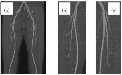
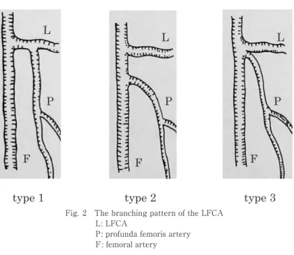
今回の MDCT 画像では 38 症例,76 肢全てで大 腿深動脈,LFCA およびその分枝の描出が可能で あった(Fig. 1).LFCA の分枝形態は 3 種類に分 類でき,type 1 は大腿深動脈から LFCA が分岐す るもの,type 2 は大腿深動脈分岐部より近位で LFCA が大腿動脈から直接分岐するもの,type 3 は LFCA と大腿深動脈が大腿動脈の共通幹から分 岐するものであった(Table 1,Fig. 2).男女とも に type 1 が最も多く,全体の 71.1%であった.左 右とも type 1 が最も多く全体の 57.9%であり,左 右の type が同一であったものは全体の 68.4%で あった(Table 2).
最も大腿外側体表近傍まで走行していた血管を評 価したところ 3 群に分類できた.group 1 は LFCA 下行枝が優位なもの,group 2 は LFCA 下行枝と 大腿深動脈終枝のいずれも同程度のもの,group 3 は大腿深動脈終枝が優位なものであった(Table 3).男女とも group 1 が最も多く,全体の 68.4%で あ っ た. 左 右 と も group 1 が 最 も 多 く, 全 体 の 55.3%であり,左右の group が同一であったものは 68.4%であった(Table 4).
type 別 の group の 割 合 で は 男 女 と も type 1,
group 1 が最も多く,全体の 47.4%であり type 2 で は group 3 が type 3 では group 2 が認められなかっ た(Table 5).
上前腸骨棘と大腿動脈から大腿深動脈の分枝する 点までの距離を Fig. 3 に示す.全体の平均は 103.3 mm(59.5 〜 153.9 mm), 男 性 の 平 均 は 107.6 mm
(78.9 〜 153.9 mm), 女 性 の 平 均 は 95.0 mm(59.5
〜 141.8 mm)で男性は女性と比較して有意に距離 が長かった.大腿動脈から大腿深動脈が分枝する点 と大腿深動脈から LFCA が分枝する点までの距離 を Fig. 4 に 示 す. 全 体 の 平 均 は 21.8 mm(0 〜 73.2 mm),男性の平均は 18.8 mm(0 〜 62.0 mm),
女性の平均は 27.5 mm(0 〜 73.2 mm)で,女性は 男性と比較して有意に距離が長かった.また,上前 腸骨棘から LFCA 分岐部までの距離を Fig. 5 に示 す. 全 体 の 平 均 は 114.3 mm(73.5 〜 166.1 mm),
Table 1 The gender difference of the branching pattern of the LFCA
male female total
type 1 32(64.0%) 22(84.6%) 54(71.1%)
type 2 11(22.0%) 3(11.5%) 14(18.4%)
type 3 7(14.0%) 1(3.9%) 8(10.5%)
total 50 26 76
type 1: the LFCA is branched from the profunda femo- ris artery
type 2: the LFCA is branched independently from the femoral artery
type 3: the LFCA and the profunda femoris artery is branched from common trunk of the femoral ar- tery
Fig. 1 MDCT image of the LFCA and branches
(a)MIP(maximum intensity projection)image (b)right LFCA (c)left LFCA
男性の平均は 113.0 mm(86.0 〜 166.1 mm),女性 の平均は 111.8 mm(73.5 〜 158.7 mm)で,男女間 に有意差はみられなかった.
考 察
MDCT は複数の検出器を用いることで,従来の 検出器が 1 列の CT と比較して短時間に広範囲の撮 影ができ,より薄いスライス画像から詳細な 3 次元 画像の作成が可能で心臓血管領域や抹消動脈疾患の 検査などに用いられている.近年深下腹壁動脈や胸 背動脈の皮膚穿通枝を用いた再建手術などの術前検 査に用いる報告2,3)が散見されるが,われわれが検
Table 3 The group of main vessel toward the lateral
thigh region
male female total
group 1 34(68.0%) 18(69.2%) 52(68.4%)
group 2 15(30.0%) 6(23.1%) 21(27.6%)
group 3 1(2.0%) 2(7.7%) 3(4.0%)
total 50 26 76
group 1: descending branch of the LFCA is dominant group 2: descending branch of the LFCA and the branch
of the profunda femoris artery are same group 3: the branch of the profunda femoris artery is
dominant
Table 2 The comparison of the branching pattern of the LFCA according to the right and left thigh
right left total
type 1 type 1 22(57.9%)
type 1 type 2 4(10.6%)
type 1 type 3 3(7.9%)
type 2 type 1 2(5.3%)
type 2 type 2 3(7.9%)
type 2 type 3 1(2.6%)
type 3 type 1 1(2.6%)
type 3 type 2 1(2.6%)
type 3 type 3 1(2.6%)
Table 4 The comparison of the group of the main vessel toward the lateral thigh according to the right and left thigh
right left total
group 1 group 1 21(55.3%)
group 1 group 2 4(10.5%)
group 1 group 3 0
group 2 group 1 6(15.8%)
group 2 group 2 4(10.5%)
group 2 group 3 0
group 3 group 1 0
group 3 group 2 2(5.3%)
group 3 group 3 1(2.6%)
Fig. 2 The branching pattern of the LFCA L: LFCA
P: profunda femoris artery
F: femoral artery
索した限りでは LFCA の解剖学的評価を MDCT で 行った報告はみられなかった.
LFCA は通常大腿深動脈の外側から分岐すると
されている4)がおよそ 20%の頻度で直接大腿動脈 から分岐するという報告もある5).われわれの研究 では解剖書に記載されている典型的な分枝形態
(type 1)を含めた 3 種類に分類できた.直接大腿 動脈から分岐する割合は以前の報告とほぼ同様で,
新たに大腿深動脈と同時に分岐するパターンが確認 できた.
また,大腿外側体表面へむけて走行する血管が前 外側大腿皮弁の栄養血管となる皮膚穿通枝を分岐し ていると考えられ,本研究では LFCA の分枝パ ターンによらず LFCA 下行枝が最も多かったが,
大腿深動脈が優位に分布する症例も存在(group 3)
した.この場合 ALT flap の栄養血管である皮膚穿 通枝は大腿深動脈から分岐していると考えられた.
この group 3 は type 3 において 25.0%とまれでは ないことが示唆された.また LFCA 下行枝欠損例
Table 5 The number of group of each type
male female total
type 1
group 1 21(42.0%) 15(57.7%) 36(47.4%)
group 2 11(22.0%) 6(23.1%) 17(22.4%)
group 3 0 1(3.8%) 1(1.3%)
type 2
group 1 7(14.0%) 3(11.6%) 10(13.2%)
group 2 4(8.0%) 0 4(5.3%)
group 3 0 0 0
type 3
group 1 6(12.0%) 0 6(7.8%)
group 2 0 0 0
group 3 1(2.0%) 1(3.8%) 2(2.6%)
total 50 26 76
Fig. 3 The distance from the anterior superior iliac spine to the profunda femoris artery
Fig. 4 The distance from profunda femoris artery to the LFCA
Fig. 5 The distance from anterior superior iliac
spine to the LFCA
の報告6‑9)がみられ,この場合外側広筋の筋枝また は内側に偏位した太い無名枝が下行枝を代償してい るとされるが,本研究ではみられなかった.しかし group 3 や LFCA 下行枝欠損例では,大腿深動脈 の外側広筋筋枝から大腿外側への皮膚穿通枝が分岐 していると考えられ,ALT flap 挙上時の血管茎が 下行枝からの皮膚穿通枝を用いた場合と比較して短 くなることが術前に判断でき,また外側広筋への栄 養血管を確認し温存して皮膚穿通枝のみを用いて皮 弁を挙上することが可能であると考えられ,術前計 画に有用であると考えられた.さらに type や group が左右で同一である割合はおよそ 70%程度にすぎ ず,左右のいずれから皮弁を挙上するかを選択する 際にも有用であると考えられた.
われわれの研究では全ての穿通枝を描出すること はできなかったが,直径 2 mm 程度の血管が描出可 能であった.青ら6)によると大腿外側面に分布する LFCA の皮膚穿通枝として,外側下行枝,外側広 筋枝が主であるとされ,これらの穿通枝は動脈 1 本 に対し伴走静脈 2 本が付随するとされ,動脈相後期 から静脈相早期にかけて穿通枝に伴走する静脈まで 造影し,上前腸骨棘から LFCA 分岐部までの距離 に男女差がないことから撮影条件や造影剤注入速度 の調節を行うことで皮膚穿通枝領域まで造影を行う ことが可能であると示唆された.
今回の研究では平均年齢が 72.7 歳と高齢で,腹 部大動脈での石灰化や動脈硬化性病変が強い状態の 症例が多かったが,全例で LFCA およびその分枝 の描出が可能であった.血管造影と比較して経静脈 的に可能で短時間に 3 次元的な解剖学的評価が可能 であり,造影効果の強い血管を皮弁挙上時に選択す ることでより手術の安全性が高まることが示唆され た.われわれの研究では 16 列 CT 装置を用いたが,
検出器の多列化により穿通枝領域をさらに詳細に描 出できると考えられ,今後造影プロトコールの確立
を含めてさらなる発展が期待された.
本研究を行うにあたり,総合南東北病院形成外科部長 斎藤昌美氏に多大なご協力をいただきましたことに深謝 いたします.
文 献
1) Song YG, Chen GZ and Song YL : The free thigh flap : a new free flap concept based on theseptocutaneous artery. 37:
149‑159, 1984.
2) Rozen WM, Phillips TJ, Ashcon MW, : Pre- operative imaging for DIEA perforator flaps: a comparative study of computed tomographic angiography and doppler ultrasound.
112:9‑16, 2008.
3) Mun GH, Kim HJ, Cha MK, : Impact of per- forator mapping using multidetector-row com- puted tomographic angiography on free thora- codorsal artery perforator flap transfer.
122:1079‑1088, 2008.
4) 平沢 興,岡本道雄:外腸骨動脈.解剖学第 2 巻 脈管学・神経系,改訂第 11 版,pp. 92‑96,
金原出版,東京,2001.
5) Lang J, Wachsmuth W:大腿の動脈.ランツ下 肢臨床解剖学(山田致知,津山直一監訳),pp.
42,医学書院,東京,1979.
6) 青 雅一:外側大腿回旋動脈系及びその皮膚穿 通 枝 の 解 剖 学 的 変 異. 日 マ イ ク ロ 会 誌 15:
155‑163,2002.
7) Ao M, Asagoe K, Maeta M, : Combined an- terior thigh flaps and vascularized fibular graft for reconstruction of massive composite oro- mandibular defect. 51:350‑
355, 1998.
8) Ao M, Uno K, Maeta M, : De-epithelialized anterior (anterolateral and anteromedial) thigh flaps for dead space filling and contour correc- tion in head and neck reconstruction.
52:261‑267, 1999.
9) 光嶋 勲,稲川喜一,森口隆彦:再建材料とそ
の採取法 皮弁―血管付き:前外側大腿皮弁.
耳鼻・頭頸外科 71:28‑35, 1999.
A ANATOMIC EVALUATION OF THE LATERAL FEMORAL CIRCUMFLEX ARTERY SYSTEM BY USING
MULTI DETECTOR-ROW CT
Kazunari HARAGUCHI, Satoshi KADOTA and Yoshiaki HOSAKA The Department of Plastic and Reconstructive Surgery,
Showa University School of Medicine
Abstract Flaps that are pedicled by perforators of the lateral femoral circumflex artery (LFCA)
system have many advantages, including the transplantation of large and reliable skin with long pedicles and a large diameter, and little invasion of the donor sites. However, preoperative planning has been dif- ficult because the perforators have many anatomic variations. We used multi detector-row CT for ana- tomical evaluation of the lateral femoral circumflex artery system. The patterns of LFCA from the main vessels were classified into three types and vessels coursing toward the lateral thigh region were classi- fied into three groups. The distance from the anterior superior iliac spine to the lateral femoral circum- flex artery showed no significant difference between men and women. We were able to evaluate vessels with a 2-mm diameter in the lateral femoral circumflex artery system, indicating that accurate evaluation and low invasive examination of the lateral femoral circumflex artery system, including the perforator area, can be achieved by adjusting the image conditions and the injection rate of the contrast dye.
Key words
: lateral femoral circumflex artery, multidetector-row CT (MDCT), antero lateral thigh flap〔受付:1 月 12 日,受理:1 月 21 日,2010〕