Mochida
第
4回腫瘍・血液内科勉強会
リンパ腫
の
診断と治療
平成
27年9月3日
加古川西市民病院 腫瘍・血液内科
岡村篤夫
Mochida
多能性造血幹細胞 リンパ系幹細胞 骨髄系幹細胞 顆粒球系幹細胞 赤芽球系幹細胞 巨核球系幹細胞 骨髄芽球 単芽球 前単球 前骨髄球 骨髄球 後骨髄球 網状赤血球 巨核球 赤血球 血小板 単球 網状血小板 好中球 好塩基球 好酸球 マクロファージ 樹状細胞骨髄
末梢血
組織
CML
MDS
M0
M4
M5
M1
M2
M3
M7
M6
AML
血球の分化と造血器腫瘍
①
Mochida
骨髄系幹細胞 多能性造血幹細胞 リンパ系幹細胞 形質細胞Pro-T
細胞V
Pro-B
細胞V
Pre-B
細胞V
未成熟B
細胞V
naiveB
細胞V
濾胞中心B
細胞V
成熟B
細胞Pre-T
細胞NK
前駆細胞 胸腺T
細胞 成熟T
細胞 成熟ヘルパーT
細胞 成熟サプレッサーT
細胞 成熟NK/T
細胞骨髄
末梢血
リンパ節
/リンパ組織
胸腺
骨髄腫
悪性リンパ腫
ALL
FL
LPL
CLL MCL DLBCL
BurkiB
PTCL/ATL
NK/T
LBL
血球の分化と造血器腫瘍
②
Mochida
リンパ球系悪性腫瘍
主に骨髄で増殖
主にリンパ節・リンパ組織で
腫瘤を形成
白血病
悪性リンパ腫
ホジキンリンパ腫
非ホジキンリンパ腫
Mochida
悪性リンパ腫の疫学
毎年
10
万人に
15
人
程度が発症している
。
加古川の人口は約
27万人…
国立がん研究センターがん対策情報センターHPより抜粋年齢調整罹患率(男性)
! 悪性リンパ腫の罹患数は年間約25,000例(2011年)。
! 罹患率/死亡率とも男性のほうが高く、いずれも50歳以降急増する。
2005年
2011年
減少 増加 増加 増加 減少 増加 増加(死亡率も)Mochida
国立がん研究センターがん対策情報センターHPより抜粋年齢調整罹患率(女性)
2005年
2011年
増加 減少 減少 増加(死亡率も) 増加 増加 増加 増加(死亡率も)Mochida
! 初期例では、
リンパ節の腫脹
が特徴であるが、ほとんど
自覚
症状はない
。
そのため、無症状のまま進行し、複数部位に
広がることも少なくない。
! 進行例では全身のリンパ節腫脹のほかに、
発熱
、
体重減少
、
盗汗
の3症状(
B
症状
)が見られる。
! 臓器にリンパ腫が広がると、以下のような臓器障害の症状が
みられる。
胸部(肺や気道など)
咳が出やすくなり、呼吸困難を伴う
腹部
腹部以下(下肢)のむくみ、尿路障害
肝臓
黄疸、腹水
骨
疼痛
悪性リンパ腫の症状
Mochida
!
ホジキンリンパ腫
- 英国医師トーマス・ホジキンにより命名
- 日本では、
悪性リンパ腫のうち10%程度
- 発症年齢:20歳代と50-60歳代に多い(二峰性)
- 日本人では欧米人に比べ少ない
- 予後:
比較的良好
(根治が治療目標)
!
非ホジキン細胞
- ホジキンリンパ腫以外の悪性リンパ腫
- 日本では、
悪性リンパ腫のうち90%程度
- 50-60歳代に多い
- 予後:悪性度により様々
悪性リンパ腫の種類
Mochida
!
ホジキンリンパ腫
は主に頸部リンパ節に初発、隣接したリンパ
節に進展。
!
非ホジキンリンパ腫
は初発の60%は体内の様々なリンパ節に、
40%はリンパ節以外の臓器に初発し、あらゆる臓器に進展。
ホジキンリンパ腫
非ホジキンリンパ腫
リンパ節(とくに
頸部)に初発する
リンパ節の初発が
多い(60%)
リンパ節以外の
初発もある(40%)
隣接したリンパ節
に進展(連続性)
リンパ節以外の
初発は少ない
あらゆる部位に進展
しうる(非連続性)
リンパ腫発症部位の特徴
Mochida
Ann Arbor分類 のB症状! 一般診察
- 問診でリンパ節の腫脹に気づいた時期と大きさの変化、
その他の自覚症状の有無を確認。
・ 2週間以内にリンパ節腫脹が消退→反応性病変の可能性が高い。
・ 4-6週間続く場合、増大速度が速い場合、新たなリンパ節腫脹が
出現した場合は、悪性リンパ腫を疑い生検を考慮。
・ 数ヵ月にわたって不変の場合や、自然縮小した場合は、低悪性度
リンパ腫の可能性が考えられる。
-
体重減少
、
発熱
、
盗汗(
B
症状)
などの全身症状も参考となる。
- 触診にて、
リンパ節腫脹
の有無と硬さ、大きさ、痛み
(圧痛)の有無を確認。
- 口腔内のリンパ組織の視診なども行う。
悪性リンパ腫の診断
(
1
)
Mochida
! 悪性リンパ腫の各検査における主な確認事項
検査
確認内容
末梢血液検査
生化学検査
貧血の有無、リンパ球増加・減少、
LDH
上昇、
Ca上昇、β
2
ミクログロブリンの上昇など
胸部X線検査 リンパ節腫大
微生物検査
オウム病クラミジア、ピロリ菌、EBウイルスなど
免疫学的検査
抗HTLV-1抗体、HIV抗体、
可溶性IL-2受容体
の上昇
悪性リンパ腫の診断
(
2
)
Mochida
! 一般診察にて悪性リンパ腫を疑った場合、リンパ節
生検で確定診断を行う。
血液検査
血球数、生化学検査など
リンパ節生検
リンパ節の切除による病理学的検査
超音波検査
リンパ節の形や数、
大きさを検査
骨髄検査
悪性リンパ腫の骨髄への
浸潤の有無を検査
PET
検査
病変の広がりを検出する
検査
CT検査
リンパ節や脾臓の
腫れなどを検査
脳脊髄液検査
悪性リンパ腫の中枢神経
への浸潤の有無を検査
消化管内視鏡検査
悪性リンパ腫の消化管
への浸潤の有無を検査
確定診断
! あわせて以下のような検査により、臨床病期の判定を行う。
悪性リンパ腫の診断
(
3
)
Mochida
前駆細胞リンパ系腫瘍 Bリンパ芽球白血病/リンパ腫(B-LBL) 特異的遺伝子異常を伴うBリンパ芽球白血病/リンパ腫(B-LBL) 成熟B細胞腫瘍 慢性リンパ性白血病/小リンパ球リンパ腫(CLL/SLL) B細胞前リンパ球白血病(B-PLL) 脾辺縁帯B細胞リンパ主(SMZL) ヘアリー細胞白血病(HCL) 脾B細胞リンパ腫/白血病、分類不能型 リンパ形質細胞リンパ腫(LPL) 重鎖症 形質細胞腫瘍(plasmacytoma) 骨孤在形質細胞腫瘍 骨外性形質細胞腫瘍 MALT関連節外性辺縁帯B細胞リンパ腫(MALT) 節性辺緑帯B細胞リンパ腫(NMZL) 濾胞性リンパ腫(grade 1,2,3a,3b)(FL) マントル細胞リンパ腫(MCL) びまん性大細胞型B細胞リンパ腫、非特異型(DLBCL、NOS) 慢性炎症関連びまん性大細胞B細胞リンパ腫 リンパ腫様肉芽腫症 原発性縦隔大細胞型B細胞リンパ腫 血管内大細胞型B細胞リンパ腫 ALK陽性大細胞型B細胞リンパ腫 形質細胞芽性リンパ腫 HHV8関連多中心性Castleman病起因大細胞型B細胞リンパ腫 原発性滲出液リンパ腫 バーキットリンパ腫(BL) B細胞リンパ腫、分類不能型(DLBCLとバーキットリンパ腫の中間型) B細胞リンパ腫、分類不能型(DLBCLと古典的ホジキンリンパ腫の中間型) ホジキンリンパ腫 結節性リンパ球優位型ホジキンリンパ腫(NLPHL) 古典的ホジキンリンパ腫 結節硬化型ホジキンリンパ腫(NSH) リンパ球豊富ホジキンリンパ腫(LPCHL) Tリンパ芽球白血病/リンパ腫(T-LBL) 成熟T細胞ならびにNK細胞腫瘍 T細胞前リンパ球白血病(TPLL) T細胞大顆粒リンパ球白血病(LGL) 慢性NK細胞リンパ球増殖性疾患 侵攻性NK細胞白血病(NK leukemia) 小児の全身性EBV陽性T細胞リンパ球増殖性疾患 種痘状水泡様リンパ腫 成人T細胞白血病/リンパ腫(ATLL) 節外性NK/T細胞リンパ腫/鼻型(nasal NK) 腸管症型T細胞リンパ腫 肝脾型T細胞リンパ腫 皮下脂肪織炎様T細胞リンパ腫 菌状息肉腫(MF) Sezary症候群(SS) 原発性皮膚CD30陽性T細胞リンパ球増殖性疾患 原発性皮膚γ・δ型T細胞リンパ腫 原発性皮膚CD8陽性侵攻性表皮向性細胞障害性T細胞リンパ腫 原発性皮膚CD4陽性小型/中型T細胞リンパ腫 末梢性T細胞リンパ腫、非特異型(PTCL-u) 血管免疫芽球性T細胞リンパ腫(AILT) 未分化大細胞リンパ腫、ALK陽性(ALK+ ALCL) 未分化大細胞リンパ腫、ALK陰性(ALK- ALCL) 芽球NK細胞リンパ腫 混合細胞型ホジキンリンパ腫(MCHL) リンパ球減少型ホジキンリンパ腫(LDHL)悪性リンパ腫の
WHO
分類第
4
版(
2008
年)
Mochida
治療目標
延命
治癒
低悪性度
中悪性度
高悪性度
年単位
無治療での
生存期間
週単位
月単位
小細胞性
リンパ形質細胞性
(≒原発性マクログロブリン血症)
有毛細胞白血病
MALT
濾胞性(
Grade1/2/3a)
(形質細胞腫
/骨髄腫)
マントル細胞
濾胞性(
Grade3b)
びまん性大細胞型
縦隔大細胞型
リンパ芽球性
バーキット型
形質細胞性白血病
前リンパ球性白血病
末梢
T細胞性
血管免疫芽球性
NK/T細胞性鼻型
未分化大細胞型
大顆粒リンパ球性
菌状息肉症
慢性型成人
T細胞性
リンパ芽球性
成人
T細胞性
T細胞性(
20%
)
WHO
分類
B細胞性(
75%
)
組織悪性度による非ホジキンリンパ腫の分類
Mochida
悪性リンパ腫の診断
(
4
)
! B細胞系非ホジキンリンパ腫の表面抗原解析による病型鑑別
CD5 CD10 CD11c CD19
CD20
CD23 CD25 CD34 CD56
Bリンパ芽球性リンパ腫
+/-
+
*
+/-
慢性リンパ性白血病
/
小リンパ球リンパ腫
+
-
+/-
+
+
+
リンパ形質細胞性リンパ腫
-
-
+/-
+
+
+/-
濾胞性リンパ腫
-
+/-
-
+
+
-/+
マントル細胞リンパ腫
+
-
-
+
+
-
形質細胞腫
-/+
-
-
+/-
びまん性大細胞型
B細胞
リンパ腫
-/+ -/+
+
+
-
バーキットリンパ腫
-/+
+
+
+
Mochida
悪性リンパ腫の診断
(
5
)
! B細胞系非ホジキンリンパ腫の代表的な染色体異常
染色体異常
関連する遺伝子
頻度
慢性リンパ性白血病
/
小リンパ球リンパ腫
+12
20%
del(13)(q14)
RB1
50%
del(11)(q22-23)
20%
t(14;19)(q32;q13.1)
IgH
BCL3
リンパ形質細胞性リンパ腫
t(9;14)(q13;q32)
PAX5
IgH
〜
50%
MALTリンパ腫
t(11;18)(q21;q21)
API2
MALT1
15-60%
t(14;18)(q32;q21)
IgH
MALT1
10-30%
濾胞性リンパ腫
t(14;18)(q32;q21)
IgH
BCL2
70-95%
t(2;18)(q12;q21)
κ-chain
BCL2
t(18;22)(q21;q11)
BCL2
λ-chain
マントル細胞リンパ腫
t(11;14)(q21;q21)
Cyclin D1
IgH
〜
100%
びまん性大細胞型
B細胞リンパ腫
t(14;18)(q32;q21)
IgH
BCL2
20-30%
bcl-6を含む3q27領域の異常が30%にみられる
バーキットリンパ腫
t(8;14)(q24;q32)
MYC
IgH
〜
80%
t(2;8)(q11;q24)
κ-chain
MYC
〜
15%
t(8;22)(q24;q11)
MYC
λ-chain
〜
10%
Mochida
! 国内で新たに非ホジキンリンパ腫と診断される患者数は、
- 低悪性度の濾胞性リンパ腫、MALTリンパ腫がそれぞれ18%、4%
- 中悪性度のびまん性大細胞型B細胞リンパ腫が最も多く33%
! わが国ではとくに濾胞性リンパ腫の増加が著しいと言われている。
Pathology International: 58:174-182 2008 濾胞性リンパ腫 18% MALTリンパ腫 4% CLL 1.4%B
細胞リンパ腫
65
%
びまん性大細胞型B細胞 リンパ腫 33% マントル細胞リンパ腫 3% 前駆Bリンパ芽球性・バーキット リンパ腫 0.9% その他 5% T/NK細胞リンパ腫 25% ホジキンリンパ腫 7% その他 3%低悪性度
中悪性度
高悪性度
非ホジキンリンパ腫の病型別患者数
Mochida
臨床病期
! Ann Arbor分類(Cotswolds修正案)
I期
II期
III期
IV期
節外病変があればEを付記。 巨大腫瘤はXを付記。 ・最大径 ≥10cm ・胸郭横径の1/3以上 全身症状 A : 症状なし B : 以下のいずれかの症状を伴う場合 (1) 初診から半年以内に、 10%以上の体重減少 (2) 38℃以上の原因不明の発熱 (3) 盗汗 →いわゆるB症状1リンパ節領域(I)、1リンパ節外領域あるいは部位
(IE)の限局的侵襲。
横隔膜で境した片側に留まる2リンパ節領域以上
の侵襲(II
2〜)。
または1リンパ節外領域あるいは部位の限局的侵
襲とその同側リンパ節領域の侵襲(IIE)。
横隔膜の上下にわたる複数のリンパ節領域の侵襲
(III)。
またはこれに1リンパ節外領域あるいは部位の限局
的侵襲(IIIE)、または脾臓への侵襲(IIIS)、あるいは
この両方(IIIES)。
リンパ節病変の有無にかかわりなく、1または複数
のリンパ節外領域あるいは部位へのびまん性浸潤
(IV)。
Mochida
• 治療目標:
根治
• 臨床病期(限局期or進行期)で治療方針を決定する。
(組織型に依らない)
• 化学療法(
ABVD
療法)および
放射線
療法が極めて有効。
A ドキソルビシン
B ブレオマイシン
V ビンブラスチン
D ダカルバジン
ホジキンリンパ腫の治療
I / IIA期
ABVD療法
4コース
局所放射線
照射
IIB - IV期
ABVD療法
6 - 8コース
局所放射線照射 (巨大病変に対し)自家移植
(< 65歳)
局所放射線 照射救援療法
非寛解・再発
≥ 部分寛解
Mochida
予後良好限局期ホジキンリンパ腫に対する最近の傾向
Risk Factor
GHSG基準
NCCN基準
赤沈・B症状
>50mm
B症状ありなら>30mm
>50mm
あるいはB症状あり
縦隔腫瘤
胸郭横径>1/3
縦隔横径>1/3
リンパ節領域
>2領域
>3領域
節外病変
あり
巨大腫瘤
>10cm
いずれのrisk factorも見られない限局期病変に対しては、
ABVD 2
コース
+
局所放射線照射(
20-30Gy
)
Mochida
ホジキンリンパ腫の予後
限局期:長期生存
≥ 90%
晩期毒性
1.
2次発癌(AML:5-9年、固形癌:15年以上)
2. 心毒性
3. 不妊
1. Alb<4g/dl
2. Hb<10.5g/dl
3. 男性
4. Ⅳ期
5. 45才以上
6. 白血球>15000/µl
7. リンパ球<600/µl (白血球の8%)
進行期ホジキンリンパ腫の予後予測因子(IPS)
観察期間(月)
全
生
存
率
(
%
)
進行期:score3以下(80%)
≥70%
score4以上(20%)
50%
Mochida
薬剤名
商品名(一般名)
投与量
投与経路
Day1
Day15
A
アドリアシン
®(ドキソルビシン)
25mg/m
2点滴静注
↓
↓
B
ブレオ
®(ブレオマイシン)
10mg/m
2(MAX 15mg)
ボーラス
↓
↓
V
エクザール
®(ビンブラスチン)
6mg/m
2(MAX 10mg)
ボーラス
↓
↓
D
ダカルバジン
®(ダカルバジン)
375mg/m
2点滴静注
↓
↓
1
日目
と
15
日目
に投与して1コースとし、
4週毎
に繰り返し、
6~8コース行う。
ABVD療法
Mochida
副作用症状
影響している薬剤出現時期
Day
1
2 3 4 5 6 7 8 9 10 11 12 13 14 15 16 17 18 19 20 21 22 23 24 25 26 27 28
血管外漏出
DXR,VLB
血管痛・血管
炎
DTIC
悪心・嘔吐 DTIC,DXR,BLM,VLB
食欲不振 DTIC,DXR,BLM,VLB
骨髄抑制
DXR
DTIC
脱毛
DXR
DXR:アドリアシン
®VLB:エクザール
®DTIC:ダカルバジン
®BLM:ブレオ
®有害事象の発現時期
ただしG-CSFは出来るだけ用いない(BLMの肺毒性↑)
遮光が必要
Mochida
ホジキンリンパ腫に用いられる救援化学療法
• DHAP
• ESHAP
• ICE
• Mini-BEAM etc.
• COPP
•
Brentuximab vedotin
(アドセトリス
®
)
抗CD30モノクローナル抗体+微小管阻害薬(MMAE)
•
Nivolumab(オプジーボ
®
)
救援化学療法に感受性のある
<65
才の患者では
、
自家移植
併用大量化学療法を考慮する
。
自家移植併用大量化学療法
<65歳
>65歳 or 合併症あり
Mochida
免疫チェックポイント阻害薬
抗CTLA-4抗体:樹状細胞(抗原提示細胞)上のB7との結合を阻害
→ Ipilimumab(ヤーボイ
®)
抗PD-1抗体:がん細胞上のPD-L1との結合を阻害
→
Nivolumab(オプジーボ
®)・・・
将来ホジキンリンパ腫にも適応拡大されるか
?
がん細胞を攻撃する
T
細胞の働きにブレーキをかける機構を阻害する抗体医薬
(≒がん細胞に対する免疫寛容を弱める働き)
Mochida
非ホジキンリンパ腫の治療
生
存
率
罹病期間
0
5
10
15
20 (年)
中悪性度リンパ腫
低悪性度リンパ腫
中悪性度リンパ腫 経過が速く早急に治療が必要。
いったん治療に反応すれば
治癒が望める
。
→
治療目標は根治
低悪性度リンパ腫 慢性に経過し、治療には一時的に反応。
再発と寛解を繰り返し
治癒は望めない
。
→
治療目標は延命・共存
(例. びまん性大細胞型B細胞リンパ腫) (例. 濾胞性リンパ腫)Mochida
濾胞性リンパ腫に対する治療
増悪
*I / II期
巨大病変なし局所放射線
照射
無治療・経過観察
化学療法
R-CHOP
R-ベンダムスチン
R単独 etc
.II期
巨大病変ありIII / IV期
GELF
Low
High
無症状
有症状
自家移植(
< 65歳)
同種移植(ミニ)
放射線照射
化学療法
R-CHOP
R-ベンダムスチン
R-フルダラビン
イブリツモマブチウキタセン
etc.
地固め療法として
≥ 部分寛解の
ハイリスク患者
増悪
*組織学的悪性度の進展が見られれば
中高悪性度リンパ腫に準じた治療を行う。
n 進行期に対する治療として、抗ヒトCD20モノクローナル抗体
リツキシマブ(
R:
リツキサン
®)
併用化学療法の有用性は認められているが、標準的な治療レジ
メンは確立されていない。
n 10年後にも約10%で再発する。
n 経過中、5年で約10%に
組織悪性度の進展
(中悪性度リンパ腫)がみられる。
Mochida
n GELF基準
濾胞性リンパ腫患者が、治療を始めることを考えるべき基準
(
治療開始前の予後予測
)
濾胞性リンパ腫の予後予測モデル
(
1
)
1. 節性病変(≥3cm)≥3領域
2. ≥7cmの節性・節外性病変
3. B症状
4. 脾腫
5. 胸水または腹水
6. 白血球 <1000/μl and/or 血小板 <10万/μl
7. 末梢血中腫瘍細胞数 >5000/μl
→ 以上を一つでも満たさなければ
、
無治療・経過観察可能
Mochida
無治療・経過観察の意義
For personal use. Only reproduce with permission from The Lancet
chemotherapy. Their actuarial chance of not dying of lymphoma nor needing chemotherapy (with non-lymphoma deaths censored) was 19% (95% CI 13–27) (figure 3). The histology slides from these 19 patients were re-reviewed. Low-grade lymphoma was confirmed in 18. One case, previously reported as a small-cell follicular lymphoma, was noted on review to be chronic thyroiditis. Of the eight cases originally reported as a
ARTICLES
THE LANCET • Vol 362 • August 16, 2003 • www.thelancet.com 519
but not overall survival. Histological subtype had no effect on overall survival. In a multivariate analysis (table 3), only stage, age, and ESR significantly affected overall survival (p=0·03, <0·0001, and 0·03, respectively) and cause-specific survival (p=0·008, 0·002, and 0·01, respectively).
110 patients (73%) in the observation group subsequently received chemotherapy. The median time to first systemic treatment was 2·6 years for the entire observation group. 26 (17%) patients in the observation group died without receiving chemotherapy. Six died of cardiovascular disease, five from infection (four of whom had bronchopneumonia), and ten from solid tumours. One patient was lost to follow-up and died of lymphoma, and another died of meningitis, possibly of lymphomatous origin, although this was not proven. In three patients the precise cause of death was not documented, but lymphoma was recorded as a cause of death on the death certificate. For the subsequent analysis, we therefore assumed that five patients died of lymphoma without receiving chemotherapy. Median time from entry into the study to death for these 26 patients who died without receiving systemic treatment for lymphoma was 4·7 years (range 0·5–7·9), and the median age at the time of death was 73 years (61–85). A further 13 (9%) patients are alive and have not yet had chemotherapy. Two patients in this group were lost to up after 8·0 and 10·2 years of follow-up, respectively. When last seen, both these patients were well with no evidence of active lymphoma.
10 years after randomisation, 19 patients in the observation group were alive and had not had
Figure 3: Time to first systemic treatment for patients in the observation group
At 10 years, 19% (95% CI 13–27) of patients either did not need chemotherapy or died of lymphoma (non-lymphoma deaths were censored). 100 80 60 40 20 0 0 4 8 Time (years) Patients at risk Observation
Patients not needing chemotherapy (%) 12 16 20
n=151
24
49 24 18 6 1 0 A
Number included in analysis=225*
Univariate Multivariate
p value Relative risk (95% CI) p value Relative risk (95% CI) Age <0·0001 2·35 (1·74–3·19) <0·0001 2·08 (1·52–2·86) Stage 0·004 1·56 (1·15–2·12) 0·03 1·37 (1·00–1·87) Haemoglobin 0·003 2·31 (1·40–3·83) 0·21 1·43 (0·84–2·45) ESR 0·0002 1·99 (1·14–2·81) 0·03 1·41 (0·98–2·04) Albumin 0·006 1·60 (1·15–2·21) 0·28 1·21 (0·86–1·70) Treatment 0·78 1·04 (0·78–1·40) 0·97 1·01 (0·75–1·36) Number included in analysis=275†
Univariate Multivariate
p value Relative risk (95% CI) p value Relative risk (95% CI) Age <0·0001 2·40 (1·82–3·16) <0·0001 2·19 (1·65–2·90) Stage 0·005 1·48 (1·13–1·96) 0·03 1·37 (1·04–1·80) ESR <0·0001 1·98 (1·45–2·70) 0·004 1·58 (1·15–2·17) B
Number included in analysis=225*
Univariate Multivariate
p value Relative risk (95% CI) p value Relative risk (95% CI) Age 0·001 1·80 (1·27–2·56) 0·002 1·59 (1·10–2·28) Stage 0·004 1·69 (1·18–2·43) 0·008 1·62 (1·13–2·33) Haemoglobin 0·02 2·22 (1·22–4·04) 0·06 1·87 (1·02–3·44) ESR 0·002 2·00 (1·34–2·99) 0·01 1·72 (1·13–2·61) Albumin 0·02 1·61 (1·10–2·35) 0·08 1·43 (0·97–2·11) Treatment 0·34 1·18 (0·84–1·68) 0·28 1·21 (0·85–1·71) Number included in analysis=275†
Univariate Multivariate
p value Relative risk (95% CI) p value Relative risk (95% CI) Age 0·0005 1·78 (1·29–2·45) 0·005 1·57 (1·13–2·19) Stage 0·002 1·67 (1·20–2·33) 0·008 1·56 (1·12–2·17) ESR 0·0002 2·09 (1·46–3·00) 0·002 1·78 (1·23–2·58)
*Patients with data for all variables. †Patients with data for age, stage, and ESR.
Table 3: Multivariate and univariate analyses of overall (A) and cause-specific survival (B)
治療開始までの期間中央値は
2.6
年
1. 搔痒 or B症状
2. 3ヶ月以内の急激な進行
3. 生命を脅かす臓器浸潤
4. 血球減少(Hb<10g/dl, WBC<3000/µl,
Plt<10万/µl)を伴う骨髄浸潤
Ardeshna KM, et al. Lancet. 2003
For personal use. Only reproduce with permission from The Lancet
ARTICLES
518 THE LANCET • Vol 362 • August 16, 2003 • www.thelancet.com
group, did not get the treatment allocated to him (figure 1). The analysis was done on an intention-to-treat basis. All exploratory subgroup analyses have been reported. Since chlorambucil therapy was well established at the start of the trial, and the side-effects of the drug known, side-effects relating to the treatment were not formally recorded.
Complete remission was defined in this trial as the complete disappearance of all disease manifestations and the reversal of all previously abnormal investigations maintained for at least 3 months after discontinuing chemotherapy. Patients had to have a normal bone marrow examination result at reassessment if they had bone marrow disease at presentation to be defined as a complete remission. Partial remission was defined as a reduction of at least 50% of the product of the perpendicular measurements of disease, and lesser responses were categorised as non-responders. Overall survival was defined as time from randomisation to death from any cause. Patients who are still alive were censored at the time they were last known to be alive. Cause-specific survival was defined as time from randomisation to death from lymphoma or immediate treatment related toxicity. Patients who are still alive were censored at the time they were last known to be alive.
Statistical analysis
We used the Kaplan-Meier method to calculate survival curves, and the log-rank test to determine significance.
The !2 test with Yates’ correction was used to determine
the difference between categorical variables in 2"2 tables, otherwise the !2 test was used alone. Multivariate analysis
was done with Cox’s proportional hazards model. Role of the funding source
This trial was funded by the Lymphoma Research Trust. The funding source had no role in the trial design, data collection, data analysis, data interpretation, writing of the manuscript, or the decision to submit the report.
Results
The overall survival was similar for both groups. The actuarial 5-year, 10-year, and 15-year overall survival rates were 57% (95% CI 49–64), 35% (28–43), and 21% (16–29), respectively, for the chlorambucil group, and 58% (50–65), 34% (27–42), and 22% (16–30), respectively, for the observation group. Median overall survival for the chlorambucil group was 5·9 years (range 0–17·8) and for the observation group 6·7 years (0·5–18·9) (p=0·84, !2=0·04, hazard ratio 1·026 [95% CI
0·798–1·319) (figure 2). Table 2 shows the causes of death in both groups.
In accord with the median age of patients entered into this trial and the long follow-up, 36 patients developed second malignancies and 25 of these died from the malignancy (table 2). Of those who died, there was one case of chronic myeloid leukaemia, one of acute myeloid leukaemia, a further unclassified case of leukaemia, and 33 solid tumours. The frequency of second malignant diseases did not differ between the two groups (p=0·2). There were 20 solid tumours in the observation group and two cases of leukaemia, whereas in the chlorambucil group there were 13 solid tumours and one case of leukaemia.
In view of the high number of non-lymphomatous deaths, cause-specific survival was determined, and deaths unrelated to lymphoma or the early effects of treatment were censored. Cause-specific survival was again similar in both groups of the trial (p=0·44, hazard ratio 1·125 [95% CI 0·835–1·517]) (figure 2). The actuarial 5-year, 10-year, and 15-year cause-specific survival rates were 67% (95% CI 59–74), 47% (39–56), and 31% (23–41), respectively, for the chlorambucil group, and 69% (61–76), 46% (38–55), and 37% (29–46), respectively, for the observation group. Median cause-specific survival was 9 years (range 0–17·8) for the chlorambucil group and 9·1 years (0·67–18·9) for the observation group.
In a univariate analysis (table 3), ESR 20 mm/h or less, haemoglobin more than 120 g/L, albumin more than 400 g/L, stage III disease, and age less than 60 years conferred a significant advantage in overall survival (p=0·0002, 0·003, 0·006, 0·004, and <0·0001, respectively). The same factors predicted for improved cause-specific survival (p=0·002, 0·02, 0·02, 0·004, and 0·001, respectively). Bone marrow involvement in stage IV disease significantly worsened cause-specific survival
100 A 80 60 40 20 0 Cumulative survival (%) 100 B 80 60 40 20 0 0 4 8 Time (years) Patients at risk Chlorambucil Observation Cumulative survival (%) 12 16 20 24 105 103 6065 4243 1517 20 00 Chlorambucil (n=158) Observation (n=151) Chlorambucil (n=158) Observation (n=151)
Figure 2: Overall and cause-specific survival
(A) Overall survival of patients in both groups. !2=0·04, p=0·84. Hazard
ratio 1·026 (95% CI 0·798–1·319). 10-year survival 34% (27–42) in observation group and 35% (28–43) in chlorambucil group.
(B) Cause-specific survival of patients in both groups. !2=0·6, p=0·44.
Hazard ratio 1·125 (0·835–1·517). 10-year survival 46% (38–55) in observation group and 47% (39–56) in chlorambucil group.
Observation Chlorambucil Non-Hodgkin lymphoma 84 (5) 90
Leukaemia 2 0
Second solid cancer 14 (10) 9
Cardiovascular disease 10 (6) 10
Infection 8 (5) 6
Other 1* 2†
Unknown 1 5
Numbers in parentheses indicate patients who died without receiving systemic treatment. *Renal failure and †dementia and suicide (by poisoning).
Table 2: Causes of death
For personal use. Only reproduce with permission from The Lancet
ARTICLES
518 THE LANCET • Vol 362 • August 16, 2003 • www.thelancet.com
group, did not get the treatment allocated to him (figure 1). The analysis was done on an intention-to-treat basis. All exploratory subgroup analyses have been reported. Since chlorambucil therapy was well established at the start of the trial, and the side-effects of the drug known, side-effects relating to the treatment were not formally recorded.
Complete remission was defined in this trial as the complete disappearance of all disease manifestations and the reversal of all previously abnormal investigations maintained for at least 3 months after discontinuing chemotherapy. Patients had to have a normal bone marrow examination result at reassessment if they had bone marrow disease at presentation to be defined as a complete remission. Partial remission was defined as a reduction of at least 50% of the product of the perpendicular measurements of disease, and lesser responses were categorised as non-responders. Overall survival was defined as time from randomisation to death from any cause. Patients who are still alive were censored at the time they were last known to be alive. Cause-specific survival was defined as time from randomisation to death from lymphoma or immediate treatment related toxicity. Patients who are still alive were censored at the time they were last known to be alive.
Statistical analysis
We used the Kaplan-Meier method to calculate survival curves, and the log-rank test to determine significance.
The !2 test with Yates’ correction was used to determine
the difference between categorical variables in 2"2 tables, otherwise the !2 test was used alone. Multivariate analysis
was done with Cox’s proportional hazards model. Role of the funding source
This trial was funded by the Lymphoma Research Trust. The funding source had no role in the trial design, data collection, data analysis, data interpretation, writing of the manuscript, or the decision to submit the report.
Results
The overall survival was similar for both groups. The actuarial 5-year, 10-year, and 15-year overall survival rates were 57% (95% CI 49–64), 35% (28–43), and 21% (16–29), respectively, for the chlorambucil group, and 58% (50–65), 34% (27–42), and 22% (16–30), respectively, for the observation group. Median overall survival for the chlorambucil group was 5·9 years (range 0–17·8) and for the observation group 6·7 years (0·5–18·9) (p=0·84, !2=0·04, hazard ratio 1·026 [95% CI
0·798–1·319) (figure 2). Table 2 shows the causes of death in both groups.
In accord with the median age of patients entered into this trial and the long follow-up, 36 patients developed second malignancies and 25 of these died from the malignancy (table 2). Of those who died, there was one case of chronic myeloid leukaemia, one of acute myeloid leukaemia, a further unclassified case of leukaemia, and 33 solid tumours. The frequency of second malignant diseases did not differ between the two groups (p=0·2). There were 20 solid tumours in the observation group and two cases of leukaemia, whereas in the chlorambucil group there were 13 solid tumours and one case of leukaemia.
In view of the high number of non-lymphomatous deaths, cause-specific survival was determined, and deaths unrelated to lymphoma or the early effects of treatment were censored. Cause-specific survival was again similar in both groups of the trial (p=0·44, hazard ratio 1·125 [95% CI 0·835–1·517]) (figure 2). The actuarial 5-year, 10-year, and 15-year cause-specific survival rates were 67% (95% CI 59–74), 47% (39–56), and 31% (23–41), respectively, for the chlorambucil group, and 69% (61–76), 46% (38–55), and 37% (29–46), respectively, for the observation group. Median cause-specific survival was 9 years (range 0–17·8) for the chlorambucil group and 9·1 years (0·67–18·9) for the observation group.
In a univariate analysis (table 3), ESR 20 mm/h or less, haemoglobin more than 120 g/L, albumin more than 400 g/L, stage III disease, and age less than 60 years conferred a significant advantage in overall survival (p=0·0002, 0·003, 0·006, 0·004, and <0·0001, respectively). The same factors predicted for improved cause-specific survival (p=0·002, 0·02, 0·02, 0·004, and 0·001, respectively). Bone marrow involvement in stage IV disease significantly worsened cause-specific survival
100 A 80 60 40 20 0 Cumulative survival (%) 100 B 80 60 40 20 0 0 4 8 Time (years) Patients at risk Chlorambucil Observation Cumulative survival (%) 12 16 20 24 105 103 6065 4342 1715 20 00 Chlorambucil (n=158) Observation (n=151) Chlorambucil (n=158) Observation (n=151)
Figure 2: Overall and cause-specific survival
(A) Overall survival of patients in both groups. !2=0·04, p=0·84. Hazard
ratio 1·026 (95% CI 0·798–1·319). 10-year survival 34% (27–42) in observation group and 35% (28–43) in chlorambucil group.
(B) Cause-specific survival of patients in both groups. !2=0·6, p=0·44.
Hazard ratio 1·125 (0·835–1·517). 10-year survival 46% (38–55) in observation group and 47% (39–56) in chlorambucil group.
Observation Chlorambucil Non-Hodgkin lymphoma 84 (5) 90
Leukaemia 2 0 Second solid cancer 14 (10) 9 Cardiovascular disease 10 (6) 10 Infection 8 (5) 6 Other 1* 2† Unknown 1 5
Numbers in parentheses indicate patients who died without receiving systemic treatment. *Renal failure and †dementia and suicide (by poisoning).
Table 2: Causes of death
OS中央値は6年前後
For personal use. Only reproduce with permission from The Lancet
ARTICLES
520 THE LANCET • Vol 362 • August 16, 2003 • www.thelancet.com
follicular lymphoma, six were reviewed as follicular, one as small lymphocytic lymphoma, and one as reactive (thyroiditis). Of the ten cases originally called lymphocytic well-differentiated lymphomas, six were re-classified as small lymphocytic lymphoma, and three as marginal zone lymphoma. In one the type of low-grade lymphoma was not classifiable. Of the 19 cases, one remained unclassifiable.
Of the 13 patients who still are alive without receiving systemic treatment, five have had local radiotherapy and do not have clinically detectable disease. Seven were not given any local radiotherapy and have had spontaneous clinical remissions, although they have not had repeated bone marrow examinations and CT scans over the years of follow-up, so minor residual disease cannot be excluded. These spontaneous remissions have been sustained in four patients. Two have subsequently had clinical disease recurrence, whereas the third has relapsed twice and had two further spontaneous remissions but currently has detectable disease. In total, four patients had disease clinically detectable at their last clinic visit.
Analysis of factors predicting for the need for chemotherapy revealed that such patients were more likely to be younger (p=0·003) and male (p=0·03) (figure 4; A, B). With respect to age, an unplanned examination of various intervals suggests that the main distinction is between patients younger than 70 years and those older than 70 years. The likelihood of not receiving chemotherapy or dying of lymphoma after 10 years of the trial was 40% (95% CI 18–67) in the 20 patients who were older than 70, compared with 16% (10–20) in the 131 younger than 70 years. Although the chances of not receiving chemotherapy at 10 years were higher in patients originally categorised as having lymphocytic well-differentiated lymphoma, as opposed to follicular lymphoma (37% [95% CI 23–53) vs 13% [7–23], respectively), the difference was not significant (figure 4, C). All 11 patients with low-grade lymphomas that were not classified as either follicular or lymphocytic well differentiated, needed treatment. Stage of disease, haemoglobin concentration, ESR, and albumin concentration did not predict for the ultimate need for chemotherapy. 40 patients (26%) in the observation group needed systemic treatment within a year of randomisation. These patients did not differ in their characteristics (age, sex, histology, ESR, albumin concentration, and haemoglobin concentration) from the others in the same group (data not shown).
In the chlorambucil group, 100 patients (63%) had complete remission, 43 (27%) partial remission, and 13 (8%) did not respond to treatment. Of those who achieved complete remission, the median time until death from lymphoma or need for next chemotherapy (disease-free survival) was 6·1 years (range 1–19·8). By comparison, in the observation group, of the 110 patients in whom chemotherapy was started, 30 (27%) had complete remission, 54 (49%) partial remission, and 23 (22%) did not respond to treatment. Of those who achieved a complete remission, the median disease-free survival was 7·3 years (range 0·8–19·1). A direct comparison of the response rates and the disease-free survival after chemotherapy between the two groups is not valid, however, because not all patients in the observation group received chemotherapy. A more appropriate analysis is a comparison of the time to initiation of second-line chemotherapy or death from lymphoma. Figure 5 shows that more patients in the observation group did not need a second course of systemic therapy or die from non-Hodgkin lymphoma at
any time, indicating a substantial advantage of the observation group at this stage of treatment. The median time from randomisation to initiation of second line chemotherapy was 66 months (6–242) in the observation group and 43 months (0–238) in the immediate chemotherapy group. 100 A 80 60 40 20 0 0 4 8 Time (years) Patients at risk !70 years !70 years, n=20
Patients not needing chemotherapy (%) 12 16 20 24
23 15 11 6 1 0 "60 years "60 years, n=75 17 4 3 0 0 0 61–70 years 61–70 years, n=56 9 5 4 0 0 0 100 B 80 60 40 20 0 0 4 8 Time (years) Patients at risk Women Women, n=72
Patients not needing chemotherapy (%) 12 16 20 24
21 12 7 2 0 0 Men Men, n=79 28 12 11 4 1 0 100 C 80 60 40 20 0 0 4 8 Time (years) Patients at risk Lymphocytic Lymphocytic, n=39
Patients not needing chemotherapy (%) 12 16 20 24
30 13 7 3 1 0 Follicular
Follicular, n=101
17 10 10 3 0 0
Figure 4: Factors predicting need for chemotherapy in the observation group
(A) age #2=7·31, p=0·03. (B) sex #2=4·43, p=0·04. (C) histological
subtype #2=2·13, p=0·14.
Mochida
n 濾胞性リンパ腫では、
治療効果の予後予測
モデルとして、FLIPI (Follicular
Lymphoma IPI)が提唱されている。
濾胞性リンパ腫の予後予測モデル
(
2
)
(月)
生
存
率
予後因子 予後不良因子 年齢 60歳以上 血清LDH 正常上限を超える 病期 ⅢまたはⅣ ヘモグロビン 12g/dL未満 リンパ節領域数 5以上 予後因子 予後不良因子 年齢 60歳以上 β2ミクログロブリン 正常上限を超える リンパ節最大径 >6cm ヘモグロビン 12g/dL未満 骨髄浸潤 ありLow:0,1 Intermediate:2 High:3~5
FLIPI (治療後の全生存期間の予測)
Low:0 Intermediate:1,2 High:3~5
Mochida
濾胞性リンパ腫に用いられる化学療法
•
R-CHOP
• R-CVP
•
R-ベンダムスチン
• R単独 etc.
•
イブリツモマブチウキセタン(ゼヴァリン
®)
イットリウム
90標識抗ヒトCD20モノクローナル抗体
•
R単独 etc.
•
R-ベンダムスチン
• R-CHOP/R-CVP
• R-フルダラビン(フルダラビン含有レジメン), クラドリビン
•
イブリツモマブチウキセタン(ゼヴァリン
®)
イットリウム
90標識抗ヒトCD20モノクローナル抗体
• R単独 etc.
• 自家移植併用大量化学療法
• 同種(ミニ)移植
1
st-line
1
st-line
後の地固め療法
2
nd-line以降
2
nd-line後の地固め療法(65歳以下のハイリスク患者)
Mochida
alkylating agents (eg, cyclophosphamide, melphalan, carmustine)
be-cause of a substance-specific interaction between bendamustine and
DNA. Bendamustine may also be associated with a relatively slower
repair of DNA damage than with other alkylating agents, and may be
more stable than other nitrogen mustards, including
cyclophospha-mide and chlorambucil.
Bendamustine does not show cross-resistance with other
cy-totoxic drugs, and is active in primary NHL cells refractory to
conventional chemotherapeutic agents such as cyclophosphamide,
doxorubicin, and etoposide.
13,16Using the National Cancer Institute’s
COMPARE analysis, Leoni et al
13demonstrated that
cyclophospha-mide, chlorambucil, and melphalan exhibited a high degree of
corre-lation, whereas the sensitivity pattern of bendamustine did not
correlate with any other agent tested, suggesting a different
mechanis-tic profile. Leoni et al exposed primary NHL lymphocytes from
pa-tients refractory to chemotherapy to various concentrations of
bendamustine and a fixed dose of cyclophosphamide.
16While high
concentrations of bendamustine (200 !mol/L) resulted in near 100%
growth inhibition, 20 !mol/L still resulted in more than 40% growth
inhibition. In contrast to cyclophosphamide, bendamustine
demon-strated activity against all samples, including those that were
cyclo-phosphamide resistant. Most recently, bendamustine has been shown
to differ from other alkylating agents by activation of DNA-damage
stress responses and apoptosis, inhibition of mitotic checkpoints,
in-duction of mitotic catastrophe, and activation of a base excisions DNA
repair pathway rather than an alkyltransferase DNA repair
mecha-nism.
13Treatment of lymphoma cells with bendamustine initiates a
p53-dependent stress pathway resulting in activation of intrinsic
apo-ptosis, with an increase in Bax protein expression. Bendamustine also
downregulated genes important in mitotic checkpoint regulation. The
unique structure of bendamustine, and these observations, suggest
that bendamustine has the potential for activity in patients with
lym-phoma relapsing after or resistant to alkylating agent– based therapy.
Chow et al
17studied lymphoma cell lines in vitro and in ex vivo
cells from patients with leukemic progression of lymphoma to clarify
interactions between bendamustine and other chemotherapeutic
agents. Antagonism was observed with mitoxantrone and
doxorubi-cin, whereas bendamustine and cladribine exhibited in vitro synergy.
Synergism between bendamustine and rituximab was demonstrated
in severe combined immunodeficiency mice with Daudi xenografts,
18while adding rituximab reduces the dose of bendamustine required to
induce apoptosis in CD20-positive DOHH-2 and WSU-NHL cell
lines and ex vivo B-cell CLL cells.
19These preclinical observations
supported clinical studies combining the two agents.
20,21CLINICAL STUDIES
For over 30 years, bendamustine was used in the former German
Democratic Republic as monotherapy in NHL, CLL, MM, Hodgkin’s
lymphoma, and breast cancer. Unfortunately, few validated study
results exist from this early period. Following the German
reunifica-tion, bendamustine was approved for the treatment of patients with
indolent NHL, CLL, MM (Table 1), and breast cancer, and study
groups began to initiate trials of bendamustine to assess its value in
these cancers.
Phase I and Pharmacokinetics
Bendamustine is primarily metabolized to mono- and dihydroxy
metabolites, with a gamma-OH-bendamustine and
N-desmethyl-bendamustine with cytotoxic activity formed by the CYP1A2
oxida-tive pathway.
22,23Bendamustine has been administered in a variety of
doses and schedules. In early studies, single doses of 150 mg/m
2were
delivered on days 1 and 2.
24Scho¨ffski et al
25conducted a phase I trial
with intravenous bendamustine in patients with solid tumors starting
at 80 mg/m
2weekly and determined 60 mg/m
2to be the phase II dose
using this schedule. Scho¨ffski et al
26identified a maximum tolerated
dose of 160 mg/m
2on a day 1 and 8 of an every-4-weeks cycle.
Rasschaert et al
27escalated bendamustine from 160 mg/m
2by
incre-ments of 20 mg/m
2. At 280 mg/m
2, grade 4 thrombocytopenia, grade
3 fatigue, and grade 2 cardiotoxicity were encountered, the latter
considered dose limiting. They recommended 260 mg/m
2every 3
weeks for subsequent trials. When delivered on days 1 and 2 every 3
weeks, the maximum tolerated dose was 180 mg/m
2, and
thrombocy-topenia was dose limiting.
28Limited pharmacokinetic data are available for bendamustine.
Rasschaert et al
28delivered the drug once every 3 weeks and found a
time to maximum serum concentration of 35 minutes with a mean
elimination half-life of 49.1 minutes, volume of distribution of 18.31
m
2and a clearance of 265 mL min/m
2, with no evidence for dose
dependency. The amount detected in the urine was highly variable.
The pharmacokinetic profile (PK) of bendamustine administered on
days 1 and 2 every 3 weeks produced virtually identical results,
sug-gesting a lack of schedule dependency.
28Owen et al
29conducted a
population pharmacokinetic analysis of bendamustine in patients
with indolent NHL treated with 120 mg/m
2day 1 and 2 every 3 weeks.
Plasma concentrations declined in a triphasic manner, with a rapid
distribution phase, an intermediate phase, and a terminal decline.
They determined the intermediate terminal half-life of 40 minutes to
be the most pharmacologically relevant since the initial phases
ac-counted for 99% of the bendamustine area under the curve.
Maxi-mum serum concentration was 6 !g/mL. Accumulation was not
expected; thus, single-dose PK reflected multidosing schedules. Of
interest was that neither mild to moderate renal nor mild liver
impair-ment altered pharmacokinetics.
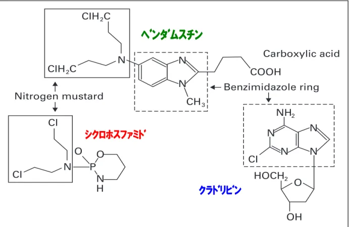
Bendamustine
Benzimidazole ring Nitrogen mustard Cladribine Cyclophosphamide CIH2C CIH2 2 C N CI CI N O OH CH3 COOH N N NH2 HOCH CI N N N O O P N H N Carboxylic acidFig 1. Chemical structure of bendamustine, cyclophosphamide and cladribine.
Cl, chlorine; H, hydrogen; N, nitrogen; O, oxygen; P, phosphorus.
Bendamustine
www.jco.org © 2009 by American Society of Clinical Oncology 1493
133.30.190.14
Information downloaded from jco.ascopubs.org and provided by at Kobe University Medical Library on June 11, 2011 from Copyright © 2009 American Society of Clinical Oncology. All rights reserved.
ベンダムスチン
クラドリビン
シクロホスファミド
ベンダムスチン(トレアキシン
®
)
アルキル化薬のナイトロジェンマスタードの化学構造と代謝拮抗薬である
プリンアナログ様化学構造を併せ持つ化合物
Mochida
R-
ベンダムスチン
vs. R-CHOP
Rummel MJ, et al. Lancet. 2013
Mochida
! 以下に限局期/進行期における標準治療を示す。
びまん性大細胞型
B
細胞リンパ腫に対する治療
• CD20陽性B細胞であれば、
リツキシマブ(R: リツキサン
®)
を併用する。
L群 :低危険度群 LI群:低-中危険度群
HI群:中-高危険度群 H群 :高危険度群
I / II期
巨大病変なしR-CHOP療法
3コース�
局所放射線
照射
II期
巨大病変ありIII / IV期
IPI
L 〜HI
H
R-CHOP療法
6〜8コース
自家移植(
< 65歳)
自家移植(
< 65歳)
非寛解・再発
同種移植
??
救援療法
≥ 部分寛解
?
or
(
or R-CHOP療法6コース)�
Mochida
! IPI(International Prognostic Index)は、中悪性度B細胞リンパ腫に対する
治療効果の予後予測
モデルとして提唱されている。
予後因子
予後不良因子
年齢
>60歳
血清LDH
正常上限を超える
PS
2-4
病期
ⅢまたはⅣ
節外性病変
2以上
0-1 : 低危険度(L)
2 : 低-中危険度(LI)
3 : 中-高危険度(HI)
4-5 : 高危険度(H)
Shipp MA, et al. N Engl J Med 1993; 329: 987-994.