Introduction
The number of patients diagnosed with dementia con- tinues to increase as the population ages. Dementia with Lewy bodies(DLB)is a neurodegenerative disease that was first reported in 1976 by Kosaka et al.1)and is the second most common form of dementia, after Alzhei- mer’s disease(AD). An essential requirement for DLB diagnosis is a progressive cognitive decline that is of sufficient magnitude to interfere with normal social or
occupational functions, or with usual daily activities.
DLB has major symptoms such as visual hallucinations, parkinsonism, fluctuating cognition, and REM sleep be- havior disorder(RBD), and also includes sensitivity to antipsychotic agents or severe autonomic dysfunction as common clinical features.
A clinical diagnosis of DLB is made according to in- ternational clinical diagnostic criteria2). DLB is known to include depressive symptoms in the prodromal stage3);there are reports that depressive symptoms are more common in DLB patients than in AD patients, and
Relationship Between Changes in Cerebral Blood Flow and Neuropsychiatric Symptoms in Dementia with Lewy Bodies
Hitoshi IIDA1), Koji OGOMORI1,2), Yasuo KUWABARA3), Shigeki NAGAMACHI4), Hiroaki KAWASAKI1)
1)Department of Psychiatry, Faculty of Medicine, Fukuoka University
2)Fukuoka International University of Health and Welfare
3)Fukuoka Seamen’s Insurance Health Care and Research Center
4)Department of Radiology, Faculty of Medicine, Fukuoka University
Abstract
In this study, we compared cerebral blood flow (CBF)images in patients with dementia with Lewy bodies(DLB)with those in the normal control and Alzheimer’s disease(AD)groups, and investigated the relationship between DLB and neuropsychiatric symptoms. The subjects in the probable DLB group were 28 outpatients who fulfilled the diagnostic criteria for DLB and underwent singlephoton emission computed tomography (SPECT). CBF was also evaluated in 32 healthy subjects and 37 patients with AD for comparison between groups, and to evaluate the presence of fluctuating cognition, visual halluci- nations, parkinsonism, REM sleep behavior disorder (RBD), and depression in the DLB group. Com- pared with the AD group, there was a significant decrease in CBF in the medial occipital lobe in the DLB group. Compared with the DLB group, there was a significant decrease in CBF in the medial temporal lobe in the AD group. A significant decrease in CBF was also observed in the right middle temporal gy- rus in the DLB group with visual hallucinations and in the right superior temporal gyrus in the DLB group with depressive symptoms. There were no significant differences in CBF with or without fluctuating cog- nition, parkinsonism, and RBD in DLB. Statistical imaging analysis therefore suggested that visual hal- lucinations and depressive symptoms were associated with specific areas of decreased CBF. Thus, it was suggested that neuropsychiatric symptoms of DLB may be associated with specific areas of brain dysfunction.
Key words : Dementia with Lewy bodies, Cerebral blood flow SPECT, Statistical imaging analysis, Visual hallucinations, Depressive symptoms, Parkinsonism
Correspondence to:Hiroaki KAWASAKI, Department of Psychiatry, Faculty of Medicine, Fukuoka University 7451, Nanakuma, Jonanku, Fukuokashi, Fukuoka, Japan
Tel:+81928011011 (ex.3385) Fax:+81928633150 Email:hkawasaki@fukuokau.ac.jp
40%of DLB patients have major depression episodes4)5). In addition to depressive symptoms, DLB presents a va- riety of psychiatric symptoms such as hallucinations, de- lusions, and agitation. Therefore, DLB causes a num- ber of impairments in daily living6), leading to a decline in quality of life and an increasing burden on care- givers7).
Furthermore, geriatric medical care and management are required because DLB is often combined with falls, fractures, aspiration, and pneumonia8). However, the appearance of psychiatric symptoms is inconsistent and their occurrence is difficult to predict. In the early stages of disease, it is especially difficult to clinically distinguish DLB patients from AD patients because of their overlapping clinical and pathological features.
To differentiate DLB from other dementias, it is impor- tant to clarify the differences between DLB and AD, which is the most common type of dementia. In addi- tion, predicting the manifestation of psychiatric symp- toms in DLB is important for early psychological inter- vention and for the evaluation of drug treatment options.
In the diagnostic criteria of DLB, neuroimaging find- ings are important. Among the diagnostic criteria, do- pamine transporter (DAT)scintigraphy and Metaiodo- benzylguanidine(MIBG)myocardial scintigraphy have become indicative biomarkers that directly contribute to diagnosis. As supportive biomarkers, brain magnetic res- onance imaging(MRI)/computed tomography(CT)and Singlephoton emission computed tomography(SPECT) /Fluorine18 2fluoro2deoxyDglucose(FDG)posi- tron emission tomography(PET)are included2). How- ever, in Japan at least, indicative biomarkers such as DAT scintigraphy and MIBG myocardial scintigraphy are expensive, and a cheaper and simpler biomarker is required.
SPECT is a functional brain imaging study that is comparatively inexpensive to perform. Cerebral blood flow(CBF)is determined by the synaptic density and activity of each brain region when there are no struc- tural abnormalities in the cerebral blood vessels9). Be- cause sites with reduced cerebral nerve cell function are depicted as sites with reduced blood flow, a differential diagnosis of dementia can be performed using differ- ences in the characteristic patterns of bloodflowre- duced sites for each disease. The characteristic CBF reduction in DLB is seen as hypoperfusion in the occipi- tal lobe, including the primary visual cortex. In AD, the CBF decrease begins in the posterior cingulate gy-
rus and precuneus before spreading to the parietal lobe, temporal lobe, and frontal lobe as the disease prog- resses. However, the primary sensory/motor cortex, occipital visual cortex, thalamus, basal ganglia, and cerebellum are not easily damaged in AD, and the blood flow in these regions is relatively maintained. This is an important diagnostic point. A metaanalysis re- ported that the sensitivity and specificity of SPECT in distinguishing between DLB and AD is 70.2% and 76.2%, respectively(DLB:hypoperfusion in the occipi- tal or bilateral parietal lobes;AD:hypoperfusion in the unilateral or bilateral temporal/parietal lobes)10). However, although many studies have compared CBF between dementia diseases such as DLB and AD, there are limited studies comparing CBF in relation to differ- ences in symptoms. Many studies have reported the re- lationship between visual hallucinations and CBF, including systematic reviews that conclude that visual hallucinations are related to hypoperfusion and hypome- tabolism in the occipital lobe11). However, few studies have compared the relationship between CBF and other neuropsychiatric symptoms of DLB.
In this study, we aimed to accurately distinguish be- tween DLB and AD patients by directly comparing CBF of patients with DLB to that of patients with AD and measuring the difference. Furthermore, by comparing the relationship between neuropsychiatric symptoms and CBF of DLB, we aimed to clarify imagingrelated factors related to neuropsychiatric symptoms of DLB.
Subjects and methods
We recruited patients with DLB or AD who under- went consultation at the Department of Psychiatry of Fukuoka University Hospital from 2011 to 2017. Pa- tients with DLB fulfilled the diagnostic criteria for prob- able DLB2)and patients with AD fulfilled the National Institute of Neurologic, Communicative Disorders and Stroke AD and Related Disorders Association(NINCDS ADRDA)diagnostic criteria for probable AD12)were
included. All patients received a medical history inter- view and neurological examinations by geriatric psy- chiatry specialist or dementia specialist and were exam- ined using minimental state examinations(MMSE)and SPECT. Their diagnoses were made by the consensus of a panel composed of psychiatrists, neurologists, and a nuclear medicine specialist. We excluded subjects who were taking acetylcholinesterase inhibitors, had his-
tories of psychiatric diseases, or had obvious cerebro- vascular lesions in MRI or CT scans. Patients with vascular stenosis or stroke were excluded because their asymmetric cerebral perfusion profile may have out- weighed the degenerative process and confounded the results. The study participants and their legal represen- tative gave written consent for their participation in this study. In total, 28 DLB patients and 37 AD patients were selected.
Geriatric psychiatric specialist or dementia specialist identified neuropsychiatric symptoms such as fluctuat- ing cognition, visual hallucinations, parkinsonism, RBD, and depression in patients with DLB. To com- pare with the DLB group for MMSE and CBF, we se- lected 32 agematched normal controls(NC)from a normal database in the National Center of Neurology and Psychiatry. This study was approved by the Institu- tional Review Board of Fukuoka University(Ethical re- view number 2018M028).
All participants with DLB and AD received SPECT with Tc99m ethylcysteinate dimer(Tc99m ECD). The SPECT scan started 5 min after the administration of 600 MBq of Tc99m ECD, and data were collected for 20 min using a 3head gamma camera(PRIZM 3000 IRIX, Philips). The SPECT images were reconstructed using the filtered back projection(FBP)method using a Butterworth filter(order 8, cutoff level 0.22 cycles/
pixel). Attenuation correction was performed using Chang’s method. Image analysis was performed using statistical parametric mapping(SPM)12 software(Func- tional Imaging Laboratory, Wellcome Trust Centre for Neuroimaging, Institute of Neurology, UCL, UK)run- ning in MATLAB R2015a(The MathWorks, Natick, MA, USA).
All participants with segmentation errors were ex- cluded by visual inspection. All of the images were
spatially normalized into the MNI standard template
(Montreal Neurological Institute, McGill University, Montreal, Canada)prior to statistical analysis. Spa- tially normalized images were smoothed by convolution using an isotopic Gaussian kernel with a 12 mm full width at half maximum. Statistical comparisons with- in subjects were performed on a voxelbyvoxel basis using twoway ANOVA(full factorial analysis on SPM12), generating SPM(t)maps. The resulting maps of Fstatistics and tstatistics were created using a height threshold at p<0.01(F test)andp<0.001(f test), uncorrected for multiple comparisons. The extent threshold of 400 voxels was also used when considering whether the cluster was significant. The twotailed paired ttest was used to compare clinical features at the initial examination with those at the second examina- tion in each of the two groups, while the twotailed non paired t test was used to compare the clinical and
demographic characteristics between the two groups.
Results
Demographic and cognitive findings of all partici- pants with DLB, AD, and NC are shown in Table 1.
MMSE scores were significantly lower in the DLB and AD groups than in the NC group.
The demographic summary and neuropsychiatric symptoms of DLB are shown in Table 2. The age at on- set was 70.9±6.1 years(range 6081), and the mean length of education was 12.3±3.2 years(range 120). The percentages of core symptoms were 67.9%for fluc- tuating cognition, 60.7% for visual hallucinations, 82.1% for parkinsonism, and 35.7%for RBD. 25.0% of DLB group had 3 core symptoms that are fluctuating cognition, visual hallucinations, and parkinsonism.
21.4%of DLB group had 2 core symptoms that are fluc-
Table 1 Demographic features of study participants
AD NC
DLB
37 32
28 N
29/8 20/12
19/9 Sex(F/M)
73.0±8.6(5485) 72.6±3.4(6878)
72.3±6.5(6282) Age, mean±SD(range), y
21.3±4.5*(1228) 29.8±0.4(2930)
24.1±4.7*(1330) MMSE score, mean±SD(range)
*p<0.05:TukeyKramer test compared with NC(twosided).
DLB, dementia with Lewy bodies;NC, normal controls;AD, Alzheimer’s disease;MMSE, minimental state examination;SPECT, singlephoton emission computed tomography.
tuating cognition and parkinsonism. Depressive symp- toms were as frequent as 64.3%.
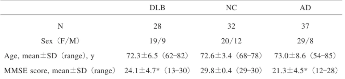
Next, we showed changes in CBF obtained by SPECT image analysis. When comparing between NC and DLB, significant hypoperfusion was observed in a wide range of cerebral cortex areas in the DLB(Fig. 1).
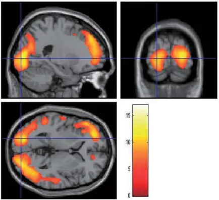
Comparing AD and DLB groups, there was signifi- cant hypoperfusion in the medial occipital lobe in the DLB compared with AD(Fig. 2a). In addition, the AD group had significant hypoperfusion around the hip- pocampus and in the posterior cingulate gyrus and pre- cuneus compared with DLB(Fig. 2b).
Next, we measured changes in CBF in the DLB group
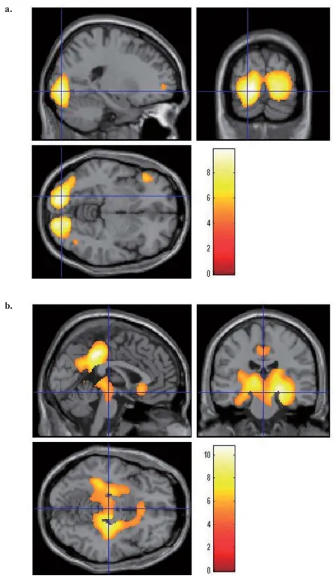
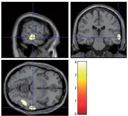
with or without fluctuating cognition, visual hallucina- tions, parkinsonism, RBD, or depressive symptoms. A significant hypoperfusion was observed in the right mid- dle temporal gyrus in the group with visual hallucina- tions compared with the group without visual hallucina- tions(Fig. 3). In addition, when comparing CBF with and without depressive symptoms, there was a signifi- cant hypoperfusion in the right superior temporal gyrus in the group with depressive symptoms(Fig. 4). There were no significant differences in CBF with or without fluctuating cognition, parkinsonism, and RBD in DLB, however.
Table 2 Demographic summary of DLB subjects(n=28)
70.9±6.1(6081)
Age at disease onset, mean±SD(range), y
12.3±3.2(120)
Education, mean±SD(range), y Fluctuating cognition, % 67.9 Visual hallucination, % 60.7 Parkinsonism, % 82.1
REM sleep behavior disorder, % 35.7 Depression, % 64.3
Fig. 1 :Brain regions with hypoperfusion in patients with DLB(n=28)compared with NC (n=32)using SPM analysis of cerebral perfusion SPECT.
Patients with DLB showed a hypoperfusion in a wide range of cerebral cortex included posterior, frontal and parietal lobe (Voxel level:p<0.001 uncorrect- ed;cluster level:p<0.05 FWE corrected). The color bar represents the range of the T value.
Fig. 2:Brain regions with hypoperfusion in patients with DLB(n=28)com- pared with AD (n=37) using SPM analysis of cerebral perfusion SPECT.
a. Patients with DLB showed a hypoperfusion in the medial occipital lobe
(Voxel level:p<0.001 uncorrected;cluster level:p<0.05 FWE cor- rected). The color bar represents the range of the T value.
b. Patients with AD showed a hypoperfusion around the hippocampus and in the posterior cingulate gyrus and precuneus(Voxel level:p<0.001 uncor- rected;cluster level:p<0.05 FWE corrected). The color bar represents the range of the T value.
b.
a.
Fig. 4:Brain regions with hypoperfusion in patients with depressive symptoms
(n=18)compared with patients without depressive symptoms (n=10) in DLB using SPM analysis of cerebral perfusion SPECT.
Patients with depressive symptoms in DLB showed a hypoperfusion in the right superior temporal gyrus (Voxel level:p<0.001 uncorrected;cluster level: p<0.05 FWE corrected). The color bar represents the range of the T value.
Fig. 3:Brain regions with hypoperfusion in patients with visual hallucinations (n= 17)compared with patients without visual hallucinations (n=11)in DLB using SPM analysis of cerebral perfusion SPECT.
Patients with visual hallucinations in DLB showed a hypoperfusion in the right middle temporal gyrus (Voxel level:p<0.001 uncorrected;cluster level: p<0.05 FWE corrected). The color bar represents the range of the T value.
Discussion
This study revealed differences between DLB and NC or AD groups in SPECT imaging and demonstrated changes in CBF with different neuropsychiatric symp- toms of DLB.
In patients who met the clinical criteria for DLB, statistical analysis of SPECT images revealed hypoper- fusion in the occipital lobes of DLB compared with AD, while AD had hypoperfusion in the posterior cingu- late gyrus and precuneus compared with DLB;this pat- tern of decreased CBF has been shown in previous stud- ies13)17).
With visual hallucinations, there was a significant de- crease in CBF in the right middle temporal gyrus. The medial temporal gyrus is involved in different processes such as distance recognition, face recognition, and word recognition during reading tasks18). The lower two thirds of the temporal gyrus(middle and inferior tempo- ral gyrus)are involved in morphological vision and perception. Disruption of this area may result in color blindness, image agnosia, and prosopagnosia. Lanctot et al. showed that CBF in the right middle temporal gy- rus was relatively low in AD in association with aggres- sion19). Although visual hallucinations are known to be associated with reduced occipital lobe blood flow in DLB11), the results of our study with and without visual hallucinations of DLB patients suggest that another mechanism may be involved in visual hallucinations.
Hypoperfusion in the medial temporal gyrus may cause visual illusions and misidentification, which may mani- fest as visual hallucinations. In addition, when visual hallucinations occur, patients are often clinically aggres- sive, which suggests that the appearance of aggression may promote visual hallucinations.
With depressive symptoms, there was a significant de- crease in CBF in the right superior temporal gyrus in our result. The right superior temporal gyrus consists of the primary auditory cortex and auditory areas20)and is involved in emotional processing and social cogni- tion21).
A metaanalysis of fMRI studies of depression noted that the right superior temporal gyrus is one of the most consistently identified areas involved in its patho- physiology22). It is also reported that smaller volumes of the left hippocampus and right superior temporal gy- rus on MRI in patients with depression are related to
longer durations of depressive symptoms23), suggesting a close relationship between the right superior temporal gyrus and depressive symptoms. In addition, fMRI studies have shown that depressed patients with AD have reduced functional connectivity between the hypo- thalamus and the superior temporal gyrus24). In PD pa- tients with depressive symptoms, decreased CBF is also observed in the right superior temporal gyrus and right medial orbitofrontal cortex25), supporting the results of this study. Abnormal superior temporal gyrus activity was observed in the background of patients with depres- sive symptoms, suggesting that depressive symptoms in DLB also show similar changes.
The present study has several limitations. First, the sample size was small, and the statistical power was therefore low. Future studies will need to use larger sample sizes to increase statistical power. Second, neu- ropsychiatric symptoms were assessed only for their presence and absence, and symptom degree was not as- sessed using a rating scale. In addition, although symp- toms were evaluated by a trained psychiatrist, they may have been underestimated or overestimated. Thus, we will consider using an evaluation scale in the future.
Third, a group of patients with major depressive disorder
(MDD)should be included in future studies. A com- parison of patients with MDD and DLB will provide more detailed information about the pathophysiology of depression in DLB patients.
Conclusions
In patients with DLB, SPECT imaging and statistical analysis revealed areas of CBF that differed in the pres- ence or absence of neuropsychiatric symptoms. It was suggested that neuropsychiatric symptoms of DLB may be associated with specific areas of brain dysfunction.
Funding sources
This research received no specific grant from any funding agency in the public, commercial, or notfor profit sectors.
Acknowledgements
We thank Dr. Kiyotaka Nemoto(Department of Psy- chiatry, Faculty of Medicine, University of Tsukuba)for his technical assistance. In addition, we thank Lisa
Kreiner, PhD, and Bronwen Gardner, PhD, from Edanz Group(www.edanzediting.com/ac)for editing a draft of this manuscript.
The authors declare no conflict of interest.
References
1)Kosaka K, Oyanagi S, Matsushita M, Hori A:Pre- senile dementia with Alzheimer, Pickand Lewy body changes. Acta Neuropathol Nov 15;36(3): 221233, 1976.
2)McKeith IG, Boeve BF, Dickson DW, Halliday G, Taylor JP, Weintraub D, Aarsland D, Galvin J, At- tems J, Ballard CG, Bayston A, Beach TG, Blanc F, Bohnen N, Bonanni L, Bras J, Brundin P, Burn D, ChenPlotkin A, Duda JE, ElAgnaf O, Feldman H, Ferman TJ, Ffytche D, Fujishiro H, Galasko D, Goldman JG, Gomperts SN, GraffRadford NR, Honig LS, Iranzo A, Kantarci K, Kaufer D, Kukull W, Lee VMY, Leverenz JB, Lewis S, Lippa C, Lunde A, Masellis M, Masliah E, McLean P, Mol- lenhauer B, Montine TJ, Moreno E, Mori E, Mur- ray M, O’Brien JT, Orimo S, Postuma RB, Ramaswamy S, Ross OA, Salmon DP, Singleton A, Taylor A, Thomas A, Tiraboschi P, Toledo JB, Tro- janowski JQ, Tsuang D, Walker Z, Yamada M, Ko- saka K:Diagnosis and management of dementia with Lewy bodies:Fourth consensus report of the DLB Consortium. Neurology Jul 4;89(1):88 100, 2017.
3)Simard M, van Reekum R, Cohen T:A review of the cognitive and behavioral symptoms in dementia with Lewy bodies. J Neuropsychiatry Clin Neuro- sci Fall;12(4):425450, 2000.
4)Ballard C, Holmes C, McKeith I, Neill D, O’Brien J, Cairns N, Lantos P, Perry E, Ince P, Perry R: Psychiatric morbidity in dementia with Lewy bodi- es:a prospective clinical and neuropathological comparative study with Alzheimer’s disease. Am J Psychiatry Jul;156(7):10391045, 1999.
5)Geser F, Wenning GK, Poewe W, McKeith I:How to diagnose dementia with Lewy bodies:state of the art. Mov Disord Aug;20 Suppl 12:S1120, 2005.
6)Namioka N, Hanyu H, Hatanaka H, Fukasawa R, Sakurai H, Iwamoto T:Comprehensive geriatric assessment in elderly patients with dementia. Ger-
iatr Gerontol Int Jan;15(1):2733, 2015.
7)Borroni B, Agosti C, Padovani A:Behavioral and psychological symptoms in dementia with Lewy bodies(DLB):frequency and relationship with dis- ease severity and motor impairment. Arch Geron- tol Geriatr JanFeb;46(1):101106, 2008.
8)Hanyu H, Sato T, Hirao K, Kanetaka H, Sakurai H, Iwamoto T:Differences in clinical course between dementia with Lewy bodies and Alzheimer’s dis- ease. Eur J Neurol Feb;16(2):212217, 2000.
9)Isaias IU, Antonini A:Singlephoton emission computed tomography in diagnosis and differential diagnosis of Parkinson’s disease. Neurodegener Dis;7(5):319329, 2010.
10)Yeo JM, Lim X, Khan Z, Pal S:Systematic review of the diagnostic utility of SPECT imaging in de- mentia. Eur Arch Psychiatry Clin Neurosci Oct; 263(7):539552, 2013.
11)Pezzoli S, Cagnin A, Bandmann O, Venneri A: Structural and Functional Neuroimaging of Visual Hallucinations in Lewy Body Disease:A System- atic Literature Review. Brain Sci Jul 15;7(7), 2017.
12)McKhann G, Drachman D, Folstein M, Katzman R, Price D, Stadlan EM:Clinical diagnosis of Alz- heimer’s disease:report of the NINCDSADRDA Work Group under the auspices of Department of Health and Human Services Task Force on Alz- heimer’s Disease. Neurology Jul;34(7):939944, 1984.
13)Ishii K, Yamaji S, Kitagaki H, Imamura T, Hirono N, Mori E:Regional cerebral blood flow differ- ence between dementia with Lewy bodies and AD.
Neurology Jul 22;53(2):413416, 1999.
14)Lobotesis K, Fenwick JD, Phipps A, Ryman A, Swann A, Ballard C, McKeith IG, O’Brien JT:Oc- cipital hypoperfusion on SPECT in dementia with Lewy bodies but not AD. Neurology Mar 13;56
(5):643649, 2001.
15)Pasquier J, Michel BF, BrenotRossi I, HassanSeb- bag N, Sauvan R, Gastaut JL:Value of(99m)Tc ECD SPET for the diagnosis of dementia with
Lewy bodies. Eur J Nucl Med Mol Imaging Oct; 29(10):13421348, 2002.
16)Hanyu H, Shimizu S, Hirao K, Kanetaka H, Sakurai H, Iwamoto T, Koizumi K, Abe K:Differentiation of dementia with Lewy bodies from Alzheimer’s disease using MiniMental State Examination and
brain perfusion SPECT. J Neurol Sci Dec 1;250
(12):97102, 2006.
17)Goto H, Ishii K, Uemura T, Miyamoto N, Yoshi- kawa T, Shimada K, Ohkawa S:Differential diag- nosis of dementia with Lewy Bodies and Alzheimer Disease using combined MR imaging and brain perfusion singlephoton emission tomography.
AJNR Am J Neuroradiol Apr;31(4):720725, 2010.
18)Acheson DJ, Hagoort P:Stimulating the brain’s language network:syntactic ambiguity resolution after TMS to the inferior frontal gyrus and middle temporal gyrus. J Cogn Neurosci Oct;25(10): 16641677, 2013.
19)Lanctot KL, Herrmann N, Nadkarni NK, Leibo- vitch FS, Caldwell CB, Black SE:Medial tempo- ral hypoperfusion and aggression in Alzheimer disease. Arch Neurol Nov;61(11):17311737, 2004.
20)Hou Z, Sui Y, Song X, Yuan Y:Disrupted Inter- hemispheric Synchrony in Default Mode Network Underlying the Impairment of Cognitive Flexibility in LateOnset Depression. Front Aging Neurosci Sep 27;8:230, 2016.
21)Arnsten AF, Rubia K:Neurobiological circuits regulating attention, cognitive control, motivation,
and emotion:disruptions in neurodevelopmental psychiatric disorders. J Am Acad Child Adolesc Psychiatry Apr;51(4):356367, 2012.
22) Fitzgerald PB, Laird AR, Maller J, Daskalakis ZJ: A metaanalytic study of changes in brain activa- tion in depression. Hum Brain Mapp Jun;29(6): 683695, 2008.
23)Caetano SC, Hatch JP, Brambilla P, Sassi RB, Nico- letti M, Mallinger AG, Frank E, Kupfer DJ, Kesha- van MS, Soares JC:Anatomical MRI study of hip- pocampus and amygdala in patients with current and remitted major depression. Psychiatry Res Dec 15;132(2):141147, 2004.
24)Liu X, Chen W, Tu Y, Hou H, Huang X, Chen X, Guo Z, Bai G, Chen W:The Abnormal Functional Connectivity between the Hypothalamus and the Temporal Gyrus Underlying Depression in Alz- heimer’s Disease Patients. Front Aging Neurosci Feb 13;10:37, 2018.
25)Kim YD, Jeong HS, Song IU, Chung YA, Nam- gung E, Kim YD:Brain perfusion alterations in de- pressed patients with Parkinson’s disease. Ann Nucl Med Dec;30(10):731737, 2016.
(令和 1.9.2受付,令和 1.10.1受理)
「The authors declare no conflict of interest.」