577 ミニ特集「肺抗酸菌症の画像」
MAC 症以外の非結核性抗酸菌症画像所見
倉島 篤行
要旨:MAC以外の非結核性抗酸菌で Mycobacterium kansasiiをのぞいては一般に先行基礎疾患への寄 生的性格が強く菌種固有の画像所見の把握は困難である。ここでは先行基礎疾患を認めがたい状態で の画像所見を提示した。M. kansasii は多かれ少なかれ結核症と類似するがやや異なった傾向も見られ る。M. fortuitum では非特異的な均等影所見を示し,M. xenopi,M. gordonae 症例は薄壁の孤立空洞を 呈し,M. abscessus 症例は樹枝状分岐陰影の広範囲な散布が特徴的であった。気道壁肥厚所見は結核 症例ではあまり見られない非結核性抗酸菌症での特徴的所見の一つとして考えられる。
キ ー ワ ー ズ: 非 結 核 性 抗 酸 菌 症,Mycobacterium kansasii,Mycobacterium fortuitum,Mycobacterium xenopi,Mycobacterium gordonae,Mycobacterium abscessus
一般に多くの非結核性抗酸菌のヒトに対する病原性は 結核菌よりかなり弱く,先行する肺疾患のない健常肺野 にいきなり病変を形成するより,既存の気道または肺構 造破壊部に寄生的,日和見的な病変を形成していく傾向 が強い。そのような場合の画像所見は,当然先行する空 洞や気管支拡張などの既存構造に規制されたり,あるい はそれを修飾するという形態で表現され,その起炎菌種 固有の特徴をその画像の中に見るのは困難である。 したがって本稿では,2008 年診断規準に合致し,免 疫不全がなく,先行する基礎肺疾患のない例に起きた MAC 以外の非結核性抗酸菌症画像について記載する。 なお,MAC 以外の非結核性抗酸菌症では Mycobacte-rium kansasii 以外については例数も少なく,提示した症 例がその菌種特有の画像所見なのかどうか,あるいはそ の菌種として典型的な画像であるのかを明示することは 困難である。 1 .Mycobacterium kansasii による胸部 X 線画像 1977年に下出が著した「日本における M.kansasii症」 では 72例を集積し,20代から40代の既往肺疾患のない 男性に多く,画像的には上葉右側優位の肺野型空洞が多 いとしている1)が,これらは今日も同様であり,多くの 外国文献でも同様の特徴が指摘されている2)。 健常若年者の肺に好発するという点では M. kansasii 症 は非結核性抗酸菌症の中では比較的肺結核症に近い性格 をもっている。
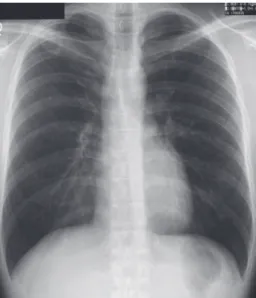
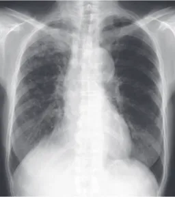
Evansなどは HIV陰性の M.kansasii 症28例と人種,性, 年齢をマッチさせた肺結核症 56例の画像を起炎菌種情 報を明らかにせず複数の放射線科医が読影した結果を報 告しているが,M. kansasii 症では胸水貯留を伴うことが なく, 1 セグメント以上の均等影が少なく,下肺野分布 が少なく,片側性であり,かつそれは右側であるという 点で有意差があったが,全体として画像所見のみで M. kansasii症固有の所見を指摘することはできないとして いる3)。 Fig. 1は上記研究に一致する典型的な M.kansasii症例, 39 歳男性の胸部単純 X 線画像である。右上肺野にかな りな大きさの空洞を推測させるが,他の肺野には散布病 巣は認めがたい。 Fig. 2は当時行われていた断層撮影であるが,多発空 洞であり周辺の均等影は少ないことが判る。 Fig. 3 は CT 画像であるが,空洞壁は比較的薄く周辺 の散布病巣はわずかしか認められない。 Fig. 4は性,年齢,病巣部位,性状を同じくした肺結 核症の画像であるが,これと比較すると M. kansaii 画像 の傾向がより明らかになる。しかしこれは M. kansasii 症 結核予防会複十字病院 連絡先 : 倉島篤行,結核予防会複十字病院,〒204_8522 東京 都清瀬市松山 3_1_24 (E-mail: kurashimaa@fukujuji.org) (Received 8 Jun. 2009)
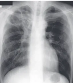
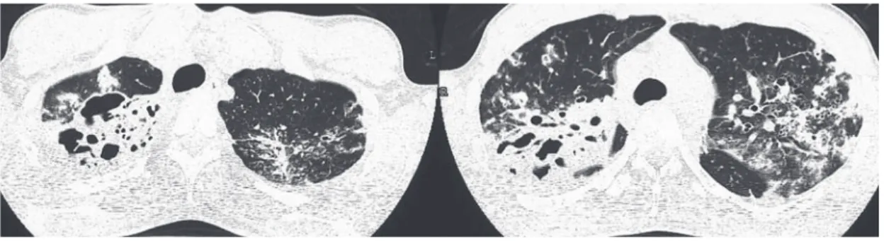
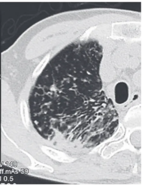
としての一般的な傾向を把握するのに有用であるが,以 下に非典型例の画像を提示する。 Fig. 5は特別の免疫不全のない32歳男性の M.kansasii 胸部単純 X 線画像であるが,右上肺野には不均等な浸 潤影中に小空洞が散在し,左肺野には主として上葉に広 範囲な均等影を認める。 Fig. 6は同一例の CT画像であるが,多発小空洞,air-bronchogram を有する均等影,気管支壁肥厚とそれに随 伴する均等影,牽引性気管支拡張所見,区域性の GGO 所見,小葉中心性の微細結節影など多彩な所見が見られ ている。 抗酸菌症画像所見をめぐる金言「典型の認識は重要だ が,それに嵌るな」は依然有効である。
Fig. 4 The plain film of equivalent tuberculosis case.
Fig. 2 Tomography of the same case. Thin wall cavities without satellite lesions are shown in the right upper lobe.
Fig. 3 CT scan of the same case.
Fig. 5 Mycobacterium kansasii pulmonary infection in 32-year-old-man.
Fig. 1 Mycobacerium kansasii pulmonary infection in 39-year-old man.
NTM Other than MAC XP Findings / A. Kurashima 579 2 .Mycobacterium fortuitum による胸部 X 腺画像 M. fortuitum は古くから使われてきた非結核性抗酸菌 を発育速度,コロニー着色性,生化学的性状などから分 類するいわゆる Runyon 分類では M.abscessus などとと もにⅣ群の迅速(結核菌や M. avium に比べてという意味 である)発育菌に位置づけられるが,臨床例の頻度は M. abscessus が圧倒的で M. fortuitum はかなり少ない。ま た M. fortuitum 自体単一の菌種というより亜種を含むグ ループとして把握されている4)。 土壌や水中に常在するが,人の肺感染症の場合,多く は肺結核治癒後や COPD などの先行基礎疾患の上に成 立する日和見感染的傾向が強いとされている。 わが国で最初のまとまった報告である 1981 年の国立 療養所非定型抗酸菌症共同研究班の報告でも,40 歳以 上の男性にやや多く,塵肺や肺結核など先行基礎疾患に 続発するものが多いとしている5)。 外国報告例では,既往肺疾患のない場合は若年発症例 が多く,その場合 achalasiaなど頻回の嘔吐を繰り返す食 道,胃疾患との関連が注意されており,消化管に常在す る菌が誤嚥により肺感染を誘起する機序が推定されてい る6)。 Fig. 7は90歳男性の胸部単純 X線画像所見である。過 去喫煙歴あり,数年前肺結核として化学療法歴あり,経 過観察中再度排菌陽性として紹介された。両側肺尖部に は胸膜に接した irregularな凹凸や索状影や結節影のまば らな散布を見る。下行大動脈と横隔膜が交差する部位に 不明瞭な陰影がある。 Fig. 8はその CT所見である。左 S10領域に気道構造に 沿った拡がりをもつ濃淡さまざまな airbronchogram を 伴った限局性均等影が見られている。対側肺中葉や下葉 に小葉中心性の辺縁不明瞭な結節影の散布が見られ,高 齢に伴う誤嚥の存在が疑われる。 なお M. fortuitum は NQ 系薬剤がきわめて有効である が,単剤投与ではなく耐性出現抑制のため CAM, doxy-cycline などの他の薬剤との併用が勧められている7)。
Fig. 6 CT scan from the same case showes cavities, segmental consolidations, centrilobular nodules, traction bronchiectasis, ground-glass opacity.
Fig. 7 Mycobacterium fortuitum pulmonary infection in 90 year-old-man. Irregular mass shadow is visible at the area of adjacent to the lower thoracic aorta.
Fig. 8 CT scan shows irregular consolidation around the left lower lobe bronchus. Centrilobular nodules are scattered sur-rounding the consolidation and the right lower lobe.
3 .Mycobacterium xenopi による胸部 X 線画像 M. xenopi は 45℃という高い至適発育温度の非結核性 抗酸菌で,病院の給湯系などから分離されることがある 菌である。 本症はわが国や米国ではあまり頻度が高くないが,カ ナダ,ヨーロッパなどでは MAC に次いで高頻度であ り8),クロアチアからは最も多い菌種という報告もあ る9)。一般的に COPDに続発することが多く,高齢男性 に多く,上葉の空洞形成で見つかることが多いとされて いる10)。先行する肺疾患のない場合はより若年での報告 が多い。 Fig. 9は34歳男性の胸部 X線単純画像である。特に先 行疾患の認めない肺野で右肺尖部よりやや下方に薄壁の 空洞陰影を認める。 同例の CT画像である Fig. 10では右上肺野に比較的薄 壁の孤立性の空洞を認め,周囲には散布病巣は見られて いない。空洞背側に内壁から膨隆した部分が見られる が,これの正確な性状は不明である。しかし結核を含め た抗酸菌症のごく早期の孤立小空洞ではよく見られる所 見であり("reversed magatama sign"と命名してもよいか もしれない),抗酸菌症で形成された壊死組織が軟化融 解し誘導気管支から排除されつつある状況を表すと推定 される。 提示例は山崎などが報告した 40歳男性例11)と酷似し た画像であり,先行肺疾患のない場合の一つの典型かも しれない。 4 .Mycobacterium gordonae による胸部 X 線画像 M. gordonae は環境中の貯留水に特に多く,病院など では内視鏡洗浄用水などのいわゆる contamination の大 きな理由になっているが,一般にヒトには非病原性菌と して考えられている。 筆者は気管支肺胞洗浄の細胞沈渣ペレットを再浮遊さ せるリン酸緩衝液が本菌で汚染されていた経験がある。 したがって本菌種がヒト検体から分離された場合,直 ちに起炎菌として扱うのではなく,colonization 例とし て慎重に経過を観察すべきである。 実際に肺結核治癒経過中に本菌が偶発微量排菌として 散発することは稀ではない。しかし呼吸器疾患画像経過 と一致した排菌経過を示し,起炎菌としてのみ解釈可能 な場合も見られる。 M. gordonae も遺伝子学的にはいくつかの亜種的な heterogeneity があるとされ12),環境中の菌と疾患からの 分離菌との異同は今後検討されるべき課題である。 Fig. 9 Mycobacterium xenopi pulmonary infection in
34-year-old man. Thin wall cavity at the right upper area is visible.
Fig. 10 CT scan of the same case shows the solitary cavity with intraluminal debris.
Fig. 11 Mycobacterium gordonae pulmonary infection in 57-year-old man.
NTM Other than MAC XP Findings / A. Kurashima 581 Fig. 11 は左背部の違和感で受診した 57 歳男性の胸部 単純 X 線画像である。左上肺野に薄壁の空洞病変が見 られ,Fig. 12の CTでは周囲に淡い毛羽立ちを伴う薄壁 の空洞が見られている。この時点で 2 回の M.gordonae が培養で検出され,本例は無治療経過観察のみで Fig. 13 のように治癒状態に達している。 なお M. gordonae は DDHによる同定は必ずしも正確で はないとされているが,本例の菌は 16S rRNA および rpoB 遺伝子解析でも M. gordonae と確認された。 5 .Mycobacterium abscessus による胸部画像所見 M. abscessus は以前は M. chelonae に属する亜種であっ たが,1986 年に束村などが 211 菌株の rapidly growers の 数値分析(116項目の検討で90%以上の一致率で同一菌 種とする)で M. chelonae と M. abscessus は異なる菌種の 可能性を指摘した13)。 1992年に楠などが DNA相同性が97%以下であること から独立した菌種への昇格を提案し国際的に認められ た14)。非結核性抗酸菌の中で最も多薬剤に耐性を示す菌 である。
Griffith などによれば迅速発育菌群(Rapidly Growing
Mycobacteria = RGM)の中での大多数(82%)は本菌で あり,先行する基礎肺疾患が認められる場合が多い(68 %)としており,基礎疾患としては陳旧性肺結核や他の 菌種による非結核性抗酸菌症,Cystic fibrosis,気管支拡 張症,慢性嘔吐を伴う食道胃疾患などを指摘している。 Fig. 15 CT scan of the same case shows wide distribution of tree-in-bud pattern with bronchiectasis.
Fig. 12 CT scan of the same case shows thin wall cavity with surrounding hazy opacity.
Fig. 13 CT scan of the same case 6 months later shows scar formation.
Fig. 14 Mycobecterium abscessus pulmonary infection in 61-year-old woman.
他方,基礎疾患のない中高年女性に見られる MAC症類 似の病型も認めている15)。 Fig. 14は61歳女性の M.abscessus症胸部単純 X線画像 である。右上肺野に斑状の均等影が融合し右側全肺野に 多数の微小散布巣が密に散布している。左肺野は右側ほ どではないがやはり小病巣が散布している。Fig. 15は同 例の CT画像である。上段の右 S2領域では濃厚な不整塊 状影が見られているが,表示されていない別のスライス では壁の厚い空洞が存在する。周囲では拡張した気道と 多数の散布病巣が見られている。上葉の空洞所見は M. abscessus 症の特異的な所見ではないが,Griffith など が指摘するように M. abscessus 症では多く見られる所見 の一つである。下段では小葉大の均等性陰影や小葉中心 性の tree-in-bud pattern,気道壁肥厚性陰影がきわめて広 範囲に分布している。 これら様々な陰影の諸要素は M. abscessus 症特有のも のではなく,M. avium complex症など非結核性抗酸菌症 一般に共通する画像要素である。しかし M. abscessus 症 が他の非結核性抗酸菌症と異なるのは,M. abscessus 症 の場合,疾患初期から広範囲に分布する点である。 Han D な ど は 12 例 の M.abscessus 症 画 像 を 検 討 し, tree-in-bud pattern は本症の 90% で見られ,これらが上葉 優位かつ両側に見られ,MAC症で画像所見が中葉舌区 領域に多く見られることと異なり,全 lobe にあまねく 分布することが特徴であると指摘している16)。 Fig. 16は本例の右上葉 CT画像であるが,ここでは右 背側の不整均等影に向かういわゆる tram lineが多数見ら れる。これらは数珠状を示す気管支拡張所見とは異な り,起始部から末梢まで全長にわたり同じ程度の壁肥厚 文 献 1 ) 下 出 久 雄: 日 本 に お け る M. kansasii 症. 結 核. 1977 ; 52 : 577 _ 585.
2 ) Christensen EE, Dietz GW, Ahn CH, et al.: Radiographic manifestations of pulmonary mycobacterium kansasii infec-tions. AJR. 1978 ; 131 : 985 _ 993.
3 ) Evans AJ, Crisp AJ, Hubbard RB, et al. : Pulmonary
myco-bacterium kansasii infection : comparison of radiological appearances with pulmonary tuberculosis. Thorax. 1996 ; 51 : 1243 _ 1247.
4 ) Brown-Elliott BA, Griffith DE, Wallace RJ Jr. : Newly described or emerging human species of non tuberculous mycobacteria. Mycobacterial Infections. 2002 ; 16 : 187 _ 220.
5 ) 国立療養所非定型抗酸菌症共同研究班 : Mycobacterium
fortuitum 呼吸器感染症の臨床像. 結核. 1981 ; 56 : 587 _ 593.
6 ) Hadjiliadis D, Adlakha A, Prakashu UB : Rapidly growing mycobacterial lung infection in association with esophageal disorders. Mayo Clin Proc. 1999 ; 74 : 744 _ 745.
7 ) Griffith DE, Aksamit T, Brown-Elliott BA, et al. on behalf of the ATS Mycobacterial Diseases Subcommittee : An Official ATS/IDSA Statement : Diagnosis, Treatment, and Prevention of Nontuberculous Mycobacterial Diseases. Am J Respir Crit Care Med. 2007 ; 175 : 367 _ 416.
8 ) Dailloux M, Abalain ML, Laurain C : Respiratory infections associated with nontuberculous mycobacteria in non-HIV patients. Eur Respir J. 2006 ; 28: 1211 _ 1215.
9 ) Marušić A, Katalinić-Janković V, Popović-Grle S, et al :
Mycobacterium xenopi pulmonary disease – Epidemiology and clinical features in non-immunocompromised patients. Journal of Infection. 2009 ; 58 : 108 _ 112.
10) Wittram C, Weisbrod GL: Mycobacterium xenopi Pulmonary Infection : Evaluation with CT. Journal of Computer Assist-ed Tomography. 1998 ; 22 : 225 _ 228.
11) 山崎泰宏, 藤内 智, 松本博之, 他:Mycobacterium xenopi 肺感染症の 2例. 日呼吸会誌. 2003 ; 41 : 556_560 12) Itoh S, Kazumi Y, Abe C, et al. : Heterogeneity of RNA
polymerase gene (rpoB) sequence of Mycobacterium
gor-donae clinical isolate identification with a DNA probe kit and by conventional methods. Journal of clinical micro- biology. 2003 ; 41 : 1656 _ 1663.
13) Tsukamura M, Ichiyama S : Numerical classification of rapidly growing nonphotochromogenic mycobacteria.
Mi-Fig. 16 CT scan of the same case shows bronchial wall thickening without irregular dilatation.
NTM Other than MAC XP Findings / A. Kurashima 583
crobiol Immnol. 1986 ; 30 : 863 _ 882.
14) Kusunoki S, Ezaki T : Proposal of Mycobacterium
peregri-num sp. nov., nom. rev., and elevation of Mycobacterium
chelonae subsp. abscessus (Kubica et al.) to species status :
Mycobacterium abscessus comb. nov. International Journal of Systematic Bacteriology. 1992 ; 42 : 240 _ 245.
15) Griffith DE, Girard WM, Wallace RJ Jr. : Clinical features
of pulmonary disease caused by rapidly growing mycobacte-ria : an analysis of 154 patients. Am Rev Respir Dis. 1993 ; 147 : 1271.
16) Han D, Lee KS, Yi CA, et al. : Radiographic and CT find-ings of nontuberculous mycobacterial pulmonary infection caused by Mycobactrium abscessus. AJR. 2003 ; 181 : 513 _ 517.
−−−−−−−− Current Topics : Medical Images of Pulmonary Mycobacterioses −−−−−−−−
RADIOGRAPHIC FINDINGS OF
PULMONARY NONTUBERCULOUS MYCOBACTERIOSIS OTHER THAN
MYCOBACTERIUM AVIUM
COMPLEX
Atsuyuki KURASHIMA Abstract Almost all nontuberculous mycobacteria (NTM)
cause opportunistic infection. Therefore, the radiographic findings of NTM have a tendency of nonspecific patterns modifying the predisposing conditions or diseases and we could not extract species specific radiographic characteriza-tions in that situacharacteriza-tions. In this review, the NTM cases without predisposing conditions or diseases are submitted essentially. Mycobacterium kansasii cases show more or less the same patterns with TB cases. Mycobacterium fortuitum case shows nonspecific consolidations. Mycobacterium xenopi case shows solitary cavity in the upper lobe area. Mycobacterium gor-donae case shows the same cavitary pattern. Mycobacterium abscessus case shows widely scattered tree-in-bud appearance foci.
Weak virulence NTM like as Mycobacterium xenopi or Mycobacterium gordonae may form solitary cavity without
predisposing conditions.
The pattern of bronchial wall thickening seems to be one of the specific findings with NTM pulmonary infection.
Key words: Nontuberculous mycobacteriosis, Mycobacte-rium kansasii, Mycobacterium fortuitum, Mycobacterium xenopi, Mycobacterium gordonae, Mycobacterium abscessus Clinical research adviser of Fukujuji Hospital, Japan Anti-Tuberculosis Association, Adviser of Research Institute of Tuberculsois, Japan Anti-Tuberculosis Association.
Correspondence to : Atsuyuki Kurashima, Clinical research adviser of Fukujuji Hospital, Japan Anti-Tuberculosis Associ-ation, 3 _ 1 _ 24, Matsuyama, Kiyose-shi, Tokyo 204 _ 8522 Japan. (E-mail : kurashimaa@fukujuji.org)