Article
Targeting Excessive EZH1 and EZH2 Activities for Abnormal Histone Methylation and Transcription Network in Malignant Lymphomas
Graphical Abstract
Highlights
d
Lymphoma transcriptome is under control of global H3K27me3 accumulation
d
EZH1 and EZH2 are druggable targets in EZH2
WT/WTand EZH2
WT/Mumalignancies
d
Inactivation of chromatin-associated genes triggers EZH1/2 perturbation
d
Targeting epigenome for pre-cancerous states is achieved by EZH1/2 inhibition
Authors
Makoto Yamagishi, Makoto Hori, Dai Fujikawa, ..., Kazushi Araki, Toshiki Watanabe, Kaoru Uchimaru
Correspondence
[email protected] (M.Y.), [email protected] (K.U.)
In Brief
A mechanism-based, effective strategy for controlling oncogenic H3K27me3 remains an open question. Yamagishi et al. provide the scientific rationale for dual targeting of EZH1+EZH2 in
malignancies overexpressing EZH2, such as ATL, PTCL, and DLBCL, or harboring mutations in histone-modifying genes, as well as in pre-cancerous cells
epigenomically perturbed by oncovirus infection.
Yamagishi et al., 2019, Cell Reports 29 , 2321–2337 November 19, 2019 ª 2019 The Author(s).
https://doi.org/10.1016/j.celrep.2019.10.083
Cell Reports
Article
Targeting Excessive EZH1 and EZH2 Activities
for Abnormal Histone Methylation and Transcription Network in Malignant Lymphomas
Makoto Yamagishi,
1,17,* Makoto Hori,
1Dai Fujikawa,
2Takeo Ohsugi,
3Daisuke Honma,
4Nobuaki Adachi,
5Harutaka Katano,
6Tsunekazu Hishima,
7Seiichiro Kobayashi,
8Kazumi Nakano,
1Makoto Nakashima,
1Masako Iwanaga,
9Atae Utsunomiya,
10Yuetsu Tanaka,
11Seiji Okada,
12Kunihiro Tsukasaki,
13Kensei Tobinai,
14Kazushi Araki,
15Toshiki Watanabe,
16and Kaoru Uchimaru
1,*
1
Department of Computational Biology and Medical Sciences, Graduate School of Frontier Sciences, The University of Tokyo, Tokyo, Japan
2
Animal Models and Retroviral Vaccines Section, Vaccine Branch, Center for Cancer Research, National Cancer Institute, NIH, Bethesda, MD, USA
3
Department of Laboratory Animal Science, School of Veterinary Medicine, Rakuno Gakuen University, Hokkaido, Japan
4
Oncology Laboratories, Daiichi Sankyo, Co., Tokyo, Japan
5
Biomarker Department, Daiichi Sankyo, Co., Tokyo, Japan
6
Department of Pathology, National Institute of Infectious Diseases, Tokyo, Japan
7
Department of Pathology, Tokyo Metropolitan Cancer and Infectious Diseases Center Komagome Hospital, Tokyo, Japan
8
Division of Molecular Therapy, Institute of Medical Science, The University of Tokyo, Tokyo, Japan
9
Department of Clinical Epidemiology, Graduate School of Biomedical Sciences, Nagasaki University, Nagasaki, Japan
10
Department of Hematology, Imamura General Hospital, Kagoshima, Japan
11
Graduate School and Faculty of Medicine, University of the Ryukyus, Okinawa, Japan
12
Joint Research Center for Human Retrovirus Infection, Graduate School of Medical Sciences, Kumamoto University, Kumamoto, Japan
13
Department of Hematology, International Medical Center, Saitama Medical University, Saitama, Japan
14
Department of Hematology, National Cancer Center Hospital, Tokyo, Japan
15
Oncology Clinical Development Department, Daiichi Sankyo Co., Tokyo, Japan
16
Future Center Initiative, The University of Tokyo, Tokyo, Japan
17
Lead Contact
*Correspondence: [email protected] (M.Y.), [email protected] (K.U.) https://doi.org/10.1016/j.celrep.2019.10.083
SUMMARY
Although global H3K27me3 reprogramming is a hall- mark of cancer, no effective therapeutic strategy for H3K27me3-high malignancies harboring EZH2
WT/WThas yet been established. We explore epigenome and transcriptome in EZH2
WT/WTand EZH2
WT/Muaggressive lymphomas and show that mutual inter- ference and compensatory function of co-ex- pressed EZH1 and EZH2 rearrange their own genome-wide distribution, thereby establishing restricted chromatin and gene expression signa- tures. Direct comparison of leading compounds in- troduces potency and a mechanism of action of the EZH1/2 dual inhibitor (valemetostat). The syn- thetic lethality is observed in all lymphoma models and primary adult T cell leukemia-lymphoma (ATL) cells. Opposing actions of EZH1/2-polycomb and SWI/SNF complexes are required for facultative het- erochromatin formation. Inactivation of chromatin- associated genes ( ARID1A , SMARCA4/BRG1 , SMARCB1/SNF5 , KDM6A/UTX , BAP1 , KMT2D/
MLL2 ) and oncovirus infection (HTLV-1, EBV) trigger EZH1/2 perturbation and H3K27me3 deposition.
Our study provides the mechanism-based rationale
for chemical dual targeting of EZH1/2 in cancer epi- genome.
INTRODUCTION
Histone H3 regulation, particularly H3 lysine 27 trimethylation (H3K27me3), is a central process for chromatin condensation and gene silencing. The suppressive histone mark is catalyzed by polycomb repressive complex 2 (PRC2), which includes either enhancer of zeste homolog 1 (EZH1) or EZH2 as an enzy- matically active core subunit, as well as other components, such as EED and SUZ12. A heterozygous gain-of-function (GoF) mu- tation of EZH2 has been observed in certain lymphoma types, contributing to increased H3K27me3 (Morin et al., 2010; Be´gue- lin et al., 2013). In contrast, many other cancer types, including the majority of malignant lymphomas and various solid tumors, also show overexpression of EZH2 and global H3K27me3 accu- mulation irrespective of EZH2 gene mutation (Comet et al., 2016;
Fujikawa et al., 2016). Epigenomic studies, particularly those ad- dressing chromatin and transcription regulation, have demon- strated that inappropriate H3K27me3 deposition is a critical determinant of the abnormal transcriptome of various cancers (Pfister and Ashworth, 2017; Yamagishi and Uchimaru, 2017).
Because H3K27me3 is an enzymatic product, this reversible
property provides a good foundation for the development
of epigenetic drugs. Several small compounds have been
A B C
D E F
G
J
H I
K
L
Figure 1. EZH1- and EZH2-Dependent H3K27me3 Accumulation
(A) ATL cell lines were transduced with shRNA lentivirus vectors. Venus-positive population transduced with shRNA series are plotted (representative data, n = 2 biological replicates).
(B)
EZH1(x axis) and
EZH2(y axis) levels in various normal cell types.
(legend continued on next page)
developed as potential EZH2 inhibitors (McCabe et al., 2012;
Knutson et al., 2013; Qi et al., 2012; Bradley et al., 2014), as well as EZH1/2 dual inhibitors (Xu et al., 2015a; Honma et al., 2017). The inhibitors of the methyltransferase activity diminish the abundance of H3K27me3 and decrease the growth of cancer cells harboring EZH2 GoF mutations. In addition, recent clinical study further reported the potential safety and efficacy of the EZH2 inhibitor against the EZH2 mutant B cell lymphomas (Ital- iano et al., 2018). However, these studies concurrently suggest that H3K27me3-high malignancies harboring EZH2
WT/WTare relatively tolerant to EZH2 inhibitors.
Only two enzymes catalyze H3K27me3 in mammalian cells.
The importance of EZH1 in chromatin regulation has also been pointed out (Shen et al., 2008; Margueron et al., 2008). A cancer panel assay in vitro and some in vivo models suggested that the EZH1/2 dual inhibitors have efficacy against several cancer types (Honma et al., 2017). However, the underlying mecha- nisms of gene silencing and therapeutic efficacies of the epige- netic drugs remain under discussion, because of a lack of data representing the dynamic action of the corresponding histone methyltransferases and other chromatin regulators. The func- tional distribution and molecular dynamics of PRC2-EZH1 and PRC2-EZH2 on the abnormal H3K27me3 pattern remain un- known, which may prevent the clinical development and appro- priate use of drugs targeting H3K27me3. In addition, molecular responses following exposure to an EZH2 inhibition remain to be elucidated. In order to precisely control ‘‘oncogenic H3K27me3’’ by the aforementioned leading compounds, drug evaluation by the unified index may also be required.
Here, we explored the regulatory mechanism of H3K27me3 in both EZH2
WT/WTand EZH2
WT/Mumalignancies. We show that the chromatin distributions of EZH1 and EZH2 are reciprocally re- programmed and tightly involved in global histone methylation in aggressive lymphomas. Direct comparisons of the several inhibi- tors suggest that dual inhibition of EZH1 and EZH2 is required for strong synthetic lethality in malignancies overexpressing EZH2 or harboring mutations to histone-modifying genes, as well as in pre- cancerous cells epigenetically perturbed by oncovirus infection.
RESULTS
Many cancers are characterized by abnormal upregulation of the H3K27me3 promoter. In adult T cell leukemia-lymphoma (ATL),
H3K27me3-mediated gene repression (Fujikawa et al., 2016) was found to be correlated with poor prognostic markers (Iwa- naga et al., 2010; Katsuya et al., 2012) (Figures S1A and S1B).
Global gene downregulation, including that of important genes for leukemogenesis and also targets of genetic lesions (Kataoka et al., 2015), was associated with H3K27me3 accumulation (Figures S1C and S1D). H3K27me3 was acquired at originally un- methylated promoters in ATL cells, suggesting neomorphic functions of the polycomb family with EZH2
WT/WT(Fujikawa et al., 2016) (Figure S1E).
EZH1- and EZH2-Dependent H3K27me3 Accumulation in ATL
We tested the functional importance of PRC1/2 components us- ing short hairpin RNA (shRNA)-mediated gene knockdown (KD) screening in two ATL-derived cell lines. Selective reduction of proliferation was observed in the PRC-defective cells (Figures 1A and S2A). In particular, EZH1 and EZH2 were found to be independently required for ATL cell proliferation (Figure S2B).
RNA sequencing (RNA-seq) data (Kundaje et al., 2015) showed that EZH1 and EZH2 genes were reciprocally regulated in phys- iological states (Figures 1B and S2C). Some cell types, such as from epithelial tissues, express low EZH1 and EZH2, and undif- ferentiated H3K27me3-high embryonic stem and progenitors express low EZH1 but high EZH2. Mature lymphocytes, such as CD4
+T cells, express high EZH1, low EZH2, and stable PRC components. KDM6B, which encodes the H3K27me3 de- methylase JMJD3, was highly expressed in almost all cell types, suggesting that basal H3K27 methylation is tightly regulated.
qRT-PCR and transcriptome data (Yamagishi et al., 2012) re- vealed that EZH1 was stably expressed and EZH2 was signifi- cantly overexpressed with promoter H3K4me3 deposition in ATL cells (Figures 1C, S2D, and S2E). KDM6B was epigenetically silenced with high H3K27me3 in ATL cells. Co-immunoprecipita- tion assays (co-IP) of primary malignant cells showed that both EZH1 and EZH2 coexisted with SUZ12, suggesting that both en- zymes are functionally active in PRC2 (Figure 1D). Although not selected as a candidate of molecular target when considering the expression changes between normal and malignant cells, functional screening suggested the importance of EZH1.
Because epigenetic control is a dynamic process, transition of the epigenetic landscape from a normal to a cancerous state could reveal the epigenetic alterations associated with disease
(C)
EZH1and
EZH2mRNA levels in normal CD4
+T cells (n = 7) and acute-type ATL cells (n = 14). *p < 10
5.
(D) Nuclear cell extracts from primary leukemic cells from acute ATL patients (n = 3) were immunoprecipitated with anti-SUZ12 antibody (+) or control IgG ( ) and analyzed using immunoblotting.
(E) Mean ChIP values of H3K27me3, EZH1, and EZH2 of all genes in ATL cells (red) and normal CD4
+T cell (black). n = 3.
(F) Differential chromatin occupancy of EZH1 and EZH2 (ChIP fold change [FC] > 2 [gain] and < 2 [loss] versus normal T cells, n = 3).
(G) Global views of the epigenetic changes (log FC mean values) at H3K27me3 altered genes (H3K27me3 ChIP abs[FC] > 1.5, 12,943 genes, 77.7% of data available, 16,654 genes).
(H) Mean H3K27me3 values of EZH1 and EZH2 targets and all genes in ATL (n = 3).
(I) Cumulative distributions of expression changes of EZH1/2 targets in acute-type ATL (n = 26) versus normal CD4
+T cells (n = 21). p < 0.0002, Kolmogorov- Smirnov test.
(J) Genome-wide DNA CpG methylation pattern in normal CD4
+versus ATL (n = 3). Averaged
bvalues and density distributions of all promoter (left) and H3K27me3-accumulated sites (right) are plotted.
(K) GO analysis of EZH1- and EZH2-bound silenced genes (H3K27me3 ChIP FC > 1.5, EZH1 or EZH2 ChIP FC > 2, expression FC < 2).
(L) PCA clustering on the basis of expression of the EZH1/2 target genes in ATL (n = 49) and normal T cells (n = 21).
See also Figures S1 and S2.
A
E
G
H
I
L M N O
J
K
B C D
F
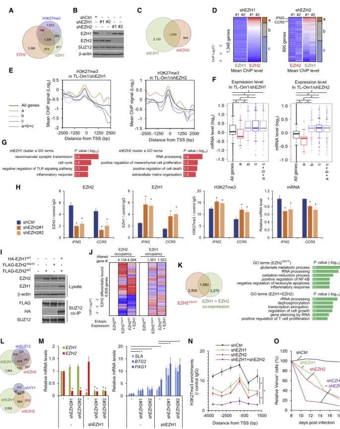
Figure 2. Reciprocal Function of EZH1 and EZH2
(A) Gene sets associated with H3K27me3, EZH1, and EZH2 in TL-Om1 cells.
(B) EZH1 and EZH2 levels in KD cells.
(C) H3K27me3-downregulated genes by EZH1 and EZH2 KD (H3K27me3 ChIP FC < 2 versus shCtrl).
(D) EZH1/2 chromatin distributions. The distribution patterns of EZH1 and EZH2 divided in three clusters.
(legend continued on next page)
characteristics. The experimental conditions of chromatin immu- noprecipitation (ChIP) were confirmed at the representative H3K27me3 target (Yamagishi et al., 2012) (Figures S2F and S2G). Using this method, we analyzed occupancy of EZH1, EZH2, H3K27me3, and H3K4me3 at all gene transcription start sites (TSSs) in freshly isolated malignant cells of patients with acute ATL (n = 3) and normal CD4
+T cells (n = 3). The mean ChIP signals of all genes showed deposition of H3K27me3, EZH1, and EZH2 in ATL cells compared with normal T cells (Fig- ure 1E). Clustering analyses identified EZH1 and EZH2 target genes (Figure 1F). The ChIP data summary is shown in Figure 1G.
More than 80% of TSS H3K27me3 upregulation was directly associated with the cis binding of EZH1 (29.1%), EZH2 (14.2%), or both (39.9%). ChIP peak analysis revealed H3K27me3 accumulation of the EZH1/2 target genes (Figures 1H, S2H, and S2I). Integration of transcriptome data (acute ATL, n = 26; normal CD4
+, n = 21) showed that abnormal binding of EZH1 and EZH2 caused transcriptional silencing (Figure 1I).
We also analyzed the methylation status of DNA CpG sites (>850,000 sites) in the same clinical cases and examined the relationship between promoter mCpG and H3K27me3. We de- tected many differentially methylated sites in ATL cells (7,408 hy- permethylated sites [ b value fold change (FC) > 0.2, p < 0.05, 4.54%], 6,925 hypomethylated sites [ b value (FC) < 0.2, p <
0.05, 4.24%]) (Figure 1J). CpG methylation status of the H3K27me3-accumulated genes was relatively low in normal cells but significantly methylated in ATL cells (1,138 hypermethy- lated sites [13.1%] versus 226 hypomethylated sites [2.60%]), suggesting that chromatin condensation is cooperated by mCpG and EZH1/2-dependent H3K27me3 (Figure 1J).
Gene Ontology (GO) analysis revealed that EZH1- and EZH2- bound silenced genes involved various cellular processes (Fig- ure 1K), including co-occupancy of EZH1 and EZH2 to silence multiple genes associated with transcriptional regulation, prolif- eration process, immune response, and cell migration. EZH1 would further directly induce H3K27me3 at genes involved with the regulation of lymphocyte activation and differentiation.
EZH2 was specifically involved in various processes, including cellular metabolism, kinase regulation, and cytokine-mediated signaling. Principal-component analysis (PCA) revealed that all clinical subtypes showed epigenetic suppression of EZH1/2 tar- gets (Figures 1L and S2J). Taken together, both EZH1 and EZH2 are important for the epigenetic landscape.
Reciprocal Function of EZH1 and EZH2
Despite the absence of quantitative change, genome-wide rear- rangement of EZH1 was preferentially observed at promoters
where the EZH2 binding pattern also changed (Figure 1G). To elucidate the mechanism of action of PRC2, we used ATL- derived TL-Om1 cells, which are dependent on EZH1/2 (Figures 1A and 2A). Specific shRNAs were used to establish EZH1- and EZH2-KD cells (Figure 2B). ChIP peak comparison detected H3K27me3 reduction in both KD cells (Figure 2C). The different effects on H3K27me3 suggested specific functions of the responsible enzymes. ChIP peak analysis of EZH1/2 chromatin distributions detected KD-mediated removal from TSS regions (Figure 2D). The distribution pattern of another enzyme corre- sponded to the depleted enzymes, which resulted in three po- tential clusters: reciprocal upregulation (cluster a), no change (cluster b), and cooperative downregulation (cluster c). To examine the functional significance of EZH1/2 redeployment, H3K27me3 levels were compared. KD of EZH1 and EZH2 gener- ally reduced H3K27me3 (Figure 2E, a+b+c) and at cooperatively downregulated regions (clusters b and c). In contrast, reciprocal upregulation of another enzyme caused higher H3K27me3 (clus- ter a). The histone modification changes modulated transcription pattern. EZH KD and H3K27me3 reduction generally induced active transcription. However, the reciprocal upregulation of H3K27me3 caused transcription silencing (Figure 2F). The compensated genes are functional in several processes, such as cell death, cell cycle, and inflammatory response (Figure 2G).
The epigenetic compensation was observed at representative loci in another cell line (Figure 2H). Thus, chromatin occupancy by EZH1 and EZH2 is critical for H3K27me3 and transcription patterns.
To test whether the EZH1/2 binding patterns are mutually interfered with each other, we ectopically expressed FLAG- tagged EZH2
WT, EZH2
Y641Fmutant (Morin et al., 2010; Be´guelin et al., 2013), or EZH2
WTwith HA-tagged EZH1 in MCF7 breast cancer cells. Immunoblots of cell lysates and SUZ12-bound complex showed elevated expression of EZH1 and EZH2, which were effectively incorporated in PRC2 (Figure 2I). ChIP peak analysis showed that EZH1 and EZH2 were redistributed when both were co-expressed or EZH2 was mutated (Figure 2J). Co- expression of EZH1 and EZH2 generated new EZH1/2 occupied loci (3,868 genes), which were partially overlapped with the tar- gets induced by EZH2 mutation (Figure 2K). Thus, the co-exis- tence of EZH1-PRC2 and EZH2-PRC2 triggers an abnormal epigenetic pattern and affects several functional processes.
Transcriptome analysis identified gene sets highly sensitive to KD and indicated unique functions of EZH1/2 in gene regulation (Figure 2L), which was consistent with the results of the cell growth assay and the chromatin analysis. KD of the PRC2 com- ponents SUZ12 and YY1 (Simon and Kingston, 2009) are
(E and F) Mean H3K27me3 ChIP levels (E) and expression levels (F) of each cluster or all genes in TL-Om1 with shEZH1 (left) or shEZH2 (right). *p < 0.01.
(G) GO analysis of cluster a.
(H) Epigenetic pattern and mRNA level at cluster a genes in ATN-1 cells with shEZH2. n = 3, mean
±SD, *p < 0.05.
(I and J) EZH2
WT, EZH2
Y641F, and EZH1+EZH2
WTwere ectopically expressed in MCF7. Functional PRC2 was confirmed by SUZ12 co-IP (I). ChIP patterns of the EZH2-differentially bound genes (EZH2 ChIP abs(FC) > 4) are shown (J).
(K) EZH1 and/or EZH2 gain gene sets (left) and results of GO analysis (right).
(L) Upregulated gene sets (expression FC > 2 versus shCtrl).
(M–O) TL-Om1 cells were transduced with retro- and lenti-viruses expressing shEZH1 and shEZH2/Venus. Expression levels ofEZH1,
EZH2, and EZH1/2 targets(M; n = 3, mean
±SD, *p < 0.05), H3K27me3 level at
SLATSS (N; n = 3, mean
±SD, *p < 0.05), and results as Venus-competitive assay (O; representative of n = 2) are shown.
See also Figure S3.
A
D
G
H I J K
E F
B C
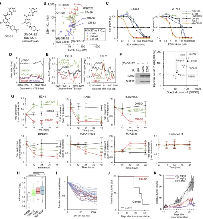
Figure 3. Chemical Dual Targeting of EZH1 and EZH2 in ATL (A) Structures of EZH1/2 dual inhibitors.
(B) IC
50values of PRC2-EZH2 (x axis) and PRC2-EZH1 (y axis) of EZH inhibitors and their derivatives are plotted. Cell-based H3K27me3 reduction activities are indicated by colors.
(C) ATL cell lines were cultured for 14 days with several dose EZH inhibitors. Dose-dependent effects on cell growth (%) are shown. n = 3, mean
±SD.
(D) Mean H3K27me3 of top 1,000 genes in TL-Om1 cells treated with DMSO, GSK126 (1,000 nM), OR-S1 (100 nM), or (R)-OR-S2 (100 nM) for 7 days. Averaged peak data from biological duplicates are shown.
(E) Mean EZH1 and EZH2 values at OR-S1 targets 3,082 genes (100 nM OR-S1 H3K27me3 FC < 2 versus 1,000 nM GSK126).
(F) Endogenous EZH2-IP with whole-cell lysates from TL-Om1 cells in the presence or absence of 100 nM (R)-OR-S2 (DS-3201b). Proteins bound to the bait were identified by immunoblotting (left) and LC-MS/MS (right). Representative of n = 2 biological replicates.
(legend continued on next page)
partially involved in gene silencing, implying that the direct inhi- bition of enzymes is required for effective epigenetic reprogram- ming. Considering the overlapped localization and function, EZH1 and EZH2 were simultaneously depleted. Compared with single KD, EZH1/2 double KD significantly reactivated the EZH1/2 co-bound targets (Figure 2M). ChIP-PCR showed that H3K27me3 was significantly reduced by double KD (Figure 2N).
The double-depleted ATL cells showed significantly reduced cell growth (Figure 2O). Collectively, EZH1 and EZH2 cooperatively form epigenetic and expression patterns, reflecting the growth of lymphoma cells.
Function of EZH1 under EZH2 Inhibition Condition Although EZH1 compensates for the genetic deletion of Ezh2 in embryonic stem cells (Shen et al., 2008), the role of EZH1 under the condition of EZH2 inhibition in somatic cells has not been ad- dressed. Standard dose treatment with the EZH2-selective in- hibitors GSK126 (McCabe et al., 2012) and E7438 (also called EPZ-6438 or tazemetostat [Knutson et al., 2014]) diminished H3K27me3 to a great extent, but residual methylation was de- tected (Figure S3A), which is consistent with the results of previ- ous studies (McCabe et al., 2012; Fujikawa et al., 2016). The GSK126-treated cells showed robust H3K27me3 downregula- tion at 964 TSSs but also non-negligible H3K27me3 upregulation at 534 TSSs (Figure S3B). The focal H3K27me3 upregulation was concomitant with increased EZH1 occupancy and decreased gene expression (Figure S3C). Enzymatic inhibition caused removal of EZH2; however, EZH1 redeployment rapidly occurred at the EZH2-depleted or EZH1/2-free loci, where removal of H3K27me3 and gene reactivation were not achieved (Figures S3C and S3D). Time course ChIP-PCR validated the compensa- tion of EZH1 (Figure S3E). EZH1 depletion effectively removed the residual H3K27me3 and significantly enhanced the efficacy of EZH2 inhibition, reflecting gene reactivation and cell growth inhibition (Figures S3F–S3I). Of note, DNA sequence suggested that YY1 may be partly involved in the EZH1/2 recruitment and their compensatory function (Figure S3D). YY1 KD attenuated the compensation of EZH1 in response to EZH2 inhibition, sug- gesting that compensatory action is partially mediated by YY1 (Figures S3J and S3K). Collectively, effective removal of H3K27me3 from EZH2
WTcells can be accomplished by dual tar- geting of EZH1/2.
Chemical Dual Targeting of EZH1/2 in ATL
We previously conducted chemical screening and derivatization of EZH inhibitors with the use of the half maximal inhibitory con- centration (IC
50) of EZH1/2 and cellular H3K27me3 (Honma et al.,
2017). In particular, OR-S1 and (R)-OR-S2 (also called DS-3201 or valemetostat) strongly and specifically inhibited EZH1 and EZH2, respectively (IC
50% 10 nM) (Figure 3A). This selectivity was also profiled with the use of panels of 34 histone methyl- transferases and 253 human protein kinases (Honma et al., 2017). To investigate the biological relevance and directly compare existing EZH inhibitors, unified experimental conditions for chemical synthesis and evaluation are required. For this pur- pose, we synthesized the EZH2-selective inhibitors E7438, GSK126, OR-S0 (Honma et al., 2017) and the EZH1/2 dual inhibitors OR-S1, (R)-OR-S2/DS-3201, and UNC1999 (Xu et al., 2015a) in house according to the chemical structures for this study (official IC
50values are shown in Figure S4A). The IC
50values against PRC2-EZH2 and PRC2-EZH1 were deter- mined in vitro. In addition, cell-based H3K27me3 reduction activities were also examined. We found that the EZH1/2 dual inhibitors OR-S1 and OR-S2 had stronger activities as S-adenosyl-L-methionine (SAM)-competitive inhibitors (Fig- ure 3B). The IC
50values of H3K27me3 were highly correlated with that of EZH1 (y axis), demonstrating that EZH1 inhibition is critical for H3K27me3 reduction under conditions of EZH2 in- hibition. Therefore, the anti-growth activities of several com- pounds (annotated in Figure 3B) were tested in an in vitro dose escalation study, which showed that the EZH1/2 dual inhibitors significantly reduced ATL cell viability compared with the others (Figure 3C; growth inhibition activity fold change 136.8, range 29.5–273.0). Differential effects between OR-S1/2 and UNC1999 were correlated with the measured IC
50value against EZH1.
We performed ChIP peak analysis for H3K27me3, EZH1, and EZH2 and showed that the dual inhibitors effectively removed H3K27me3 and also prevented unexpected gain of H3K27me3 compared with a 10-fold dose of the EZH2 inhibitor (Figures 3D and S4B). OR-S1 effectively evicted both EZH1 and EZH2 at the downstream and upstream of the H3K27me3 sites (Fig- ure 3E). In contrast, GSK126 treatment removed EZH2 only but led to a significant gain of EZH1 occupancy (p < 10
88). Liquid chromatography-tandem mass spectrometry (LC-MS/MS) anal- ysis of isolated complex by anti-EZH2 antibody showed no significant difference of PRC2 core components under treat- ment with dual inhibitors (Figure 3F). Thus, OR-S1/2 act as SAM-competitive inhibitors without complex disruption. Time course ChIP-PCR validated the removal of EZH1, EZH2, and H3K27me3 and also revealed reprogramming of the PRC1 component RING1B and its enzymatic product H2AK119ub and active chromatin mark H3K27ac by treatment with low- dose OR-S1 (Figure 3G). To obtain a global view of the impact
(G) Time course ChIP assay at
SLAloci. TL-Om1 cells were treated with DMSO, 1,000 nM GSK126, or 100 nM OR-S1. ChIP-qPCR was conducted at 0, 12, 24, and 48 h after drug treatment. Data are shown as fold enrichment of means
±SD for six regions near the TSS; *p < 0.05.
(H) Expression levels of EZH1/2 target genes (H3K27me3 ChIP FC < 2 by OR-S1/2) in TL-Om1 treated with DMSO, GSK126 (1,000 nM), OR-S1 (100 nM), or (R)-OR-S2 (100 nM). Means of biological replicates n = 2. *p < 10
14and **p < 10
71.
(I) Primary ATL cells (acute ATL, red lines, n = 12; chronic ATL, blue lines, n = 14) were cultured for 7 days in the presence of several dose of EZH inhibitors.
Residual cell numbers were analyzed using WST-8 assay. Details are provided in Figure S4I.
(J) NOD/SCID mice inoculated with TL-Om1 were orally administered with OR-S1 (200 mg/kg, daily, n = 6) or 0.5% methylcellulose (n = 6). Kaplan-Meier curve shows tumor-free survival (p value: nonparametric log rank test).
(K) After tumor engraftment (at day 14), OR-S1 were orally administered daily. Relative tumor volumes are shown (n = 6 or 7, mean
±SEM, *p < 0.05).
See also Figures S4 and S5.
of EZH1/2 dual inhibition, we analyzed ChIP signals at the H3K27me3 downregulated 4,610 genes. The occupancy of EZH2 and the compensatory redistribution of EZH1 were effec- tively inhibited, and these initial changes were further accompa- nied by the removal of H2AK119ub, feedback increase of H3K27ac and H3K4me3, and recruitment of RNA polymerase II (Figures 3E and S4C). Expression analysis revealed global reac- tivation of H3K27me3-accumulated genes. The dual inhibitors significantly reduced H3K27me3 and effectively reactivated the expression of EZH1/2 targets compared with 10-fold dose of GSK126 (Figures 3H and S4D–S4F). These results demonstrated that EZH1/2 inhibition effectively reprogrammed the epigenomic abnormalities.
We further sought to ascertain the effects of dual inhibitors in two more biological models. Dual inhibitor treatment significantly reactivated the expression of the highly H3K27me3-accumu- lated EZH1/2 target genes in an ex vivo culture of primary ATL cells (Figures S4G and S4H). The treatment reduced the viability of all tested ATL cells more effectively than the EZH2 inhibitor (Figures 3I and S4I). Furthermore, oral administration of OR-S1 completely prevented ATL cell engraftment and in vivo growth in immunodeficient mice (Figures 3J, 3K, and S4J–S4L). We confirmed significant inhibition of tumor growth and liver metas- tasis, without significant body weight loss. These data indicate that EZH1/2 dual inhibitors have strong potential for anti-ATL activity.
Integrative transcriptome and epigenome data identified epigenetic silencing of functionally important genes (Figure S5A).
EZH1/2 inhibitors effectively diminished H3K27me3 and induced gene reactivation (Figures S5B and S5C). The gene set includes SLA and PAG1 as CD3 z (CD247)-T cell receptor (TCR) signal negative regulators (Smida et al., 2007; E´rsek et al., 2012), BTG1 and BTG2 tumor suppressors (Winkler, 2010), TBX21, CCR5 for lineage specification, and EMP1 and HEG1 for cell- cell junction (Kleaveland et al., 2009). The dual inhibitors signifi- cantly induced SLA and PAG1, disrupting cell surface CD3 z , which is critical for TCR signaling pathway (Brownlie and Zamoy- ska, 2013) and may be linked to strong apoptosis (Figures S5C–S5E).
Hyperactive EZH1/2 have also been implicated in the silencing of NF- k B negative regulators (Yamagishi et al., 2012) and are suggested to associate directly with NF- k B components (Lee et al., 2011). Dual inhibitors significantly upregulated H3K27me3-accumulated microRNAs, removed RelA, RelB, and acetylated histones from NF- k B target loci, and prevented target gene expression (Figures S5F–S5K). Cellular NF- k B deactivation was achieved by low-dose EZH1/2 inhibitors (Figure S5L).
Furthermore, continuous inhibition (1 week) of EZH1/2 re- sulted in significant alterations of RNA polymerase II occu- pancy and transcriptome without H3K27me3 changes (Fig- ure S5M), which may have resulted from the effects of reprogramming of multiple transcription factors (Figure 1K).
Thus, malignant cells have a complex transcriptome network that is initially established by EZH1/2. Collectively, inhibitor- dependent H3K27me3 downregulation directly and indirectly reprograms the transcriptome, explaining the effectiveness of the EZH1/2 dual inhibitors.
Dual Targeting of EZH1/2 in Malignant Lymphomas We next probed the cell sensitivity to the dual inhibitors using a panel of malignant lymphoma cell lines that originated from EZH1
+differentiated lymphocytes, including peripheral T cell lymphoma (PTCL; including ATL), diffuse large B cell lymphoma (DLBCL; EZH2
WT/WTand EZH2
WT/Mu), Burkitt lymphoma (BL), and primary effusion lymphoma (PEL). All of the tested models showed considerable miR-31 silencing, suggesting abnormal H3K27me3 deposition (Figure S5N). In addition, MLL-rearranged leukemia was also tested because previous studies showed high sensitivity to EZH1/2 inhibitors (Neff et al., 2012; Xu et al., 2015a;
Honma et al., 2017). To directly compare the growth IC
50values, we performed in vitro dose escalation study and determined IC
50values of GSK126, OR-S1, and (R)-OR-S2 (DS-3201) by the uni- fied experimental condition. We found superior anti-growth effects of the dual inhibitors compared with EZH2-selective in- hibitor in all models (Figure 4A; growth inhibition activity fold change 29.3, range 4.99–417.9). All calculated IC
50values were less than 1,000 nM, suggesting that malignant lymphomas and MLL-rearranged leukemia are generally sensitive to the EZH1/2 inhibitors. (R)-OR-S2 potently inhibited H3K27me3 by low-dose treatment (0.1–100 nM) in the sensitive lymphoma types, demonstrating on-target effects (Figure 4B).
Rank ordering particularly indicated that ATL and other T cell- derived lymphomas are highly sensitive to EZH1/2 inhibitors.
PTCL is a category of malignancies with poor prognosis that are derived from mature T cells, for which no durable molecular targeting therapy has been established (Vose et al., 2008). We re- analyzed previous transcriptome data from clinical PTCL cases (n = 141; Iqbal et al., 2010) and found stable expression of EZH1 and overexpression of EZH2 in all types of PTCL, including PTCL-NOS, AITL, and ALCL (Figure 4C). Overexpression of EZH2 is mediated by several transcription factors, including NF- k B (Fujikawa et al., 2016). We found that TCR/CD28 pathway is critical for EZH1/2 expression (Figure 4D). Short-term activa- tion was insufficient to achieve the stable EZH2 overexpression, suggesting the requirement of constitutive activation of the TCR pathway, which is frequently detected in T cell lymphomas (Ka- taoka et al., 2015; Vallois et al., 2016). Indeed, inhibition of NF- k B reduced EZH2 expression in all PTCL cell lines (Figure 4E). EZH1 was stably and highly expressed and was not affected by NF- k B inhibition.
Further inspection of clinical cases revealed that all PTCL sub- types showed decreased expression of the EZH1/2 target genes, involving cell growth and other functional processes (Figure 1), and were differentially clustered from normal lympho- cytes, suggesting that T cell-derived lymphoma has EZH1/2- dependent epigenetic changes (Figures 4F, 4G, and S5O). The EZH1/2 dual inhibitors showed superior anti-growth effects compared with EZH2-selective inhibitors and UNC1999 in PTCL models (Figure 4H), as well as in MLL-rearranged leukemia models (Figure 4I). These results suggest that T cell lymphoma is a primary disease category for treatment with EZH1/2 inhibitors.
Cell line-based screening also suggested that germinal center (GC)-type and non-GC-type DLBCL are also potential disease entities. We collected clinical frozen samples and found that DLBCL cells highly expressed both EZH1 and EZH2 (Figure 5A).
EZH2 baseline level was low and significantly upregulated in
DLBCL, although there was no statistical significance among clinical subtypes (GC versus non-GC, Epstein-Barr virus [EBV] versus EBV+), suggesting that EZH1/2 expression is a common characteristic. Another DLBCL cohort (Tagawa et al., 2005) validated high expression levels (Figure S5P). As is the case with T cell lymphoma, DLBCL is frequently associated with hyper-activation of immune receptor signaling. We found that the proximal promoter sequence was significantly activated by B cell receptor (BCR) engagement (Figure 5B). In addition, mimetic models of BCR pathway activation, including mutations to CARD11 (Lenz et al., 2008) and MYD88 (Ngo et al., 2011), as well as inactivation of A20 (Kato et al., 2009; Compagno et al., 2009) and PTEN (Mayo et al., 2002; Perkins, 2012), showed
enhanced promoter activity, suggesting that EZH2 may be over- expressed in the majority of malignant B cells (Figure 5C).
Some DLBCL cases ( 21.7% of GCB-DLBCL) have heterozy- gous EZH2 mutation (Morin et al., 2010). To unravel the molecu- lar mechanism underlying the H3K27me3 accumulation with EZH1 and EZH2, we selected two cell models harboring EZH2
WT/WT(SUDHL8) or EZH2
Y641F/WT(WSU-DLCL2); the latter type is sensitive to EZH2 inhibition (Figure 4A). shRNA-mediated KD showed that EZH2 was essential for both models. In contrast, EZH1 KD was critical for EZH2
WT/WTcells but not for EZH2
Y641F/WTcells (Figure 5D). We next tested the sensitivity of DLBCL cells to the EZH2 inhibitor in the presence or absence of shEZH1. In addition to EZH2
WT/WTcells, EZH2
Y641F/WTcells A
C
H I
D E
B
F G
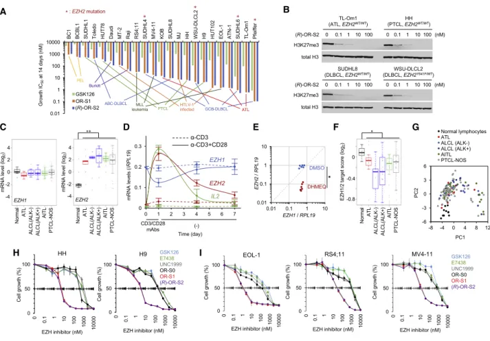
Figure 4. Targeting EZH1 and EZH2 in Malignant Lymphomas
(A) The effect of OR-S1, (R)-OR-S2, and GSK126 on 23 cell lines derived from hematological malignancies represented as the concentrations required to inhibit 50% of growth after 14 days. Means of n = 3 biological replicates; statistical significance between OR-S1/2 and GSK126 was observed in all models.
EZH2mutation is indicated by asterisk.
(B) Evaluation of H3K27me3 in four sensitive cell lines following treatment for 7 days.
(C)
EZH1and
EZH2levels in PTCL cohort (normal lymphocytes, n = 11; ATL, n = 13; ALK[ ]-anaplastic large cell lymphoma (ALCL), n = 10; ALK[+]-ALCL, n = 20;
angioimmunoblastic T cell lymphoma [AITL], n = 37; peripheral T cell lymphoma, not other specified [PTCL-NOS], n = 50). **p < 0.001.
(D) Expression changes by transient TCR signal in CD4
+T cells. Cells were washed at day 1 and maintained without monoclonal antibodies (mAbs) for 7 days.
n = 3, mean
±SD.
(E) Six T cell lymphoma cells were treated with DHMEQ (5
mg/mL) for 24 h. Quantified
EZH1and
EZH2were plotted. n = 3, *p < 0.05.
(F) Averaged levels of EZH1/2 target 307 genes (H3K27me3 ChIP FC > 2, expression FC < 2, EZH1 and/or EZH2 ChIP FC > 0 in ATL cells versus normal T cells) in PTCL cohort. *p < 0.02.
(G) PCA clustering on the basis of expression data of EZH1/2 target genes.
(H and I) PTCL (H) and MLL-fusion leukemia (I) cell lines were cultured for 14 days in the presence of several dose EZH inhibitors. n = 3, mean
±SD.
See also Figure S5.
A B C D
F E
G
J K
L M
H I
Figure 5. EZH1- and EZH2-Dependent H3K27me3 in DLBCL
(A)
EZH1and
EZH2levels in primary frozen DLBCL (GC, n = 31; non-GC, n = 35; EBV[ ], n = 45; EBV[+], n = 23) and CD19
+normal B cells (n = 7). *p < 0.01 and
**p < 10
7.
(B)
EZH2promoter activity in BJAB cells treated with anti-IgM. n = 4, mean
±SD, *p < 0.05.
(C)
EZH2promoter activity in BCR pathway-activating cells. n = 4, mean
±SD, *p < 0.05.
(D) SUDHL8 (non-GC type,
EZH2WT/WT, BCL6 /CD10 ) and WSU-DLCL2 (GC type,
EZH2WT/Y641F, BCL6
+/CD10
+) were treated with shEZH1 or shEZH2, and results as Venus-competitive assay (representative of n = 2) are shown. Functional PRC2 was detected by SUZ12 co-IP.
(E) SUDHL8 and WSU-DLCL2 with shCtrl or shEZH1 (#1, #2) were cultured for 14 days in the presence of several dose GSK126. n = 3, mean
±SD.
(F) DLBCL cell lines were cultured for 14 days in the presence of several dose EZH inhibitors. n = 3, mean
±SD.
(legend continued on next page)
showed high sensitivity upon EZH1 depletion, suggesting a compensatory action of EZH1 upon EZH2 inhibition regardless of the EZH2 mutation status (Figure 5E). Consistent with above mechanism, EZH1/2-targeting H3K27me3 inhibition and syn- thetic lethality were confirmed in both cell types (Figures 4B and 5F). The superior effect of EZH1/2 dual inhibition was further confirmed in all tested B cell malignancies (DLBCL, BL, and PEL) with both EZH2
WT/WT(six lines) and EZH2
Mu/WT(four lines) (Figure 4A).
We further explored whole expression data and identified genotype-specific dual-inhibitor-responsive gene sets (Figures 5G and 5H). EZH2
Y641F/WTcells showed high sensitivity at gene reactivation level. ChIP peak analysis after inhibitor treat- ment revealed that the gene set was under the control of H3K27me3, and OR-S1 could reduce methylation to baseline levels (Figure 5I). Treatment with OR-S1, but not GSK126, diminished H3K27me3 at the EZH1 targets (Figure 5J). Further- more, GSK126 also induced substantial methylation compen- sation with bound EZH1, but OR-S1 reduced H3K27me3 significantly and reactivated the expression (Figure 5K), sug- gesting that EZH1 inhibition is critical for targeting DLBCL epi- genome. GO analysis suggested that EZH1/2 was involved in the regulation of the several functional processes, such as cell growth and activation and the immune response, explain- ing the effective growth inhibition in both types (Figure 5L).
Finally, the transcriptome data of primary cases showed that the expression levels of the EZH1/2 targets were significantly low in both the non-GC and GC types of DLBCL (Figures 5M and S5Q). Collectively, DLBCL may also be responsive to EZH1/2 inhibitors.
Synthetic Lethality Targeting Various Cancer Epigenome
We next examined the relationship between EZH1/2-mediated histone methylation and chromatin remodeling in heterochro- matin formation. mSWI/SNF (BAF complex) is one of the most frequently mutated chromatin remodeling genes in a wide variety of cancers (Hodges et al., 2016), in which EZH2 activity has been implicated (Wilson et al., 2010; Bitler et al., 2015; Kim et al., 2015; Yamagishi and Uchimaru, 2017). We found antago- nistic functions of both EZH1 and EZH2 to BAF. Dual depletion or dual inhibition of EZH1/2 induced rapid recruitment of the BAF components ARID1A, BRG1 (SMARCA4), and SNF5 (SMARCB1), accessible chromatin formation, and stable tran- scription (Figures 6A–6D). Prolonged treatment (7 days) further enhanced chromatin accessibility and gene expression, sug- gesting that effective epigenetic reprogramming requires long- time exposure.
Genome-wide promoter analysis demonstrated global BAF pattern changes and the opposite reactions of BAF and EZH1/2 (Figures 6E and 6F). Chromatin unwinding effectively occurred upon EZH1/2 dual inhibition, which induced changes of expression (Figures 6G and 6H). The opposite action was observed in the heterochromatin formation process; namely, hy- per-activation of EZH1/2 was associated with BAF eviction (Fig- ure 6I). Conversely, BAF depletion globally recruited both EZH1 and EZH2 and silenced transcription, which could be canceled by EZH1/2 inhibition (Figures 6J–6N). These data support the notion that, in addition to EZH2, EZH1 plays a critical role in BAF-mediated chromatin remodeling.
Gene mutations of other epigenetic factors are strongly asso- ciated with cancers such as KDM6A (H3K27 demethylase) and BAP1 (histone H2A deubiquitinase), which are implicated in H3K27me3-dependent cancers (Ler et al., 2017; LaFave et al., 2015). We analyzed all the promoter regions in KDM6A- and BAP1-KD cells and found EZH1- and EZH2-dependent H3K27me3 accumulation, which could be canceled by OR-S1 treatment (Figures 7A–7D). Expression profiling identified com- mon target genes, including Th1-type chemokines, leading to innate immune evasion (Peng et al., 2015) (Figures 7E and 7F).
These data suggest that inactivation of the EZH1/2 antagonists have oncogenic roles with H3K27me3 accumulation in various cancers. Indeed, ARID1A-KD, KDM6A-KD, and BAP1-KD cells showed high sensitivity to OR-S1 (Figure 7G). The strong syn- thetic lethality was induced when both EZH1 and EZH2 were in- hibited. Furthermore, an in vitro dose escalation study showed higher antiproliferation activity of the dual inhibitors than the EZH2 inhibitor in cell models of SMARCB1 (SNF5)-mutated ma- lignant rhabdoid tumors (G-401), ARID1A-mutated ovarian clear cell carcinoma (TOV-21G), and BAP1-mutated mesothelioma (NCI-H226) (Figure 7H).
The epigenetic balance between H3K27me3 and H3K4me3 is a main determinant of transcription (Schuettengruber et al., 2007; Piunti and Shilatifard, 2016). In particular, the MLL2-con- taining trithorax group regulates H3K4 methylation, and MLL2 (KMT2D) is frequently inactivated in B cell lymphomas (Pasqua- lucci et al., 2011; Morin et al., 2011). Analysis of primary samples revealed that MLL2 transcription was silenced in DLBCL (Fig- ure 7I). Depletion of the MLL2 genes promoted EZH1/2-depen- dent cell growth in pre-tumorigenic EBV+ B cell lymphoblastoid (LCL) cells (Figures 7J and 7K). MLL2 loss led to H3K4me3 loss at 2,322 gene promoters, whereas concomitant H3K27me3 gain was observed in 1,402 genes (60.4%) with cis bindings of EZH1 and/or EZH2 (Figures 7L and 7M).
These findings suggest that the facultative heterochromatin formation involves both EZH1/EZH2 and chromatin remodeling
(G–I) SUDHL8 and WSU-DLCL2 cells were treated with GSK126 (1,000 nM), OR-S1 (100 nM), or (R)-OR-S2 (100 nM) for 7 days. Gene expression profiling identified dual-inhibitor-responsive gene sets (expression FC > 2 by OR-S1/2, *p < 10
15). Gene numbers (G) and expression levels (H) are shown. Mean H3K27me3 ChIP values of all genes (black), the inhibitor-responsive gene set with DMSO (blue) or OR-S1 (red) near the TSS are shown (I).
(J) Mean H3K27me3 ChIP values of EZH1 target 894 genes (EZH1 ChIP FC < 1.5 by 100 nM OR-S1) in WSU-DLCL2 cells treated with EZH inhibitors.
(K) Mean H3K27me3 ChIP values (left) and gene expression (right) of EZH1 compensated 495 genes (EZH1 ChIP FC > 2 by 1,000 nM GSK126).
(L) Gene Ontology ( log
10[p value]) of the dual-inhibitor-responsive gene sets.
(M) Expression levels of all genes versus EZH1/2 target genes (expression FC > 2 by EZH1/2 inhibitors in WSU-DLCL2 and SUDHL8) in primary GC- and non-GC- DLBCL cells. *p < 0.01.
See also Figure S5.
A
B
E F G
D C
H
J K
L M
N I
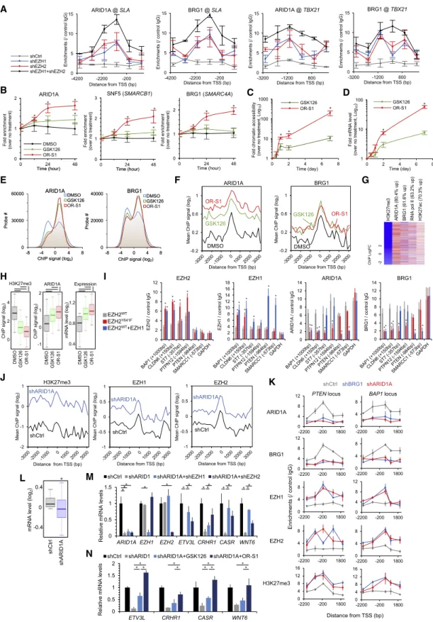
Figure 6. Importance of EZH1 and EZH2 in BAF-Mutated Cancers
(A) Chromatin occupancy of ARID1A and BRG1 in TL-Om1 with shEZH1 and/or shEZH2. n = 3, mean
±SD, p < 0.05 between single-KD versus double-KD was observed.
(legend continued on next page)
factors in (1) aberrant EZH1 + EZH2 dual-expressing tumors, like the majority of lymphomas; (2) EZH2 GoF mutated DLBCL and follicular lymphoma (FL); and (3) other cancer types with muta- tions in epigenetic factor genes, which are suggested to occur in early clonal evolution (Flavahan et al., 2017) (Figure 7N). The inactivation of genes involved in chromatin relaxation (H3K27 de- methylation, H3K4 trimethylation, H2A deubiquitination, and chromatin unwinding) caused restricted H3K27me3 accumula- tion mediated by both EZH1 and EZH2, which may be an emerging vulnerable characteristic in certain cancer types. Large numbers of regulated genes are unique in each epigenetic pathway of cancers.
Targeting Epigenetic Changes of Premalignant Cells Besides genetic and expression changes of epigenetic factors, oncovirus infection may be one of the initial drivers of epigenetic changes. We reanalyzed the transcriptome data from asymp- tomatic individuals or indolent-type ATL patients and detected EZH1- and EZH2-dependent epigenetic silencing at a part of the target loci in the human T-lymphotropic virus 1 (HTLV-1)-in- fected cells (Figures S6A–S6D). EZH1/2 inhibition diminished abnormal methylation with EZH1/2 removal in a Tax-expressing HTLV-1-infected cell model (Figures S6E and S6F). In addition, H3K27me3 ChIP peak from Tax-dependent immortalized T cells (Fujikawa et al., 2016) showed that H3K27me3 was upre- gulated at both EZH1 and EZH2 targets (Figure S6G). Higher antiproliferation activity of the dual inhibitors compared with EZH2-selective inhibitors was detected in the HTLV-1-infected cell lines (Figure 4A). To further test whether EZH1/2 dual inhibi- tion can selectively deplete the HTLV-1-infected cell population, we conducted low-dose treatment of OR-S1 and OR-S2/DS- 3201 in primary ex vivo culture. EZH1/2 inhibitors selectively depleted the HTLV-1-infected cell population in indolent ATL pa- tients (11 of 13 [84.6%]) and asymptomatic carriers (31 of 35 [88.6%]) more effectively than a 5-fold dose of the EZH2-selec- tive inhibitor (Figures S6H–S6J). These data suggest that target- ing of EZH1 and EZH2 is an effective strategy to diminish the HTLV-1-infected cell population.
EBV is a B cell-tropic oncovirus, which influences the host epigenome (Allday, 2013; Jiang et al., 2017). We found that EBV-infected LCLs expressed both EZH1 and EZH2, and the cell proliferation was highly dependent on both EZH1 and EZH2 (Figures S7A and S7B). Transcriptome and epigenome an- alyses revealed EZH1- and EZH2-dependent global H3K27me3
redistribution in EBV-infected, untransformed cells (Figures S7C–S7E). EZH1/2 inhibition significantly affected the transcrip- tome, which contributed to the several functional processes (Fig- ures S7F–S7I). In contrast, low-dose did not affect cell death and the proliferation of normal lymphocytes in vitro (Figures S7J and S7K). These data collectively suggest that two lymphotropic on- coviruses, HTLV-1 and EBV, reprogrammed the global EZH1/2- H3K27me3 patterns and established the unique transcriptome in the non-malignant, virus-infected cell population.
DISCUSSION
Precise target molecule(s) should be identified for mechanism- based medicine. EZH2 has been recognized as the molecule responsible for excessively accumulated H3K27 methylation, which is one of the molecular hallmarks of cancers, because of GoF mutation and/or overexpression of EZH2.
In this study, we discovered the involvement of another enzyme, EZH1, in abnormal H3K27 methylation. We showed that the chromatin distribution of EZH1 is globally reprogrammed and tightly involved in the global histone methylation in aggres- sive lymphomas. Reciprocal interference and compensatory ac- tion of the EZH1 and EZH2 rearrange their own genome-wide distribution, establishing restricted chromatin and gene expres- sion signatures.
On the basis of these findings, we developed EZH1+EZH2 dual inhibitors (among them, valemetostat/DS-3201b has been used in clinical trials) and demonstrated significant H3K27me3 inhibition efficacy. LC-MS/MS showed that the SAM-competi- tive dual inhibitors do not alter PRC2 core complex composition, but chromatin occupancy of EZH1 and EZH2 are decreased, which may potentiate and sustain the drug effect. Considering that trimethylated H3K27 itself is an active interaction site of PRC2 and stimulates PRC2 activity (Margueron et al., 2009), one possible mechanism is that diminishment of trimethylation mark might lead to weak interaction with PRC2.
In exploring mechanisms of action of the inhibitors, we also showed that both EZH1 and EZH2 play critical roles in the chro- matin regulation together with multiple histone modifiers and chromatin regulatory factors. Strong synthetic lethality was observed in cancer cells harboring mutations to histone-modi- fying genes, as well as in pre-cancerous cells epigenetically perturbed by oncovirus infection. In addition to EZH1/2 dysregu- lation, inactivation of genes involved in chromatin relaxation and
(B–D) Time course epigenetic alteration at
SLAloci. TL-Om1 cells were treated with DMSO, 1,000 nM GSK126, or 100 nM OR-S1. ChIP assay were conducted for 48 h (B). Time course evaluation of chromatin accessibility (C; n = 3) and transcription (D; n = 3) were also conducted for 7 days. Mean
±SD, *p < 0.05.
(E and F) TL-Om1 cells were treated with EZH inhibitors for 7 days. Normalized all ChIP signals (E) and mean values of ARID1A and BRG1 at H3K27me3 decreased 3,086 loci (F) are shown.
(G) Epigenetic reprogramming by 100 nM OR-S1 treatment at H3K27me3 decreased loci.
(H) H3K27me3 (left) and ARID1A (middle) ChIP values and expression levels of EZH1/2 target genes (right). *p < 10
15.
(I) MCF7 cells were transfected with FLAG-EZH2
WT, FLAG-EZH2
Y641F, or FLAG-EZH2
WT+ HA-EZH1. Promoter binding pattern at EZH1/2 targets and
GDPDHloci was analyzed using ChIP-PCR assay. n = 3, mean
±SD, *p < 0.05 versus EZH2
WTexpression.
(J) MCF7/shARID1A were analyzed using ChIP-chip. EZH1 and EZH2 recruitments at H3K27me3 upregulated loci (H3K27me3 FC > 2 by ARID1A KD, 2,376 genes) were induced by ARID1A KD.
(K) Epigenetic patterns at
PTENand
BAP1loci. n = 3, mean
±SD.
(L) Expression levels of H3K27me3 upregulated genes. *p < 10
36.
(M and N) Expression levels of ARID1A-EZH1/2 targets. Gene silencing induced by ARID1A KD was canceled by shEZH1 and shEZH2 (M) or 100 nM OR-S1 (N).
n = 3, mean
±SD, *p < 0.05.
A
B
C
E D
F
G
I J
H
K
L M
N
(legend on next page)
virus infection appear to be vulnerable characteristics (Fig- ure 7N). Further clinical epigenomics in primary tumors by using high-sensitive assays may be helpful to establish mechanism- based medicine and develop accurate biomarkers.
Regarding the diversity of genetic lesions in lymphomas, the developmental pathway seems to be initiated by EZH1/2 and epigenetically imprinted in the non-malignant, virus-infected population throughout disease development. This finding could provide new preemptive therapeutic strategies for the elimina- tion of malignant cells and their founder population in human ma- lignancies. Repeated dosing did not cause any critical or severe toxicity in vitro and in vivo (Honma et al., 2017), suggesting that (pre)malignant cells are more highly addicted by EZH1/2-depen- dent epigenome than normal cells.
This study provides convincing rationale for dual targeting of EZH1+EZH2 and suggests the validity of drugs for treating ma- lignant lymphomas and premalignant clones. On the basis of these results, a phase 1 clinical trial against T- and B cell non- Hodgkin’s lymphomas including ATL is now under way, and clinical safety and promising activity against lymphomas have been suggested (Maruyama et al., 2018, Am. Soc. Hematol., conference).
STAR + METHODS
Detailed methods are provided in the online version of this paper and include the following:
d
KEY RESOURCES TABLE
d
LEAD CONTACT AND MATERIALS AVAILABILITY
d
EXPERIMENTAL MODEL AND SUBJECT DETAILS
BCell culture
B
Clinical samples
BChemicals
d
METHOD DETAILS
B
Cell-free assay to detect methyltransferase activities
BCell-based assay to detect H3K27me3 reduction
BLiquid chromatography-tandem mass spectrometry
(LC-MS/MS)
BFlow cytometry
B
Chromatin immunoprecipitation (ChIP) assay
BChromatin accessibility assay
B
RNA isolation and RT-PCR analysis
BVectors
B
NF- k B analysis
B
Immunoblotting and immunoprecipitation
BQuantification of HTLV-1 proviral load
BCell growth assay
B
In vivo xenograft studies
BChIP-chip experiments
BGene expression analysis
BDNA methylation analysis
BGEO gene expression data
d
QUANTIFICATION AND STATISTICAL ANALYSIS
d
DATA AND CODE AVAILABILITY
SUPPLEMENTAL INFORMATIONSupplemental Information can be found online at https://doi.org/10.1016/j.
celrep.2019.10.083.
ACKNOWLEDGMENTS
We thank staff members at all collaborating institutions and the central office of the JSPFAD. We thank Drs. Hiroyuki Miyoshi and Atsushi Miyawaki for providing the Venus-encoding lentivirus vectors. We thank Dr. Kazuo Ume- zawa for providing the NF-
kB inhibitor DHMEQ. This research was supported by AMED under grants JP18ak0101086 (M.Y., K.U., T.W.), JP17fk0108112 (K.U., T.W.), JP17im0210101 (K.U., K.A.), and JP17fk0410208 (K.U., S.O., H.K.) and JSPS KAKENHI grants JP15K06907 (M.Y.), JP16H05323 (M.Y), and JP18K08317 (M.Y).
AUTHOR CONTRIBUTIONS
M.Y. conceived and supervised the project; designed and performed almost all experiments, including the gene expression profile, ChIP experiments,
in vitrocell culture, and drug evaluation; performed data analysis; and wrote the paper. M.H. performed ChIP-chip for EZH1 and EZH2 in ATL clinical sam- ples. D.F. established the ChIP-chip experiments and supervised data anal- ysis. T.O. performed
in vivostudies. D.H. and N.A. produced EZH1/2 inhibitors and analyzed chemical properties. H.K. and T.H. prepared DLBCL clinical samples and provided advice. S.K. and K.N. developed the CADM1/CD7- based flow cytometry method, provided clinical samples, and obtained gene expression data. M.N. prepared ATL clinical samples. M.I. maintained the JSPFAD and analyzed epidemiological data. A.U. provided clinical samples and gave advice. Y.T. provided anti-Tax antibody. S.O. organized DLBCL study and provided advice. K. Tsukasaki and K. Tobinai managed ATL clinical study and provided advice. K.A. managed the EZH1/2 dual inhibitor project and provided advice. T.W. established the ATL cohort study and supervised
Figure 7. Importance of EZH1 and EZH2 in KDM6A , BAP1 , and MLL2 Mutated Cancer Types
(A–C) Proportion of EZH1/2-bindings at H3K27me3 gain genes triggered by depletion of
ARID1A(2,369 genes),
KDM6A(2,509 genes), and
BAP1(2,219 genes) (A). KD efficiencies and mean ChIP values at H3K27me3 upregulated loci are shown (B and C). n = 3, mean
±SD, *p < 0.05.
(D) MCF7/shCtrl, /shKDM6A, and /shBAP1 were treated with EZH inhibitors for 7 days, and H3K27me3 were quantified. n = 3, mean
±SD, *p < 0.05.
(E) Gene sets downregulated by depletion of
ARID1A,KDM6A, andBAP1(expression FC < 2).
(F)
CXCL9and
CXCL10levels. n = 3, mean
±SD, *p < 0.05.
(G) Cell growth (%) at day 14 treated with EZH inhibitors. n = 3, mean
±SD, *p < 0.05.
(H) Dose-dependent effects on cell growth (%) for 14 days. n = 3, mean
±SD.
(I)
MLL2mRNA level in DLBCL samples. *p < 0.05.
(J) Venus-competitive assay in LCLs transduced with shRNA lentivirus vectors. n = 3, mean
±SD, *p < 0.05.
(K) LCL/shMLL2 cells were treated with EZH inhibitors (10, 100, and 1,000 nM). Venus
+rate (day 7 to day 14) is shown. n = 3, mean
±SD, *p < 0.05 versus shMLL2 without inhibitors.
(L)
MLL2depletion led to H3K4me3 loss at 2,322 genes in LCL. Proportion of concomitant H3K27me3 gain (60.4%) with
cisbindings of EZH1/2 is shown.
(M) Mean ChIP values at H3K4me3 downregulated loci (H3K4me3 FC < 1.5 in MLL2 KD cells, 2,322 genes).
(N) A schematic view illustrating modes of H3K27me3 accumulation.
See also Figures S6 and S7.
the ATL study. K.U. conceived and supervised the project and wrote the paper.
All authors discussed the results and commented on the manuscript.
DECLARATION OF INTERESTS
D.H., N.A., and K.A. are employees of Daiichi Sankyo Co., Ltd. M.Y. and K.U.
received research funding from Daiichi Sankyo Co., Ltd. Daiichi Sankyo Co., Ltd., holds substance patents on the EZH1/2 inhibitors. The University of To- kyo and Daiichi Sankyo Co., Ltd., hold patents for application of the EZH1/2 inhibitors to ATL patients and HTLV-1-infected individuals. M.Y., T.W., D.H., and N.A. are named as inventors. All other authors declare no competing interests.
Received: April 1, 2019 Revised: September 8, 2019 Accepted: October 21, 2019 Published: November 19, 2019
REFERENCES
Allday, M.J. (2013). EBV finds a polycomb-mediated, epigenetic solution to the problem of oncogenic stress responses triggered by infection. Front. Genet.
4,212.
Be´guelin, W., Popovic, R., Teater, M., Jiang, Y., Bunting, K.L., Rosen, M., Shen, H., Yang, S.N., Wang, L., Ezponda, T., et al. (2013). EZH2 is required for germinal center formation and somatic EZH2 mutations promote lymphoid transformation. Cancer Cell
23, 677–692.Bitler, B.G., Aird, K.M., Garipov, A., Li, H., Amatangelo, M., Kossenkov, A.V., Schultz, D.C., Liu, Q., Shih, IeM., Conejo-Garcia, J.R., et al. (2015). Synthetic lethality by targeting EZH2 methyltransferase activity in ARID1A-mutated can- cers. Nat. Med.
21, 231–238.Bradley, W.D., Arora, S., Busby, J., Balasubramanian, S., Gehling, V.S., Nas- veschuk, C.G., Vaswani, R.G., Yuan, C.C., Hatton, C., Zhao, F., et al. (2014).
EZH2 inhibitor efficacy in non-Hodgkin’s lymphoma does not require suppres- sion of H3K27 monomethylation. Chem. Biol.
21, 1463–1475.Brownlie, R.J., and Zamoyska, R. (2013). T cell receptor signalling networks:
branched, diversified and bounded. Nat. Rev. Immunol.
13, 257–269.Cao, R., and Zhang, Y. (2004). SUZ12 is required for both the histone methyl- transferase activity and the silencing function of the EED-EZH2 complex. Mol.
Cell
15, 57–67.Comet, I., Riising, E.M., Leblanc, B., and Helin, K. (2016). Maintaining cell iden- tity: PRC2-mediated regulation of transcription and cancer. Nat. Rev. Cancer
16, 803–810.Compagno, M., Lim, W.K., Grunn, A., Nandula, S.V., Brahmachary, M., Shen, Q., Bertoni, F., Ponzoni, M., Scandurra, M., Califano, A., et al. (2009). Muta- tions of multiple genes cause deregulation of NF-kappaB in diffuse large B- cell lymphoma. Nature
459, 717–721.E´rsek, B., Molna´r, V., Balogh, A., Matko´, J., Cope, A.P., Buza´s, E.I., Falus, A., and Nagy, G. (2012). CD3
z-chain expression of human T lymphocytes is regu- lated by TNF via Src-like adaptor protein-dependent proteasomal degrada- tion. J. Immunol.
189, 1602–1610.Flavahan, W.A., Gaskell, E., and Bernstein, B.E. (2017). Epigenetic plasticity and the hallmarks of cancer. Science
357, eaal2380.Fujikawa, D., Nakagawa, S., Hori, M., Kurokawa, N., Soejima, A., Nakano, K., Yamochi, T., Nakashima, M., Kobayashi, S., Tanaka, Y., et al. (2016). Poly- comb-dependent epigenetic landscape in adult T-cell leukemia. Blood
127,1790–1802.
Hans, C.P., Weisenburger, D.D., Greiner, T.C., Gascoyne, R.D., Delabie, J., Ott, G., M€ uller-Hermelink, H.K., Campo, E., Braziel, R.M., Jaffe, E.S., et al.
(2004). Confirmation of the molecular classification of diffuse large B-cell lym- phoma by immunohistochemistry using a tissue microarray. Blood
103,275–282.
Hodges, C., Kirkland, J.G., and Crabtree, G.R. (2016). The many roles of BAF (mSWI/SNF) and PBAF complexes in cancer. Cold Spring Harb. Perspect.
Med.
6, a026930.Honma, D., Kanno, O., Watanabe, J., Kinoshita, J., Hirasawa, M., Nosaka, E., Shiroishi, M., Takizawa, T., Yasumatsu, I., Horiuchi, T., et al. (2017). Novel orally bioavailable EZH1/2 dual inhibitors with greater antitumor efficacy than an EZH2 selective inhibitor. Cancer Sci.
108, 2069–2078.Iqbal, J., Weisenburger, D.D., Greiner, T.C., Vose, J.M., McKeithan, T., Kucuk, C., Geng, H., Deffenbacher, K., Smith, L., Dybkaer, K., et al.; International Pe- ripheral T-Cell Lymphoma Project (2010). Molecular signatures to improve diagnosis in peripheral T-cell lymphoma and prognostication in angioimmuno- blastic T-cell lymphoma. Blood
115, 1026–1036.Italiano, A., Soria, J.C., Toulmonde, M., Michot, J.M., Lucchesi, C., Varga, A., Coindre, J.M., Blakemore, S.J., Clawson, A., Suttle, B., et al. (2018). Tazeme- tostat, an EZH2 inhibitor, in relapsed or refractory B-cell non-Hodgkin lym- phoma and advanced solid tumours: a first-in-human, open-label, phase 1 study. Lancet Oncol.
19, 649–659.Iwanaga, M., Watanabe, T., Utsunomiya, A., Okayama, A., Uchimaru, K., Koh, K.R., Ogata, M., Kikuchi, H., Sagara, Y., Uozumi, K., et al.; Joint Study on Predisposing Factors of ATL Development investigators (2010). Human T-cell leukemia virus type I (HTLV-1) proviral load and disease progression in asymptomatic HTLV-1 carriers: a nationwide prospective study in Japan.
Blood
116, 1211–1219.Jiang, S., Zhou, H., Liang, J., Gerdt, C., Wang, C., Ke, L., Schmidt, S.C.S., Nar- ita, Y., Ma, Y., Wang, S., et al. (2017). The Epstein-Barr virus regulome in lym- phoblastoid cells. Cell Host Microbe
22, 561–573.e4.Kataoka, K., Nagata, Y., Kitanaka, A., Shiraishi, Y., Shimamura, T., Yasunaga, J., Totoki, Y., Chiba, K., Sato-Otsubo, A., Nagae, G., et al. (2015). Integrated molecular analysis of adult T cell leukemia/lymphoma. Nat. Genet.
47, 1304–1315.
Kato, M., Sanada, M., Kato, I., Sato, Y., Takita, J., Takeuchi, K., Niwa, A., Chen, Y., Nakazaki, K., Nomoto, J., et al. (2009). Frequent inactivation of A20 in B-cell lymphomas. Nature
459, 712–716.Katsuya, H., Yamanaka, T., Ishitsuka, K., Utsunomiya, A., Sasaki, H., Hanada, S., Eto, T., Moriuchi, Y., Saburi, Y., Miyahara, M., et al. (2012). Prognostic index for acute- and lymphoma-type adult T-cell leukemia/lymphoma. J. Clin. Oncol.
30, 1635–1640.