Review Article
PATHOLOGICAL CHARACTERISTICS OF CHRONIC LYMPHOCYTIC LEUKEMIA/SMALL LYMPHOCYTIC LYMPHOMA
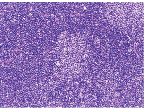
Chronic lymphocytic leukemia/small lymphocytic lym-phoma (CLL/SLL) usually exhibits heterogeneous features such as ill-defined bright nodules, termed proliferation cen-ters, with chromatin-rich background cells (Figure 1). The proliferation centers are composed of paraimmunoblasts, which are medium-sized cells, and the Ki67 index of these cells is higher than that of ordinary CLL/SLL cells. On the contrary, the majority of CLL/SLL cells are usually “small” lymphoma cells, but they are slightly larger than normal lym-phocytes (Figure 2). In some cases, paraimmunoblasts are prominent and such cases should be differentiated from fol-licular lymphomas. By lymphoma-cell morphology and immunostaining, this differential diagnosis is simple. CLL/ SLL cases may exhibit prominent paraimmunoblasts (Figure 3). Such cases should not be diagnosed as Richter syndrome because it features large blast cells; one feature of diffuse large B-cell lymphoma is the monotonous proliferation of large cells (the nucleus is more than twice the size of that of
normal lymphocytes with varying cytoplasm sizes) (Figure 4).
Bone marrow biopsy demonstrates many features. Some cases exhibit a nodular pattern of infiltration, or interstitial or mixed nodular and interstitial pattern, whereas others have
Differential diagnosis of chronic lymphocytic leukemia/
small lymphocytic lymphoma and other indolent
lymphomas, including mantle cell lymphoma
Tadashi Yoshino, Takehiro Tanaka, Yasuharu Sato
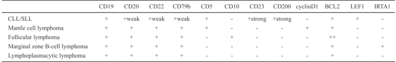
Chronic lymphocytic leukemia/small lymphocytic lymphoma (CLL/SLL) accounts for approximately 1% of all lymphomas in our department. In this article, we describe the differential diagnosis of CLL/SLL from other indolent lymphomas, with special reference to follicular lymphoma, marginal zone B-cell lymphoma, lymphoplasmacytic lymphoma, and mantle cell lymphoma, although the latter is considered to be aggressive. CLL/SLL often exhibits proliferation centers, similar to follicular lym-phoma. Immunohistological examination can easily distinguish these two lymphomas. The most important characteristic of CLL/SLL is CD5 and CD23 positivity. Mantle cell lymphoma is also CD5-positive and there are some CD23-positive cases. Such cases should be carefully distinguished from CLL/SLL. Some marginal zone lymphomas are also positive for CD5 and such cases are often disseminated. Lymphoplasmacytic lymphoma should also be a differential diagnosis for CLL/SLL. It frequently demonstrates MYD88 L265P, which is a key differential finding. By immunohistological examination, the expres-sion of lymphoid enhancer-binding factor 1 is specific for CLL/SLL and can be a good marker in the differential diagnosis.
Keywords: chronic lymphocytic leukemia/small lymphocytic lymphoma, differential diagnosis, indolent lymphoma
Received: November 22, 2019. Revised: December 25, 2019. Accepted: January 9, 2020. J-STAGE Advance Published: April 3, 2020 DOI:10.3960/jslrt.19041
Department of Pathology, Okayama University Graduate School, Okayama, Japan
Corresponding author: Tadashi Yoshino, Department of Pathology, Okayama University Graduate School of Medicine, Dentistry, and Pharmaceutical Sciences, 2-5-1,
Shikata-cho,Kita-ku, Okayama 700-8558, Japan. E-mail: [email protected] Copyright © 2020 The Japanese Society for Lymphoreticular Tissue Research
This work is licensed under a Creative Commons Attribution-NonCommercial-ShareAlike 4.0 International License.
Fig. 1. Typical features of CLL/SLL. There are two
proliferation centers in this field. If the proliferation centers are prominent, follicular lymphoma is to be differentiated.
diffuse involvement, which usually suggests more advanced disease.1
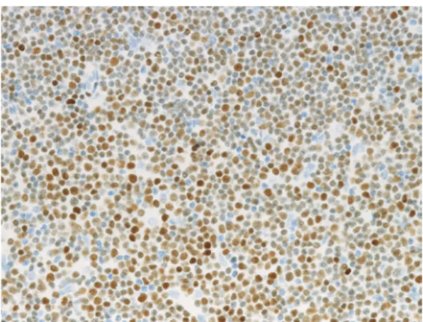
Immunohistologically, circulating leukemic B cells express CD19, and weak surface CD20, CD22, and CD79b. They are positive for CD5, and CD43, and strongly positive for CD23 and CD200. They are negative for CD10, FMC7 (usually), and cyclinD1, although cyclinD1 may be positive at proliferation centers.1 Lymphoid enhancer-binding factor
1 (LEF1) is specific to CLL/SLL and is a good marker in the differential diagnosis. Menter T et al. reported that 77/80 of CLL cases were positive for LEF1 (Figure 5), whereas only one of 38 follicular lymphoma and two of 33 marginal zone B-cell lymphoma cases were positive. The sensitivity of LEF1 for CLL is 0.96 and the specificity is 0.93.2 O’Malley
DP et al. reported that only 4-9% of MCL cases express LEF1.3 The differential diagnosis by immunostaining and
flow cytometry analysis is summarized in Table 1.
Although CLL/SLL has no specific genetic markers, most (80-90%) cases have cytogenetic abnormalities. The most common alternations are deletions in 13q14.3 and trisomy 12 or partial trisomy 12q13. Deletion in 11q22-23, 17q13, or 6q21 are less common. Deletion in 11q (ATM and BIRC3) and 17p (TP53) leads to a poorer clinical outcome, whereas isolated deletion in 13q14 is associated with a more favorable
clinical course.1
CLL, SLL, AND MONOCLONAL B-CELL LYMPHOCYTOSIS
In the 2017 WHO classification,1 monoclonal B-cell
lym-phocytosis (MBL) is defined as a monoclonal B-cell count less than 5X10⁹/L in peripheral blood of subjects who have
no associated lymphadenopathy, organomegaly, or other extramedullary involvement. MBL is classified into three categories: (1) CLL-type, (2) atypical CLL type, and (3) non-CLL type. The non-CLL type is the most common (75% of all cases), and it is characterized by B-cell markers (CD19, CD20 (weak)), CD23, and CD5, and the B-cells exhibit light chain restriction or lack surface immunoglobulin. The esti-mated incidence of CLL-type MBL is 3.5% to 12% in healthy individuals. Although all CLLs are preceded by MBL, low-count (lower than 0.5X10⁹L) MBL does not
prog-ress to CLL, whereas high-count (0.5X10⁹L or higher) MBL
has features identical to low-stage CLL and progresses to therapy-required CLL at a rate of 1-2%/year. MBL cells usually have mutated IGHV genes.
MBL has an atypical CLL phenotype positive for CD19, CD20 (bright), CD5, and surface immunoglobulin. CD23 Fig. 2. “Small-cell” component of CLL/SLL. In
com-parison with non-neoplastic lymphocytes (arrows), lymphoma cells are slightly larger than “true” non-neoplastic small lymphocytes.
Fig. 3. Paraimmunoblasts. These cells have a
promi-nent nucleoli, and are approximately 1.5-times larger than non-neoplastic lymphocytes.
Fig. 4. Richter’s syndrome. The proliferated cells
have a large nucleus that is more than twice the size of that of small lymphocytes.
Fig. 5. LEF1 expression in CLL/SLL. The lymphoma
may be negative, and such cases should be carefully excluded from those of mantle cell lymphoma and other B-cell lym-phomas. MBL with a non-CLL phenotype is characterized by negative or weak CD5 expression, and CD19 and CD20-positive B-cells. Some cases exhibit transient clonal expan-sion and are self-limited. Additional phenotypic and cytoge-netic studies are needed to exclude a specific lymphoid neoplasm.
SLL includes cases with a circulating CLL cell count of less than 5X10⁹/L and documented nodal, splenic, or other
extramedullary involvement. SLL should be differentiated from CLL-type MBL. Nodal infiltration by CLL-type cells without notable proliferation centers in individuals without lymphoadenopathy (more than 1.5 cm across) may constitute a nodal equivalent of MBL rather than SLL.1
DIFFERENTIAL DIAGNOSIS: MANTLE CELL LYMPHOMA
Mantle cell lymphoma (MCL) is also positive for CD5. Most mantle cell lymphomas are composed of intermediate lymphocytes and centrocytes exhibiting irregular nuclear contours, which is an important differential point (Figure 6). However, it may be composed of small-sized cells (Figure 7) that resemble CLL/SLL. Saksena A et al.4 reported that 103
(13%) MCL cases were weakly positive for CD23. CD23 expression in CLL/SLL is usually high. CD23-positive
MCL lymphoma has an increased leukocyte count, bone mar-row involvement, and leukemic presentation. Of note, CD23-positive MCL is more often associated with CD200 positivity and weak SOX11 expression. Although patients with CD23-positive MCL have a leukemic presentation simi-lar to CLL, their prognosis is better than that of CD23-negative patients. The expression of CD23 is closely associ-ated with CD200 expression. Ye H et al.5 reported similar
findings and referred to such cases as smoldering mantle cell lymphoma. CD200 is an important marker of CLL/SLL in the differential diagnosis from other CD5-positive indolent lymphomas. However, Hu Z et al.6 reported that
approxi-mately 4% of MCL (25 cases in their series) is positive for CD200, and most of these cases (76%) are positive for CD23. Moreover, only 24% of CD200-positive MCL cases express SOX11, 39% (9 cases) exhibit round nuclear contours, simi-lar to CLL, and 44% of (11 cases) cases are a non-nodal leu-kemic variant of MCL, which is closely related to IGHV-mutated cases. CLL/SLL can be subdivided into two groups: IGHV-unmutated and IGHV-mutated, and the patient prognosis of the former group is poorer than that of the latter group. Of MCL cases, only a small subset is IGHV-mutated, which is frequently associated with a non-nodal leukemic presentation. In conclusion, CD200-positive MCL is highly similar to CLL/SLL, and IgH-cylinD1 rearrangement is needed for the differential diagnosis.
CD200 belongs to a type I immunoglobulin gene
Fig. 7. Mantle cell lymphoma. This case is composed
of lymphoma cells, which are almost the same size as small lymphocytes, and was difficult to distinguish from small lymphocytic lymphoma without immuno-histological examination.
CD19 CD20 CD22 CD79b CD5 CD10 CD23 CD200 cyclinD1 BCL2 LEF1 IRTA1
CLL/SLL + +weak +weak +weak + - +strong +strong - + +
-Mantle cell lymphoma + + + + + - - - + + -
-Follicular lymphoma + + + + - + - - - ++ -
-Marginal zone B-cell lymphoma + + + + - - - + - +
Lymphoplasmacytic lymphoma + + + + - - - + -
-Table 1. Immunophenotype of indolent lymphomas, including mantle cell lymphoma*
*: The presented immunophenotype of each lymphoma is the most common. There are rare exceptional findings, which are described in the text.
Fig. 6. Mantle cell lymphoma with typical features.
The lymphoma cells are intermediate between small lymphocytes and medium-sized centrocytes with irreg-ular nuclei contours.
superfamily composed of a light-chain-like structure with extracellular variable and constant-like domains, and a cyto-plasmic tail. CD23 is also an important marker of CLL/ SLL. It is a low-affinity IgE receptor, and contains a C-terminal lectin-like domain, which resembles C-type car-bohydrate-recognition domains.7 CD23 has two isotypes,
CD23a and CD23b.8 As described above, some MCL cases
express low-level CD23, which is closely related to the expression of CD200. To our knowledge, the relationship between CD200 and CD23 has not been clarified.
OTHER CD5-POSITIVE LYMPHOMAS
CD5 is another important marker of CLL/SLL. CD5 was first found in mice, and CD5-positive B-cells (B-1 cells) are distinct from CD5-negative B-cells (B-2 cells).9 B-1
cells are responsible for natural antibody production and rapid immune responses. B-1 cells in humans are abundant in cord blood and the fetal spleen. CLL is thought to origi-nate from B-1 cells. Of note, CLL is characterized not only by CD5 expression, but also by an abnormal BCR repertoire encoding autoreactive and/or poly-reactive antibodies. Indeed, an increase in CD5-positive cells is observed in patients with autoimmune diseases. B-1 cells may prolifer-ate greatly with age and eventually develop into CLL.9
As described above, CD5 is usually observed in CLL and MCL, and the incidence of CLL and MCL in Japan is 1% and 2%, respectively. Their incidences in Western countries are higher, accounting for 6% of all lymphomas. This strongly suggests that Japanese (and Asian) people infrequently develop B-1 cell-related lymphomas.
Marginal zone B-cell lymphoma (MZL) is composed of mucosa-associated lymphoid tissue (MALT) lymphoma, nodal MZL, and splenic MZL. CD5 is rarely positive in MALT lymphoma. Jaso J et al.10 described 14 cases of
CD5-positive MALT lymphoma: 4 in the salivary glands, 2 in the nasopharynx, 1 each in the conjunctiva, thyroid gland, stom-ach, colon, skin, lung, kidney, and retroperitoneum. Approximately 60% of MALT lymphoma cases originate from the stomach, but CD5-positive MALT lymphoma is rare
in the stomach, making positive cases rare. CD5-positive MALT lymphoma was reported to commonly pres-ent with disseminated disease, although the prognosis is fair.
MALT lymphoma may comprise small lymphoma cells such as ocular adnexal MALT lymphoma (Figure 8) and gas-tric MALT lymphoma with t(11;18) (Figure 9).
Jaso JM et al.11 compared 7 patients with CD5-positive
nodal MZL with 66 with CD5-negative nodal MZL. Six of 7 CD5-positive nodal MZL patients exhibited wide-spread lymphoadenopathy and bone marrow involvement. They concluded that CD5-positive nodal MZL often presents dis-semination, but the patients have an indolent clinical course.
Kojima M et al.12 reported 11 patients with CD5-positive
splenic MZL with leukemic manifestation. They found that less than 20% of splenic MZL patients are CD5 positive. The clinical characteristics of the examined patients did not differ from those of CD5-negative patients. IRTA-1 is spe-cific for marginal zone B-cell lymphoma and is useful for diagnosis.13
Li Y et al.14 reported 88 patients with CD5-positive
follic-ular lymphoma. For MZL, CD5-positive patients often exhibit dissemination, bone marrow involvement, and/or leu-kemic state. In contrast, patients with CD5-positive follicu-lar lymphoma more commonly have a high international prognostic index, often develop diffuse large B-cell lym-phoma, and have a shorter median progression-free survival. These findings suggest that CD5 expression in follicular lym-phoma is closely related to aggressiveness, and the role of CD5 varies among lymphomas.
LYMPHOPLASMACYTIC LYMPHOMA (LPL) Lymphoplasmacytic lymphoma is closely associated with Waldenstrom macroglobulinemia. It is composed of small lymphomacytes intermingled with plasmacytic cells (Figures 10 and 11). The typical morphology is not difficult to differ-entiate from that of CLL/SLL; however, some cases demon-strate lymphoplasmacytoid features of a small nucleus with heterochromatin in a characteristic cartwheel or clock face arrangement and narrow cytoplasm with occasional Dutcher
Fig. 9. Gastric MALT lymphoma with t(11;18). The
lymphoma cells are uniform, and nuclear size is simi-lar to that in SLL.
Fig. 8. Ocular-adnexal MALT lymphoma. Lymphoma
bodies (Figure 12). Such cases resemble CLL/SLL. Lymphoplasmacytic lymphoma is highly positive for CD5, but not CD23. The most important characteristic of lympho-plasmacytic (including lymphoplasmacytoid) lymphoma is
MYD88 mutation.15 Lymphoplasmacytic lymphoma
involv-ing extranodal organs is often difficult to differentiate from MALT lymphoma. Both lymphoma types are associated with plasma cells with Dutcher bodies. Treon SP et al. reported that most (approximately 90%) LPL cases have MYD88 L265P.15 Similar reports have been published, and
although examination of MYD88 mutation is useful to diag-nose LPL, MYD88 L265P is not specific to LPL.16
Approximately 10% of marginal zone B-cell lymphomas exhibit this mutation. CLL/SLL also has this mutation, although rarely. Regarding diffuse large B-cell lymphoma (DLBCL), CNS, testicular, and leg-type lymphomas highly frequently exhibit MYD88 mutation,16 and CNS and
testicu-lar patients have a poor prognosis, with most cases being the activated B-cell type. We previously reported that 59% of breast DLBCL cases have MYD88 L265P, leading to a poor prognosis,17 and only 6% of gastrointestinal DLBCL has
MYD88 L265P. Most cases are the ABC type, but the prog-nosis is fair.18 These findings strongly suggest that MYD88
L265P plays a key role in the lymphomagenesis of LPL and DLBCL, and is related to the prognosis.
CONCLUSION
CLL/SLL is a rare type of leukemia/lymphoma in Japan. Therefore, differential diagnosis is difficult for pathologists. Moreover, CLL cases in Japan are not uniform and some exhibit “atypical” features. In this article, we did not describe atypical cases. However, CLL/SLL should be dif-ferentiated from other indolent types (small-sized lymphoma cells), including mantle cell lymphoma. A precise diagnosis is needed to select the most effective therapy, and although the exact role of each molecule described in this article has not been fully clarified, the molecular pathogenesis will be clarified in the near future. Newly developed drugs are expected to be used for lymphoma treatment.
ACKNOWLEDGMENT
We sincerely thank Ms. Misa Sakamoto for immunohisto-logical examination.
CONFLICT OF INTEREST
The authors declare no conflict of interest.
REFERENCES
1 Campo E, Chia P, Montserrat E, et al. Chronic lymphocytic leu-kaemia / small lymphocytic lymphoma. In: Swerdlow SH, Campo E, Harris NL et al. (eds). WHO Classification of Tumours. 4th ed, Lyon, IARC Press. 2017; pp. 216-221. 2 Menter T, Trivedi P, Ahmad R, et al. Diagnostic utility of
lym-phoid enhancer binding factor 1 immunohistochemistry in small B-cell lymphomas. Am J Clin Pathol. 2017; 147 : 292-300. 3 O’Malley DP, Lee JP, Bellizzi AM. Expression of LEF1 in
man-tle cell lymphoma. Ann Diagn Pathol. 2017; 26 : 57-59.
Fig. 10. Bone marrow involvement of
lymphoplasma-cytic lymphoma. The lymphoma cells attach to the tra-becular bone, which is thought to be specific to follicu-lar lymphoma, but lymphoplasmacytic cells show a similar feature.
Fig. 11. Lymphoplasmacytic lymphoma with MYD88
L265P. Small-sized lymphoma cells are intermingled with plasmacytic cells. Cases with plasmacytoid cells are highly similar to small lymphocytic lymphoma.
Fig. 12. Lymphoplasmacytic lymphoma. Lymphoma
cells are uniform and have Dutcher bodies. Inset is immunostaining of IgM.
4 Saksena A, Yin CC, Xu J, et al. CD23 expression in mantle cell lymphoma is associated with CD200 expression, leukemic non-nodal form, and a better prognosis. Hum Pathol. 2019; 89 : 71-80.
5 Ye H, Desai A, Zeng D, et al. Smoldering mantle cell lym-phoma. J Exp Clin Cancer Res. 2017; 36 : 185.
6 Hu Z, Sun Y, Schlette EJ, et al. CD200 expression in mantle cell lymphoma identifies a unique subgroup of patients with fre-quent IGHV mutations, absence of SOX11 expression, and an indolent clinical course. Mod Pathol. 2018; 31 : 327-336. 7 Jégouzo SAF, Feinberg H, Morrison AG, et al. CD23 is a
gly-can-binding receptor in some mammalian species. J Biol Chem. 2019; 294 : 14845-14859.
8 Kriston C, Bödör C, Szenthe K, et al. Low CD23 expression correlates with high CD38 expression and the presence of tri-somy 12 in CLL. Hematol Oncol. 2017; 35 : 58-63.
9 Baumgarth N. A hard(y) look at B-1 cell development and func-tion. J Immunol. 2017; 199 : 3387-3394.
10 Jaso J, Chen L, Li S, et al. CD5-positive mucosa-associated lymphoid tissue (MALT) lymphoma: a clinicopathologic study of 14 cases. Hum Pathol. 2012; 43 : 1436-1443.
11 Jaso JM, Yin CC, Wang SA, et al. Clinicopathologic features of CD5-positive nodal marginal zone lymphoma. Am J Clin Pathol. 2013; 140 : 693-700.
12 Kojima M, Sato E, Oshimi K, et al. Characteristics of CD5-positive splenic marginal zone lymphoma with leukemic mani-festation; clinical, flow cytometry, and histopathological find-ings of 11 cases. J Clin Exp Hematop. 2010; 50 : 107-112. 13 Falini B, Agostinelli C, Bigerna B, et al. IRTA1 is selectively
expressed in nodal and extranodal marginal zone lymphomas. Histopathology. 2012; 61 : 930-941.
14 Li Y, Hu S, Zuo Z, et al. CD5-positive follicular lymphoma: clinicopathologic correlations and outcome in 88 cases. Mod Pathol. 2015; 28 : 787-798.
15 Treon SP, Xu L, Yang G, et al. MYD88 L265P somatic muta-tion in Waldenström’s macroglobulinemia. N Engl J Med. 2012; 367 : 826-833.
16 Yu X, Li W, Deng Q, et al. MYD88 L265P mutation in lym-phoid malignancies. Cancer Res. 2018; 78 : 2457-2462. 17 Taniguchi K, Takata K, Chuang SS, et al. Frequent MYD88
L265P and CD79B mutations in primary breast diffuse large B-Cell lymphoma. Am J Surg Pathol. 2016; 40 : 324-334. 18 Nagakita K, Takata K, Taniguchi K, et al. Clinicopathological
features of 49 primary gastrointestinal diffuse large B-cell lym-phoma cases; comparison with location, cell-of-origin, and fre-quency of MYD88 L265P. Pathol Int. 2016; 66 : 444-452.