* 群馬県立心臓血管センター循環器内科 受付:13 年 12 月 15 日
最終稿受付:14 年 4 月 10 日
別刷請求先:前橋市亀泉町甲 3–12 (0 371–0004) 群馬県立心臓血管センター循環器内科
鶴 谷 英 樹 I. は じ め に
負荷心筋 SPECT にて虚血を認めないにもかか わらず,運動負荷時に虚血性心電図変化 (特に胸 部 V2–V4 誘導の ST 低下) を呈する症例を経験す る.このような現象は心筋梗塞後の患者に認めら れることが報告されている1〜4).今回,心筋虚血 の診断において広く施行されている 99mTc-MIBI 運動負荷心筋 SPECT (Single Photon Emission Com-
puted Tomography) と,近年開発された LVEF な どの心内腔体積指標の算出,および局所の壁運動 の評価を可能とする解析ソフトウエア quantitative gated SPECT (QGS)5,6) を用い,左回旋枝を責任病 変とする心筋梗塞患者における運動負荷時での
V2–V4 誘導の ST 低下の意義について,局所壁運
動異常の有無との関連を検討した.
II. 方 法
(1) 対 象
対象は左回旋枝領域を責任病変とする陳旧性心 筋梗塞患者で,1998 年 1 月から 2001 年 6 月の間 に運動負荷 99mTc-MIBI (methoxy-isobutyl-isoni- trile) 心筋 SPECT を施行した症例のうち,明らか に虚血所見を認めなかった 21 例 (男性 11 例,女
《原 著》
左回旋枝病変をもつ陳旧性側壁心筋梗塞例における 運動負荷時前胸部誘導 ST 低下の臨床的意義;
運動負荷時
99mTc-MIBI Myocardial Scintigraphy による検討
鶴谷 英樹* 外山 卓二* 磯部 直樹* 星崎 洋*
大島 茂* 谷口 興一*
要旨 本研究の目的は,左回旋枝領域の陳旧性心筋梗塞 (LCX-OMI) 患者で虚血を認めず,負荷心電 図上前胸部誘導で ST 低下をきたす原因を明らかにすることである.運動負荷 99mTc-MIBI 心筋 SPECT 上虚血を認めない LCX-OMI 患者 21 例 (男性 11 例,女性 10 例,平均年齢 62±19 歳) を,負荷心電図 上前胸部誘導で有意に ST 低下を認めた ST 低下陽性群 (n=11) と認めない ST 低下陰性群 (n=10) に分 けた.SPECT は 20 区域に分割し,左回旋枝領域の欠損スコアの合計を TDS として,また QGS 上同 欠損部位の壁運動を TWMS として算出した.また,安静時に対する負荷時の TWMS の差を ∆TWMS とし,負荷時の左室拡張末期容積 (EDV), 左室収縮末期容積 (ESV), 左室駆出率 (EF) を求めた.ST 低下陽性群では ST 低下陰性群に比し,安静時,負荷時とも TDS, EDV, ESV で有意に高値を示し,
TWM, EF では有意に低値を示した.また ∆TWMS は ST 低下陽性群で有意に低値を示した.LCX-OMI
患者で,負荷シンチ上虚血がなく負荷心電図上 V2–V4 誘導で有意に ST 低下を示す原因は,運動負荷 で誘発された後側壁領域の広範囲な壁運動障害によるものと考えられた.
(核医学 39: 477–484, 2002)
ナーゼ値が 500 IU/l 以上であった症例とし,発症 から 4 週間以上経過した慢性期患者を対象とした.
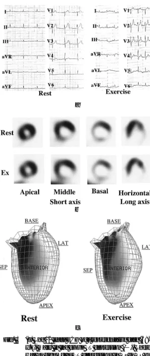
(2) 運動負荷99mTc-MIBI 心筋 SPECT 撮像と 運動負荷心電図
運動負荷 99mTc-MIBI 心筋 SPECT は,自転車エ ルゴメータによる多段階的運動負荷を行い,運動 負荷終点 1 分前に 99mTc-MIBI (296 MBq) を静注 し,静注 15 分後に負荷像を 4 時間後に 99mTc- MIBI (592 MBq) を再静注して安静時像を撮影し た (Fig. 1).また,同時に運動負荷中および負荷 後 5 分間,12 誘導心電図を記録した.負荷心電図 における判定は,水平型および下行型の 1 mm 以 上の ST 低下,または,junctional 型では J 点から 0.08 秒時点での 2 mm 以上の ST 低下を陽性とし た.
すべての SPECT 像は PRISM3000 (PICKER 社 製) を用い,360 度カメラ回転法で 72 ステップ (1 ステップ 40 秒) でデータ収集した.エネルギー ピークは 140 keV, 15% ウインドウに設定して撮 像した.データ収集は ODYSSEY-VP (PICKER 社 製) を用い,ランプフィルターにより再構成後,
Butterworth フィルター (後処理フィルター) によ りノイズカットし,心長軸垂直断層像,心長軸水 平断層像および心短軸断層像を表示した.心電図 同期では R-R 間隔を 16 等分し,収集ビートにつ いて先行 R-R 間隔のウインドウ幅を 30% とした.
(3) 心筋 SPECT の評価と QGS 法による局所 壁運動評価
心筋 SPECT は短軸断層像の心基部,中央部お よび心尖部の 3 スライスをそれぞれ 6 区域に分割 し,心長軸垂直断層像の心尖部を 2 区域に分割,
合計 20 区域において評価した (Fig. 2).アイソ トープの集積欠損程度を定性的視覚的に defect
score (DS) を用い,0 の normal uptake, 1 の mildly reduced uptake, 2 の moderately reduced uptake, 3 の severely reduced uptake-complete defect の 4 段階 で評価し,安静時,負荷時ともに欠損像を認めた 場合を梗塞と判定した.左回旋枝領域の側壁から 下壁にいたる区域 (Seg. 11, 12, 13, 14, 15, 16, 17, 18) の DS の合計を total defect score (TDS) とし た.また負荷時像および安静時像を解析ソフトウ エアである QGS を用いて収集し,左回旋枝領域 の側壁の壁運動を評価した.QGS での評価法と しては,心筋 SPECT の各 20 区域の局所壁運動を 視覚的 6 段階の wall motion score (WMS) を用い,
0 の normal wall motion, −1 の mild hypokinesis,
−2 の moderate hypokinesis,−3 の severe hypokinesis, −4 の akinesis, −5 の dyskinesis の 6 段階にスコア化し,左回旋枝領域の側壁から下 壁にいたる区域の WMS の合計を total wall motion score (TWMS) とした.また,安静時に対する負 荷時の TWM の差を ∆TWMS として算出した.
さらに,安静時および負荷時の左室拡張末期容積
(EDV),左室収縮末期容積 (ESV),左室駆出率
was obtained. Four hours later again rest 99mTc- MIBI QGS was obtained.
Fig. 2 Schemes of the segments for grading myocardial uptake of 99mTc-MIBI myocardial SPECT and for grading left ventricular wall motion by QGS method.
a
b
c
Fig. 3 (a) The 12 leads ECG of a representative case (68- y.o., male) in the group ST depression (−). There was no significant ST depressions in V2 to V4 on exercise. (b) The myocardial SPECT of the same case. The defect in LCX region was found in both image. There was no ischemia in LCX region. (c) The QGS of LAO veiw of the same case. Left hand side shows rest 99mTc-MIBI QGS and right hand side shows exercise 99mTc-MIBI QGS.
There was no change of wall motion between both QGS imagings.
a
b
c
Fig. 4 (a) The 12 leads ECG of a representative case (58- y.o., female) in the group ST depression (+). There were significant ST depressions in V2 to V5. (b) The myocardial SPECT of the same case. Defect in LCX region was found in both images, there was no ischemia in LCX region. (c) The QGS of LAO view of the same case. Left hand side shows rest
99mTc-MIBI QGS and right hand side shows exercise 99mTc-MIBI QGS. The wall motion of LCX region was disturbed in exercise 99mTc-MIBI QGS comparing rest 99mTc-MIBI QGS.
(EF) を求めた.
(4) 検討項目および統計学的処理
負荷時心電図上 V2–V4 誘導にて有意に ST 低下 を認めたものを ST 低下陽性群 (11 症例,最大 ST 低下=2.8±0.4 mm) とし,認めない例を ST 低下 陰性群 (10 例) とした (Fig. 3,4).それぞれの群 において DS, TDS, TWMS, ∆TWMS, EF, EDV お よび ESV につき比較検討した.各項目における,
各群での安静時と負荷時の有意差検定は,対応
のある t 検定で,両群間における有意差は対応の ない t 検定で行い,p<0.05 をもって有意差あり と判定した.
III. 結 果
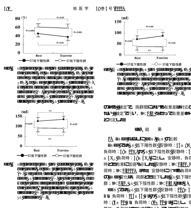
1. LVEF ならびに EDV, ESV の比較
LVEF では ST 低下陽性群 (安静時:46±9%,
負荷時:38±10%) が ST 低下陰性群 (安静時:61
±5%, 負荷時:59±6%) に比し,安静時,負荷
時とも有意に低値を示した (安静時;p<0.001,負 荷時;p<0.0001).また,安静時に対する負荷時 の LVEF 変化は,両群とも低下した (ST 低下陽性 群;p<0.01, ST 低下陰性群;p<0.05) (Fig. 5).
EDV では ST 低下陽性群 (安静時:129±47 ml,負荷時:136±56 ml) が ST 低下陰性群 (安静 時:94±20 ml,負荷時:90±22 ml) に比し,安 静時,負荷時とも有意に拡大していた (安静時,
負荷時とも;p<0.05).安静時に対する負荷時の 変化では,両群に有意差を認めなかった (Fig. 6).
ESV では,やはり ST 低下陽性群 (安静時:72
±33 ml,負荷時 87±46 ml) が ST 低下陰性群 (安 静時:38±11 ml,負荷時:37±8 ml) に比し,安 静時,負荷時とも有意に拡大していた (安静時,
負荷時とも;p<0.05).安静時に対する負荷時の 変化では,ST 低下陽性群においては,負荷時に 有意な拡大がみられたが,ST 低下陰性群では有 Fig. 5 The change of LVEF from exercise to rest QGS in
both groups. The LVEF of exercise QGS in both groups was significantly lower than that of rest QGS. The LVEF of the group ST depression (+) was significantly lower than that of the group ST depression (−) in both phases. Closed circles show the group ST depression (+), and open circles show the group ST depression (−).
Fig. 6 The change of EDV from exercise to rest QGS in both groups. The EDV of the group ST depression (+) was significantly higher than that of the group ST depression (−) in both phases. There were no significant change from exercise to rest in both groups. Closed circles shows the group ST depression (+), and open circles show the group ST depression (−).
Fig. 7 The change of ESV from exercise to rest QGS in both groups. The ESV of the group ST depression (+) was significantly higher than of the group ST depression (−) in both phases. Closed circles show the group ST depression (+), and open circles show the group ST depression (−).
意な変化を認めなかった (Fig. 7).
2. Total defect score (TDS) の比較
左回旋枝領域の心筋灌流欠損を示す TDS で は,ST 低下陽性群 (安静時:13.6±4.9, 負荷時:
14.1±5.8) は ST 低下陰性群 (安静時:5.8±2.6,
負荷時:6.7±2.5) に比し,安静時,負荷時とも有 意に高値を示した (安静時;p<0.001, 負荷時;
p<0.005).しかし,両群とも安静時,負荷時の TDS に有意な変化は認められなかった.ST 低下 陽性群の心筋梗塞範囲が,ST 低下陰性群より広 Fig. 8 The change of TDS from exercise to rest. The TDS of the group ST depression (+) was signifi- cantly higher than that of the group ST depression (−) in both phases. There was no significant change of TDS from exercise to rest in both groups.
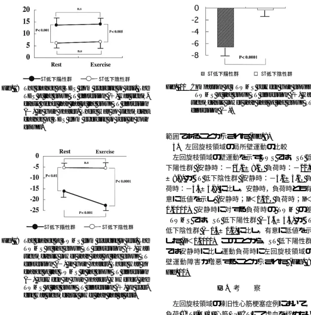
Fig. 9 The change of TWMS from exercise to rest. The TWMS of the group ST depression (+) was significantly lower than that of the group ST depression (−) in both phases. There was no change of the TWMS in the group ST depression (−) between in both phases. However, the TWMS of the group ST depression (+) on exer- cise was significantly lower than that at rest.
Fig. 10 Comparison of ∆TWMS between both groups.
∆TWMS of the group ST depression (+) was significantly lower than that of the group ST depression (−).
範囲であることが示された (Fig. 8).
3. 左回旋枝領域の局所壁運動の比較
左回旋枝領域の壁運動を示す TWS では,ST 低 下陽性群 (安静時:−16.1±8.1, 負荷時:−22.7
±9.8) が ST 低下陰性群 (安静時:−5.2±3.1, 負 荷時:−5.5±3.8) に比し,安静時,負荷時とも有 意に低値を示し (安静時;p<0.01, 負荷時;p<
0.0001),安静時に対する負荷時の TWM の差
∆TWMS では,ST 低下陽性群 (−6.6±3.5) が ST 低下陰性群 (−0.3±0.2) に比し,有意に低値を示 した (p<0.0001).このことから,ST 低下陽性群 では安静時に比し運動負荷時に左回旋枝領域の 壁運動障害が増悪することが示された (Fig. 9, Fig. 10).
IV. 考 察
左回旋枝領域の陳旧性心筋梗塞症例において,
負荷 99mTc-MIBI 心筋 SPECT にて虚血を認めない にもかかわらず,運動負荷中前胸部誘導で ST 低 下を示すことがあり,この原因について検討し た.本研究の結果から,左回旋枝領域の陳旧性心 筋梗塞患者のうち,運動負荷中に前胸部 V2–V4 誘 導で ST 低下を認める症例では,心筋 SPECT に おける側壁の欠損領域が広く,すなわち梗塞範囲 が広く,左室拡張末期容量が大きく,左室駆出率 が低値であった.さらに,安静時に比し運動負荷 時では,梗塞領域での壁運動が著明に悪化してい ることが判明した.
心筋 SPECT の診断精度において,感度は 70–
90%, 特異度は 80–95% 程度とされ,運動負荷心
電図と比べて高い診断精度であることが報告され ている8).負荷心電図において虚血性 ST 変化をき たしやすい誘導は,Blackburm ら8)によると,
V5,V6,V4,V3,aVF,II, I の順であり,運動 負荷時の虚血性 ST 低下は心筋の虚血の局在とは 無関係としている.また従来より,左前下行枝の 広範囲な陳旧性心筋梗塞患者に運動負荷をかける と,運動負荷時に前胸部誘導 ST が上昇すること が指摘されている.この理由として,運動負荷時 の左室内圧の上昇に伴い,aneurysm である前壁 心尖部の壁運動障害が関与していると考えられて
いる9〜11).Shah らの検討では12),経皮的冠動脈
形成術施行時に左回旋枝を閉塞したとき,心電図 上最大 ST 低下が V2,V3 にて有意に出現すると している.この場合の左回旋枝閉塞に対する感度 は 70%, 特異度は 96% と報告し,これら前胸部 誘導の特異的変化は,左心室後壁での ST 上昇の 鏡面像であるとしている.壁運動異常により ST 上昇をきたす機序の詳細はいまだ不明であるが,
Elhendy らの最近の報告13) によると,運動負荷時
ST 上昇を示した症例の 85% は心筋梗塞の既往が あり,それらの症例はすべて負荷心エコー図法で 壁運動異常を呈したとされている.この観点から 推察すると,本研究の広範囲側壁梗塞における運 動負荷時の前胸部誘導 ST 低下は,後壁あるいは 側壁の ST 上昇の鏡面像と考えられる.本研究の 限界として,左回旋枝領域の壁運動障害について 運動負荷時の壁運動評価を QGS で評価したが,
梗塞領域の正確な壁運動評価が得られるかという 問題が挙げられる.しかし下津らは,心筋梗塞に よる欠損例と正常あるいは軽度欠損例の 2 群間に
であったとしている15).これらの指摘から,梗塞 部位であっても,ある程度の精度をもって壁運動 評価が可能であると考えられ,本研究でも梗塞部 位の壁運動を検討した.しかしその精度にはまだ 問題も残り,今後の検討が必要である.
V. 結 論
左回旋枝領域の陳旧性心筋梗塞患者で,負荷シ ンチグラフィ上虚血がなく,負荷心電図上 V2–V4
誘導で有意に ST 低下を示す原因は,運動負荷に よって誘発された後側壁領域の広範囲な壁運動障 害によるものと考えられた.
文 献
1) Bittl JA, Levin DC: Nuclear cardiology. In Heart Disease: A Textbook of Cardiovascular Medicine (ed.
by Braunwald E) 5th ed. WB Saunders, Philadelphia, 1997: 273–316.
2) Lew AS, Weiss AT, Shah PK, Maddahi J, Peter T, Ganz W, et al: Precordial ST segment depression during acute inferior myocardial infarction: Early thallium-201 scintigraphic evidence of adjacent posterolateral or inferoseptal involvement. J Am Coll Cardiol 1985; 5: 203–209.
3) Salcedo JR, Baird MG, Chambers RJ, Beanlands DS:
Significance of reciprocal S-T segment depression in anterior precordial leads in acute inferior myocardial infarction: Concomitant left anterior descending coronary artery disease? Am J Cardiol 1981; 48:
1003–1008.
4) Abraham RD, Freedman SB, Dunn RF, Newman H, Roubin GS, Harris PJ, et al: Prediction of multivessel coronary artery disease and prognosis early after acute myocardial infarction by exercise electrocardiography and Thallium-201 myocardial perfusion scanning. Am J Cardiol 1986; 58: 423–427.
5) Germano G, Kiat H, Kavanagh PB, Moriel M, Mazzanti M, Su HT, et al: Automatic quantification of ejection fraction from gated myocardial perfusion
SPECT. J Nucl Med 1995; 36: 2138–2147.
6) DePuey EG, Nichols K, Dobrinsky C: Left ventricular ejection fraction assessed from gated technetium- 99m-sestamibi SPECT. J Nucl Med 1993; 34: 1871–
1876.
7) Detrano R, Gianrossi R, Mulvihill D, Lehmann K, Dubach P, Colombo A, et al: Exercise-induced ST segment depression in the diagnosis of multivessel coronary disease: a meta analysis. J Am Coll Cardiol 1989; 14: 1501–1508.
8) Blackburn H, Katigbak R: What electrocardiographic leads to take after exercise? Am Heart J 1964; 67:
184–185.
9) Macieira-Coelho E, Garcia-Alves M, Dionisio I, Cantinho G, Da Costa BB, Gouveia A: Role of myocardial ischemia and left ventricular wall motion abnormalities as contributory factors in the genesis of exercise-induced ST-segment elevation in Q-wave myocardial infarction. Cardiology 1999; 91: 227–230.
10) Hsu L, Rostami A, Sreeharan N, Teo KK, Kappagoda T: Evolution of the responses to exercise in left ventricular aneurysms after anterior wall acute myocardial infarction. Am J Cardiol 1988; 61: 236–
239.
11) Candell-Riera J, Santana-Boado C, Armadans-Gil L, Blanch P, Aguade S, Castell J, et al: Comparison of patients with anterior wall healed myocardial
infarction with and without exercise-induced ST- segment elevation. Am J Cardiol 1998; 81: 12–16.
12) Shah A, Wagner GS, Green CL, Crater SW, Sawchak ST, Wildermann NM, et al: Electrocardiographic differentiation of the ST-segment depression of acute myocardial injury due to the left circumflex artery occlusion from that of myocardial ischemia of nonocclusive etiologies. Am J Cardiol 1997; 80: 512–
513.
13) Elhendy A, Geleijnse ML, Roelandt JR, van Domburg RT, Cornel JH, TenCate FJ, et al: Evaluation by quantitative 99m-technetium MIBI SPECT and echocardiography of myocardial perfusion and wall motion abnormalities in patients with dobutamine- induced ST-segment elevation. Am J Cardiol 1995;
76: 441–448.
14) 下津順子,石田良雄,村川圭三,福地一樹,林田 孝平,高宮 誠,他: 99mTc-MIBI 心電図同期心筋
SPECT による左室機能自動計測の信頼性の検
討.核医学 1997; 34: 1093–1099.
15) Wahba FF, Lamb HJ, Bax JJ, Dibbets-Schneider P, Bavelaar-Croon CD, Van Der Wall EE, et al:
Assessment of regional myocardial wall motion and thickening by gated 99mTc-tetrofosmin SPECT: a comparison with magnetic resonance imaging. Nucl Med Commun 2001; 22: 663–671.
Shigeru O
SHIMAand Koichi T
ANIGUCHI Division of Cardiology, Gunma Prefectural Cardiovascular Center[Purpose] The aim of this study is to clarify the causes of exercise-induced ST-segment depression in patients with broad lateral old myocardial infraction involving LCX (LCX-OMI) without ischemia on ex- ercise scintigraphy.
[Method] Twenty one patients (M/F=11/10, age=
62±19 years) with myocardial infraction involving LCX (LCX-MI), but without fill-in on exercise and rest MIBI quantitative gated SPECT (QGS), were se- lected. They were divided into two groups of Group ST(+) (n=11, with significant ST depression (max
−2.8±0.4 mm), Group ST(−) (n=10) without ST depression. On 20 SPECT segments of both exercise and rest SPECT, we scored uptake score as DS (0=
normal to 3=defect) and wall motion as WMS (0=
normal to −5=dyskinesis) and summed DS (TDS) and WMS (TWMS) in LCX region, furthermore, cal- culated the difference of TWMS (∆TWMS {exercise
−rest}), end diastolic volume (EDV) and ejection fraction (EF) during exercise were compared between the two groups.
[Result] Group ST(+) showed significantly (p<
0.01) lower EF (35.4±9.2% vs. 60.2±6.2%), larger EDV (146±53 ml vs. 93±15 ml), higher TDS (5 vs.
7±3), lower TWMS (−25±9 vs. −6±5), further- more lower ∆TWMS (−6.9±4.0 vs. −2.0±0.8) than Group ST(−).
[Conclusion] Exercise-induced significant ST de- pression in V2–4 without ischema in LCX-MI was ob- served in patients with broad LCX-MI, low EF, and was related to impaired wall motion in LCX region.
ST depression in V2–4 was considered to appear as miller image of ST elevation at postero-inferior wall due to disturbed wall motion on exercise.
Key words: Disturbance of wall motion, Exercise induce ST-depression, Gate SPECT.