肺炎球菌による感染性大動脈瘤の 1 例および文献的考察
1)滋賀県立成人病センター臨床検査部,2)同 循環器内科,3)同 心臓血管外科,
4)同 血液・腫瘍内科,5)国立病院機構京都医療センター循環器内科
中村由紀子
1)齋藤 崇
1)西尾 久明
1)石井 充
2)5)小菅 邦彦
2)勝山 和彦
3)池口 滋
2)鈴木 孝世
4)(平成 22 年 12 月 6 日受付)
(平成 23 年 6 月 6 日受理)
Key words : Streptococcus pneumoniae, Mycotic aortic aneurysm
序 文
感染性大動脈瘤は,その頻度は大動脈瘤の 0.7〜
3.8% と少ないが,破裂の頻度が高い動脈瘤である1)2). 発生機序としては,菌血症下での動脈壁や動脈瘤壁へ の細菌感染,近接した感染巣からの波及,感染性心内 膜炎など離れた感染巣からの細菌性塞栓が考えられて いる3).起炎菌としてはサルモネラ,黄色ブドウ球菌,
大腸菌などが報告されているが,肺炎球菌の報告はま れである2).今回,肺炎球菌を起炎菌とする感染性大 動脈瘤の症例を経験したので,文献的考察を加えて報 告する.
症 例 患者:60 歳代,男性.
主訴:食欲低下,下腿浮腫.
既往歴:特記事項なし.
現病歴:来院 2 カ月前から左肩痛と腰痛を自覚し,
消炎鎮痛剤を約 1 カ月間服用していた.来院数日前か ら食欲低下と下腿浮腫を認めたため,当院消化器内科 を受診した.血液検査で肝機能障害を認めたため腹部 CT 検査を実施したところ,腹部大動脈に 5cm 径の 多房性の動脈瘤が認められ,感染性大動脈瘤が疑われ 緊急入院となった.
入院時現症:意識清明,体温 37.1℃,脈拍 120 回!
分,整,呼 吸 数 12 回!分,血 圧 109!70mmHg,心 雑 音なし,腹部は平坦・軟,圧痛なし.両下肢浮腫あり.
入院時検査所見(Table 1):血算では白血球数の増 加と好中球分画の上昇,正球性低色素性貧血を認めた.
生化学検査では軽度の肝機能障害,電解質異常を認め た.血清検査では CRP:20.94mg!dL と炎症反応の上
昇を認めた.尿所見には異常を認めなかった.
入院時胸部 X 線写真(Fig. 1):正面像(a)にて下 行大動脈辺縁の拡大(矢印頭)がみられ,側面像(b)
で下行大動脈の瘤形成(矢印)が認められた.
入院時 CT 写真(Fig. 2):胸部 CT に て 最 大 径 56 mm,約 60mm 連続する大動脈瘤が認められ,大動脈 周囲には低濃度領域があり,壁在血栓や膿瘍形成が疑 われた.
経胸壁心エコー所見:壁運動は正常で,各弁にも明 らかな疣贅は認められなかった.
入院時経過(Fig. 3)血液検査所見及び画像所見か ら胸部と腹部の感染性大動脈瘤を疑い,血液培養検査 のための検体採取後,セフトリアキソン(CTRX)2g! 日と塩酸バンコマイシン(VCM)2g!日の点滴投与を 開始した.貧血に対して赤血球輸血を実施したところ 紅班が出現したため,副腎皮質ホルモン剤を投与した.
治療開始 3 日目,入院時に採取した血液培養検体より ペニシリン感受性肺炎球菌が検出されたため,ペニシ リン G カリウム(PCG)2400 万単位!日へと変更した.
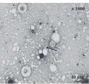
大動脈瘤破裂の危険性を考慮し,第 4 病日に腹部大動 脈瘤人工血管置換術,第 5 病日に胸部下行大動脈瘤人 工血管置換術を実施した.周術期抗菌薬としてセファ メジン(CEZ)3g!日を追加した.手術で切除した各 動脈瘤壁のグラム染色から莢膜を有するグラム陽性双 球菌が認められた(Fig. 4).動脈瘤壁の培養検査か らは菌の発育は認められなかったが,PCR 法による 遺伝子検査で肺炎球菌の特異遺伝子(lytA)を確認し た4)(Fig. 5).第 23 病日より尿量減少,浮腫,体重増 加が出現し,経胸壁心エコーで左室拡張末期径 52mm,
左室収縮末期径 45mm,左室駆出率 13% で,左室壁 運動の低下が認められた.PCG による薬剤性心不全 を疑い,第 34 病日に CTRX 2g!日へ変更した.その 症 例
別刷請求先:(〒524―8524)滋賀県守山市守山 5―4―30 滋賀県立成人病センター臨床検査部
中村由紀子
Table 1 Laboratory findings on admission
<Hematology> <Chemistry> <Serology>
WBC 13,100 /μL TP 6.9 g/dL CRP 20.94 mg/dL
Band 1 % GOT 40 IU/L
Seg 80 % GPT 77 IU/L <Urinalysis>
Eosino 2.0 % LDH 128 IU/L pH 6.5
Lymph 9.0 % ALP 2,419 IU/L Protein (±)
Mono 8.0 % γ-GTP 411 IU/L Glucose (−)
RBC 260×104/μL T-Bil 0.93 mg/dL
Hb 7.2 g/dL UA 2.4 mg/dL <Ultrasound CardioGraphy>
Ht 21.1 % BUN 11.0 mg/dL Vegetation (−)
PLT 54.8×10⁴/μL Cr 0.56 mg/dL
Na 123 mEq/L
<Coagulation> K 4.7 mEq/L
PT 13.3 sec Cl 87 mEq/L
PT (INR) 1.18 FBS 128 mg/dL
Fig. 1 Posteroanterior (a) and lateral (b) chest radiography on ad- mission showing widening descending thoracic aneurysm (arrow head and arrow).
Fig. 2 Chest computed tomography (CT) on admission showing lobulated aneurysm with low-density area around the aorta (arrow).
後経過良好であったため第 65 病日からレボフロキサ シン(LVFX)500mg!日に変更し,第 72 病日に軽快 退院となった.手術時腹部大動脈後壁の切除は困難で あり,また胸部大動脈は肺と強固に癒着していた.感 染性大動脈瘤壁の全切除は困難であり動脈瘤壁が一部 残存したため LVFX は退院後も継続した.その後白 血球数は基準値内で推移し CRP は陰性で経過したた め,抗菌薬加療は退院後約 4 カ月間で終了した.
考 察
肺炎球菌を起炎菌とする,胸部下行大動脈と腹部大 動脈の感染性大動脈瘤の 1 例を経験した.感染性大動 脈瘤は最も治療困難な動脈瘤の一つであり,破裂の頻 度の高い動脈瘤である3).感染経路は感染性心内膜炎 に続発するものが最も多いが,肺炎や骨髄炎などの周 囲感染病巣からの波及による場合もある3).
過去 20 年間(1991〜2010 年)の肺炎球菌による感
Fig. 3 Clinical course. mPSL; methylprednisolone sodium succinate, CTRX; ceftri- axone, VCM; vancomycin, CEZ; cefazoline, PCG; penicillin G, LVFX; levofloxacin.
Fig. 4 Gram stain of aortic wall (original mag- nification ×1000). Gram-postive diplococci was detected in aortic wall (arrow).
Fig. 5 PCR detection of pneu- mococcal specific gene, lytA.
Lane: M, DNA marker; TA, S.
pneumoniae isolated from tho- racic aorta; AA, S. pneumoniae isolated from abdominal aorta;
N, negative control; P, positive control.
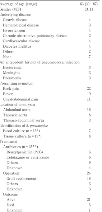
染性大動脈瘤の報告例は 27 例であり(Table 2),男 性 13 人,女性 14 人,平均年齢 63 歳(範 囲:30〜87 歳)であった.主な基礎疾患として消化器疾患,高血 圧疾患,心血管疾患,糖尿病などを有していた.その うち 9 例(33%)は 3 カ月以内に肺炎球菌を起炎菌と する肺炎や菌血症,髄膜炎の既往を有し,22 例(81%)
に背部痛の訴えが認められていた.本症例では肺炎球 菌による先行感染を示唆する症状等はなく,肺炎球菌 の感染経路は不明である.大動脈瘤は 16 例(59%)が 腹部大動脈に存在していた.
細菌培養検査は報告 27 例中,血液培養検査の記載 があったのは 15 例で,手術後の動脈瘤組織培養のそ
れは 15 例であった.前者の 7 例,後者の 8 例で肺炎 球菌が同定されていた.本症例では動脈瘤組織培養か らは肺炎球菌は同定されなかったが,グラム染色でグ ラム陽性双球菌を認めていたため PCR 法による遺伝 子解析を行い,肺炎球菌の特異遺伝子を確認した4).
治療について,まず抗菌薬投与の記載があったのが 25 例で,その内詳記されていたのが 21 例であった.
その多くが Benzylpenicillin(PCG)や Cefotaxime ま
Table 2 Characteristic in patients with mycotic aortic aneurysm caused by Streptococcus pneumoni- ae (n=27)
Average of age (range) 63 (30−87)
Gender (M:F) 13: 14
Underlying disease
Gastric disease 4
Hematological disease 3
Hypertension 4
Chronic obstructive pulmonary disease 2
Cardiovascular disease 4
Diabetes mellitus 4
Others 2
None 7
An antecedent history of pneumococcal infection
Bacteremia 3
Meningitis 3
Pneumonia 3
Presenting symptom
Back pain 22
Fever 9
Chest-abdominal pain 11
Location of aneurysm
Abdominal aorta 16
Thoracic aorta 8
Thoraco-abdominal aorta 3
Identification of S. pneumoniae
Blood culture (n=15*) 7
Tissue culture (n=15*) 8
Treatment
Antibiotics (n=25**)
Benzylpenicillin (PCG) 6
Cefotaxime or ceftriaxone 6
Others 9
Unknown 4
Operation 24
Graft replacement 18
Others 3
Unknown 3
Outcome
Alive 21
Died 5
Unknown 1
*: No of cases reported results of cultures
**: No of cases received antibiotics therapy
たは CTRX にて加療されていた.次に手術施行例は 24 例であり,人工血管置換術が 18 例と大多数であっ た.一方手術未施行例は 3 例あり,経過の詳記があっ た 2 例はいずれも手術に至る前に死亡していた.5 例 の死亡が報告され,死因の記載は 3 例あり,2 例は感 染性大動脈瘤破裂で入院後 2 日目と 17 日目に死亡,1 例は敗血症で手術後 38 日目に死亡していた.
初期治療薬として CTRX と VCM を使用した理由 は感染性大動脈瘤の起炎菌頻度から,サルモネラ,大 腸菌,メチシリン耐性黄色ブドウ球菌などが起炎菌で
ある可能性を考慮したためである.
術後 PCG 投与中に心不全を認めた.PCG 投与中止 後 1 週間で症状が軽快したため PCG による薬剤性心 不全の可能性を疑ったが,感染による高心拍出性心不 全傾向に PCG による循環血液量の増加が加わり心不 全増悪の一因になったものと考えた.そのため外来経 過観察中の経口抗菌薬としてペニシリン系を選択せず LVFX とした.
感染性大動脈瘤の抗菌薬による推奨治療期間ははっ きりとしていない.過去 20 年間の報告例でも 3 週間 から一生涯までの幅があった4)〜22).我々は感染性動脈 瘤が一部残存していることを考慮し手術後 6 カ月間の 抗菌薬治療とした.抗菌加療中止後 6 カ月以上経過し ているが,再発は認められていない.
今回,我々は肺炎球菌を起炎菌とする感染性大動脈 瘤のまれな症例を経験した.抗菌薬治療開始前に血液 培養検査を実施していたことが,起炎菌確定に有用で あった.今後,感染性大動脈瘤の起炎菌として肺炎球 菌も考慮する必要があると考えられる.
文 献
1)Cinà CS, Arena GO, Fiture AO, Clase CM, Doo- bay B:Ruptured mycotic thoracoabdominal aortic aneurysms:a report of three cases and a systematic review. J Vasc Surg 2001;33(4):
861―7.
2)Hsu PJ, Lee CH, Lee FY, Liu JW:Clinical and microbiological characteristics of mycotic aneur- isms in a medical center in southern Taiwan. J Microbiol Immunol Infect 2008;41:318―24.
3)安田慶彦,佐々木重幸:領域別症候群.別冊日
本臨床 1996;14:325―7.
4)大楠清文,江崎孝行:遺伝子検査による呼吸器
感染症の迅速診断.呼吸器科 2008;14(1):
63―74.
5)Gelabert HA, Quinones-Baldrich WJ:Mycotic aneurysm of suprarenal aorta secondary to Streptococcus pneumoniae:an unusual pathogen.
Ann Vasc Surg 1991;5(6):529―32.
6)Gomes MN, Choyke PL, Wallace RB:Infected aortic aneurysms:achanging entity. Ann Surg 1992;215(5):435―42.
7)Cordero JA Jr, Darling RC 3rd, Chang BB, Shah DM:In situ prosthetic graft replacement for mycotic thoracoabdominal aneurysms. Am Surg 1996;62(1):35―9.
8)Albrecht WE, Papasian CJ, Bamberger DM, Fiorella R:Infected abdominal aortic aneurysm due to penicillin-, ceftriaxone-, and cefotaxime- resistant Streptococcus pneumoniae. J Clin Micro- biol 1997;35(4):985―7.
9)Berdat PA, Malinverni R, Kipfer B, Carrel TP:
Homograft failure in mycotic aortic aneurysm caused byStreptococcus pneumoniae. Ann Thorac
Surg 1998;66(5):1818―9.
10)Brouwer RE, van Bockel JH, van dissel JT:
Streptococcus pneumoniae, an emerging pathogen in mycotic aneurysms? Neth J Med 1998;52
(1):16―21.
11)Lussier N, Girouard Y, Chapleau D:Mycotic aneurysm of the suprarenal aorta due to Strep- tococcus pneumoniae:case report and literature review. Can J Surg 1999;42(4):302―4.
12)Naktin J, Desimone J:Lumbar vertebral osteo- myelitis with mycotic abdominal aortic aneu- rysm caused by highly penicillin-resistantStrep- tococcus pneumoniae. J Clin Microbiol 1999;37
(12):4198―200.
13)Steig TA, Johannesen N, Schønheyder HC:
Propensity of Streptococcus pneumoniae for the aorta. Report of 3 cases. Scand J Infect Dis 2001;33(10):772―4.
14)Rabitsch W, Brugger SA, Trubel W, Keil F, Gre- inix HT, Kalhs P:Streptococcus pneumoniae my- cotic aortic aneurysm after allogeneic bone mar- row transplantation. Transplantation 2002;74
(7):1048―50.
15)Shinonaga M, Kanazawa H, Nakazawa S, Yoshiya K, Ujiie T, Yamazaki Y:Rupture of a smoldering mycotic aneurysm of the thoracic aorta into the lung. Ann Thorac Cardiovasc Surg 2002;8(3):177―9.
16)Coutu M, Blair JF:Multiple mycotic aortic aneurysms due toStreptococcus pneumoniae. Ann Vasc Surg 2003;17(5):554―7.
17)Luo CY, Ko WC, Kan CD, Lin PY, Yang YJ:In situ reconstruction of septic aortic pseudoaneu- rysm due to Salmonella or Streptococcus micro-
bial aortitis:long-term follow-up. J Vasc Surg 2003;38(5):975―82.
18)Englert C, Aebert H, Lenhart M, Solleder A, Nerlich M, Neumann C:Thoracic spondylitis from a mycotic(Streptococcus pneumoniae)aortic aneurysm:a case report. Spine 2004;29(17):
E373―5.
19)Goswami R, Cleveland KO, Gelfand MS:Evolv- ing infectious aortitis caused by Streptococcus pneumoniae. South Med J 2004;97(10):
1004―6.
20)Rondina MT, Raphael K, Pendleton R, Sande MA:Abdominal aortitis due to Streptococcus pneumoniaeand Enterobacter aerogenes:a case report and review. J Gen Intern Med 2006;21
(7):C1―3.
21)Brant-Zawadzki P, Kinikini D, Kraiss LW:Deep leg vein reconstruction for an isolated mycotic common iliac artery aneurysm in an HIV- positive patient. Vascular 2007;15(2):98―
101.
22)Dickinson KJ, Parry DJ, Sandoe JA, Gough MJ:Multiple peripheral pneumococcal mycotic aneurysms without aortic involvement : a unique case confirmed with the novel use of a molecular diagnostic technique. J Vasc Surg 2007;45(6):1253―5.
23)Nagano N, Yamamoto T, Amano A, Kikuchi K:Infected aneurysm of the aortic arch with purulent pericarditis caused by Streptococcus pneumoniae. Interact Cardiovasc Thorac Surg 2010;10(3):459―61.
Mycotic Aortic Aneurysm due toStreptococcus pneumoniae: A Case Report and Review of Literature
Yukiko NAKAMURA1), Takashi SAITO1), Hisaaki NISHIO1), Mitsuru ISHII2)5), Kunihiko KOSUGA2), Kazuhiko KATSUYAMA3), Sigeru IKEGUCHI2)& Takayo SUZUKI4)
1)Department of Clinical Laboratory,2)Department of Cardiology and3)Department of Cardiovascular Surgery, Shiga Medical Center for Adults,
4)Department of Hematology and Oncology, Shiga Medicalcenter for Adults,
5)Department of Cardiology, National Hospital Organization Kyoto Medical Center
Mycotic aortic aneurysm due toStreptococcus pneumoniae is rare. The case of we report occurred in 62- year-old man with no antecedent infection admitted for appetite loss and lower leg edema. Chest and ab- dominal computed tomography, blood culture, and gene analysis to detec arterial wall pneumococci led to a diagnosis of mycotic aortic aneurysm caused byS. pneumoniae. The man had a graft replaced and was ad- ministered antibiotics. He remains well and infection-free 12 months after surgery. We also review the litera- ture on these aortic aneurysms.
〔J.J.A. Inf. D. 85:515〜519, 2011〕