Design of Biofunctional Liposomes for Delivery
of Bioactive Molecules
著者
Yuba Eiji
内容記述
学位授与大学: Osaka Prefecture University(大阪
府立大学), 学位の種類: 博士(工学), 学位記番号:
論工第1251号, 学位授与年月日: 2010-03-31, 指導
教員: 河野健司.
Design of Biofunctional Liposomes
for Delivery of Bioactive Molecules
Eiji Yuba
February 2010
i
Contents
Chapter 1. General Introduction
1.1. Drug Delivery System~a Promising Technology for Advanced Medical Treatments
1.2. Liposomes and Their Functionalization
1.3. Design of Functional Liposomes Based on Chemistry for Drug Delivery System
1.4. Objectives and Outline of This Study 1.5. References
Chapter 2. pH-Sensitive Fusogenic Polymer-Modified Liposomes as a Carrier of Antigenic Proteins for Activation of Cellular Immunity.
2.1. Introduction
2.2. Materials and Methods 2.2.1. Materials
2.2.2. Cell Lines Culture 2.2.3. Animals
2.2.4. Generation of Murine Bone Marrow-Derived DCs 2.2.5. Preparation of Liposomes
2.2.6. Dynamic Light Scattering and Zeta Potential 2.2.7. Fusion Assay
2.2.8. Cellular Uptake 2.2.9. Microscopy
2.2.10. In Vitro Antigen Presentation Assay 2.2.11. Immunization
2.2.12. CTL Assay 2.3. Results and Discussion
2.3.1. Characterization of Liposomes 2.3.2. Fusogenic Activities of Liposomes 2.3.3. Association of Liposomes with DCs
2.3.4. Cytoplasmic Delivery of Antigenic Protein to DCs 2.3.5. Induction of Cellular Immune Responses
2.3.6. Estimation of Antigen Presentation
2.3.7. Influence of Liposome Size on Cellular Immunity Activation 2.4. Conclusion 2.5. References 1 1 3 5 8 10 13 13 15 15 15 16 16 17 17 17 18 19 19 19 20 20 20 21 22 23 26 28 29 31 33
ii
Chapter 3. Carboxylated Hyperbranched Poly(glycidol)s for Preparation of pH-Sensitive Liposomes
3.1. Introduction
3.2. Materials and Methods 3.2.1. Materials
3.2.2. Synthesis of Hyperbranched Poly(glycidol) Derivatives 3.2.3. Cell Culture
3.2.4. Precipitation pH 3.2.5. Pyrene Fluorescence
3.2.6. Preparation of Pyranine-Loaded Liposomes 3.2.7. Release of Pyranine from Liposome
3.2.8. Liposome Size Change
3.2.9. Intracellular Behavior of Liposomes 3.2.10. Fusion of Liposomes in Cell 3.3. Results and Discussion
3.3.1. Characterization of HPG Derivatives
3.3.2. Interaction of HPG Derivatives with Lipid Membrane 3.3.3. Preparation of pH-Sensitive Liposomes Using HPG 3.3.4. Cytoplasmic Delivery by Polymer-Modified Liposomes 3.3.5. Fusion of Polymer-Modified Liposomes within Cell 3.4. Conclusion
3.5. References
Chapter 4. Gene Delivery to Dendritic Cells Mediated by Complexes of Lipoplexes and pH-Sensitive Fusogenic Polymer-Modified Liposomes
4.1. Introduction
4.2. Materials and Methods 4.2.1. Materials
4.2.2. Cell Culture
4.2.3. Preparation of pH-Sensitive Polymer-Modified Liposome–Lipoplex Complexes
4.2.4. Dynamic Light Scattering and Zeta Potential 4.2.5. Transfection
4.2.6. Cellular Uptake 4.2.7. Microscopic Analysis 4.2.8. Cytotoxicity
4.2.9. MHC Class I Presentation 4.3. Results and Discussion
35 35 36 36 38 38 38 39 39 39 40 40 41 42 42 46 48 49 52 54 55 57 57 59 59 60 60 61 61 62 62 62 63 63
iii
4.3.1. Transfection of DC2.4 Cells
4.3.2. Ligand Effect on Transfection of DC2.4 Cells
4.3.3. Effect of pH-Sensitive Fusogenic Polymers on Transfection Activity of Complexes
4.3.4. Mechanism of the Efficient Transfection Activity by the MGluPG Complexes
4.3.5. Toward Application to Immunotherapy 4.4. Conclusion
4.5. References
Chapter 5. Modification of Liposome Surface with pH-Responsive
Polyampholytes for the Controlled-Release of Drugs
5.1. Introduction 5.2. Material and Methods
5.2.1. Materials
5.2.2. Synthesis of PDEAEMA and Polyampholytes of Varying DEAEMA:AA Ratios
5.2.3. Analysis of Polymers Using NMR Technique 5.2.4. Titration of Polymers
5.2.5. Encapsulation of Pyranine and Liposome Modification 5.2.6. Estimation of Lipid Concentration in Liposome
5.2.7. Pyranine Release from Liposome 5.3. Results and Discussion
5.3.1. 1H NMR Analysis of Copolymers 5.3.2. Titration of Copolymers
5.3.3. Pyranine Release from Neat and Polymer-Modified Liposomes
5.3.4. Proposed Mechanism of Contents Release Induced by Polymers
5.4. Conclusion 5.5. References
Chapter 6. pH-Sensitive Vesicles That Undergo Transition to Micelles for Intracellular Delivery
6.1. Introduction 6.2. Materials and Methods
6.2.1. Materials 6.2.2. Cell Culture
6.2.3. DL/PEG Dispersion Preparation
63 65 68 70 71 72 73 75 75 77 77 77 78 78 78 79 79 80 80 81 82 85 86 87 89 89 91 91 91 92
iv
6.2.4. Differential Scanning Calorimetry (DSC) 6.2.5. Optical Density Measurement
6.2.6. Dynamic Light Scattering (DLS)
6.2.7. Transmission Electron Microscopy (TEM) 6.2.8. Release Assay
6.2.9. Microscopy 6.3. Results and Discussion
6.3.1. DSC Analysis of DL/PEG Dispersion
6.3.2. pH-Dependent Change of Optical Density of DL/PEG Dispension
6.3.3. Structural Analysis of DL/PEG Dispersion
6.3.4. Accurate Release Contents Responding to pH-Change 6.3.5. Application to Intracellular Delivery System
6.4. Conclusion 6.5. References
Chapter 7. Preparation and Characterization of Complexes of Liposomes with Gold Nanoparticles
7.1. Introduction
7.2. Materials and Methods 7.2.1. Materials
7.2.2. Liposome Preparation
7.2.3. Preparation of the Complex of Liposomes with Au NPs 7.2.4. Characterization
7.2.5. Calcein Release from Liposomes 7.3. Results and Discussion
7.3.1. Preparation and Surface Plasmon Resonance of the Complexes of Gold Nanoparticles with Liposomes 7.3.2. Morphology of the Complexes of Gold Nanoparticles with
Liposomes
7.3.3. Preparation of the Complexes of Gold Nanoparticles with Various Liposomes
7.4. Conclusion 7.5. References
Chapter 8. General Conclusion
Acknowledgements 92 92 92 93 93 93 93 93 95 95 97 98 99 100 102 102 103 103 105 105 105 106 106 106 108 111 112 113 115 119
v
1
Chapter 1
General Introduction
1. 1. Drug Delivery System~a Promising Technology for Advanced Medical Treatments
Since the early 19th century, progress of organic chemistry led pharmaceutical industry to rise. P. Ehrlich and J. Langley suggested the concept of drug and its receptor (‘magic bullet’) in the end of 19th and early 20th century, respectively, which developed medicinal chemistry. A. Fleming first found penicillin in 1928. Since 1940s, many antibiotics have been developed, resulting in real start of drug discovery.
In 1953, Watson and Crick demonstrated the molecular structure of deoxyribonucleic acid (DNA) as ‘double helix structure’ [1]. This discovery led the rapid developments of biochemistry and molecular biology. In 1970, recombinant DNA techniques were established. And then, biotechnology-based medicine has been produced for many intractable diseases. In early 1960’s, some diseases were clarified to be induced by the pathogenetic mechanisms, and genetic factors were shown to contribute to the etiology of diseases. The fundamental understandings of diseases based on genetics generated gene therapy. Gene therapy is a therapeutic method in which can be treated by expressing the curative genes or suppressing specific genes.
Recent progress in biotechnology has revealed the function of embryonic stem cell (ES cell), which has abilities to differentiate into any types of cells constructing bodies [2]. This discovery enhanced opportunities for regeneration of missing tissue or organs. However, there are ethical difficulties regarding the use of human embryos, as well as the problem of tissue rejection following transplantation in patients. In 2006, S. Yamanaka et al. suggested the notion that could overcome all these problems, the induction of pluripotent cells from completely differentiated fibroblasts, which were called the induced pluripotent stem (iPS) cells [3, 4]. To date, many researchers have studied the functions, efficient preparation
2
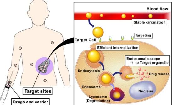
Fig. 1-1. The concept of Drug Delivery system (DDS).
methods and applications of iPS cells [5-9].
In addition to stem cell-based regenerative medicine, functional cell-based cell therapy has risen. Cell therapy is the treatment in which functional cells from patient’s tissue or blood induce curative effects. Especially, dendritic cells (DCs), which play a crucial role in the control of immune responses and activations, have attracted much attention for their application to cancer immunotherapy or vaccination [10, 11]. DCs recognize, acquire, process and present antigens (substance materials of diseases) to native and resting T cells for induction of an antigen-specific immune response. Because DCs presenting tumor-associated antigens (TAAs) can activate TAA-specific immune response, many researchers have attempted to use TAA-presenting DCs as a vaccine for cancer immunotherapy.
As described above, recent remarkable progress in drug discovery, medicinal chemistry and molecular biology to led a new class of medicine or approach to therapy such as biotechnology-based medicine, gene therapy, stem cell-based regenerative medicine and cell therapy. To achieve these therapies, it is required to transfer bioactive molecules to specific sites in body or specific cells or specific organelle in cells. Drug delivery system (DDS) has risen to fulfill these requirements (Fig. 1-1.). DDS carrier containing drugs has to
3
overcome following processes, (i) stable circulation in blood flow, (ii) reaching to target cell (targeting), (iii) efficient internalization and (iv) delivery to target organelle.
In gene therapy, curative DNA must enter the specific cells and then their nucleus, and be expressed in the cells efficiently. Because DNA can’t permeate cellular membrane spontaneously, a carrier that delivers DNA into nucleus of specific cells is necessary. Besides, ribonucleic acid interference (RNAi), which suppresses the gene expression by binding specific messenger RNA (mRNA) and preventing them from producing a protein, requires an efficient vehicle of small interfering RNAs (siRNAs) that can transfer siRNA to target cell’s cytosol [12]. To reprogram the differentiated cells to iPS cells, an efficient gene carrier is desired which can induce gene expression for a proper time period [13]. In addition, it is important for cancer immunotherapy to induce the cellular immunity. To induce the cellular immunity, the delivery of antigens (such as proteins) to cytosol of DCs is essential [10, 11].
Thus, drug discovery and technology has grown the more advanced, carrier systems which can deliver bioactive molecules to specific cells or specific organelle have become the more important. To date, numerous studies have been reported to establish the efficient drug delivery system using various materials such as biopolymers, synthetic polymers, peptides, lipids, virus, virus-derived proteins and bacteria [14-31]. Above all, lipid-based carrier system is promising due to their biocompatibility, drug-loading ability and capability for functionalization.
1. 2. Liposomes and Their Functionalization
In 1964, A. D. Bangham found that lipid suspension formed bilayer vesicles, so called “liposomes” [32, 33]. Liposomes have two regions, inner water phase and lipid membrane. Therefore liposome can encapsulate both water-soluble drugs and hydrophobic drugs. Although since the early studies of liposomes they have been used as a model of biological membrane, after finding of their potentials as a drug carrier, many researches have studied
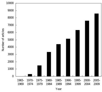
4 0 1000 2000 3000 4000 5000 6000 7000 8000 9000 10000 1965-1969 1970-1974 1975-1979 1980-1984 1985-1989 1990-1994 1995-1999 2000-2004 2005-2009 Year N u m ber o f ar ti c les
Fig 1-2. The number of articles concerning liposome searched by PubMed.
about potentials of liposomes as DDS. Fig 1-2 shows the temporal transition of the number of articles about liposome. The number of articles on liposomes has increased steadily, indicating that liposome technology is of great importance in a wide range of fields.
Liposome studies as a drug carrier have progressed markedly since appearance of the concept about ‘long-circulating liposomes’. Allen and Chonn constructed such liposomes by using a glycolipid having sialic acids because erythrocytes containing many sialic acids on their surface could circulate in blood flow by escaping from recognition of reticuloendothelial system (RES) [34]. Importantly, the concept of escape from RES has become a standard as long-circulating liposomes. In 1990, L. Huang et al. prepared poly(ethylene glycol) (PEG)-modified liposomes as a new type of long-circulating liposomes [35]. PEG forms a steric barrier against RES due to its high hydration ability. Since the appearance of PEG-modified liposomes, the utility of liposome as a DDS carrier has been showed and liposome studies have become more active. In fact, many liposome-based drug formulations have been commercialized to date, which contain anticancer drugs, antibacterial reagents, photosentitizers and so on.
5
In addition to long-circulating liposomes, many “functional” liposomes have so far been developed. For example, by conjugating antibodies or specific ligands to liposome surface, targeted liposomes could be constructed, which could bind to specific sites or cells bearing corresponding receptors [36-39]. By using lipid phase-transition or conjugation of thermo-sensitive materials, thermo-responsive liposomes have been developed [40, 41]. Because thermo-responsive liposomes can release the contents in response to mild heating, by local heating limited at disease sites, drugs are released only at local sites and selective drug effects are obtained. Another type of important functional liposomes is “pH-sensitive liposomes” [42, 43]. The circumstance of body is normally kept to be at neutral pH. On the other hands, inflammatory sites or tumor neighborhood are known to be at weakly acidic pH. Inside of endosome and lysosome, which are cellular compartments involved in the internalization and degradation of exogenous materials, is also weakly acidic environments [44]. In response to such pH-differences, pH-sensitive liposomes are destabilized and release drugs. Besides, various functional liposomes responsive to various external stimulations have been developed such as magnetic filed-sensitive “magnetoliposomes” [45], ultrasound-responsive micro bubble-containing “bubble liposomes” [46] and “photo-responsive liposomes” [47].
1. 3. Design of Functional Liposomes Based on Chemistry for Drug Delivery System
It is important to give functions to liposomes for their application to DDS. To date, many types of functional liposomes have been designed. One approach to establish the functional liposome is to use the property change of lipid itself under external stimulations. For example, Yatvin et al. prepared thermo-responsive liposomes containing dipalmitoyl phosphatidylcholine (DPPC), which has phase-transition temperature at 42 °C [40]. DPPC-containing liposomes showed content release around DPPC’s phase-transition temperature. H. Ellens et al. prepared pH-sensitive liposomes consisting of
6
non-bilayer-forming lipid, such as dioleoyl phosphatidylethanolamine (DOPE) and carboxyl group-containing amphiphilies, such as cholesteryl hemisuccinate (CHEMS) [43]. At neutral pH, deprotonated carboxyl groups promote the hydration of liposome surface and generate the electrostatic repulsion between liposomes, resulting in formation of stable liposomes. However, at acidic pH, liposomes are destabilized due to protonation of carboxyl groups.
Instead of using lipid’s properties, the conjugation of functional materials to stable liposomes is an attractive approach to prepare the functional liposomes. As a pioneering work, H. Ringsdorf et al. prepared a variety of polymer-introduced liposomes and J. Sunamoto et al. constructed polysaccharide-modified liposomes [48, 49]. Because Ringsdorf and Sunamoto developed many methods for introducing polymers or functional molecules to liposomes, their works are basis of following studies about constructing functional liposomes.
K. Kono prepared thermo-sensitive liposomes by complexation of typical thermo-responsive polymer, poly(N-isopropylacrylamide) (PNIPAM), with stable liposomes and achieved efficient drug release above polymer’s lower critical solution temperature (LCST), where polymer changes character from hydrophilic to hydrophobic [41]. In addition, PNIPAM-based copolymers containing various amounts of acrylamide units showed a variety of LCST which depends on their compositions. These polymers induced drug release from liposomes above their LCST. D.A. Tirrell et al. constructed pH-sensitive liposomes by conjugation of poly(carboxylic acid) with liposomes [50-52]. At neutral pH, deprotonated carboxyl groups don’t interact to liposome membrane. However, at acidic pH, protonated carboxyl groups destabilize liposome membrane rapidly. Because hydrophobic interaction plays an important role in the interaction of polymers with lipid membrane, more hydrophobic polymers were synthesized and incorporated to liposomes. These hydrophobic polymers exhibited stronger membrane-disrupting ability [53]. K. Kono et al. also developed pH-sensitive polymers, a series of carboxylated poly(glycidol) derivatives for pH-sensitization of liposomes [54, 55]. These polymers have a backbone structure similar to
7
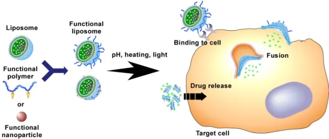
Fig. 1-3. Design of functional liposome by the conjugation of functional materials with liposome.
that of PEG and carboxyl groups on the side chains, which control interaction of the polymer backbone with lipid membranes in a pH-dependent manner. These polymer-modified liposomes are stable at neutral pH, but they exhibit considerable destabilization under mildly acidic conditions and deliver contents into cytosol by membrane fusion with endosome/lysosome membranes.
S.H. Park et al. reported the complex between Au nanoparticles (Au NPs) and liposomes [56]. Au NPs strongly absorb light in the visible region of the spectrum, due to the surface plasmon resonance (SPR), and convert the absorbed light to heat. Hence, Au NPs are considered to be useful for photothermal therapy as well as for imaging in the biomedical field. Because Au NPs are prone to aggregate, stabilization of Au NPs is important. Similarly, magnetite-incorporated “magnetoliposomes” were also developed for application to diagnosis and photothermal therapy [45].
Thus the complexation of functional materials with liposomes has benefits from the following viewpoints:(i) original liposomes before complexation are stable, and (ii)
obtained functional liposome’s properties are tunable by changing properties of conjugated materials. Hence, by using functional materials with high performance, excellent
8
1. 4. Objectives and Outline of This Thesis
As mentioned above, it has been required to construct efficient functional liposomes, which can deliver bioactive molecules to specific sites, specific cells and specific cellular compartments efficiently for the establishment of advanced medical technology. The conjugation of functional materials to liposomes is a smart approach for producing functional liposome, which can overcome extra- or intracellular barriers against efficient delivery of bioactive molecules. In this study, highly potent pH-sensitive or photo-sensitive functional liposomes were developed by the conjugation of functional materials to liposomes.
This doctoral thesis consists of 8 chapters. The outline of each chapter is summarized as follows:
Chapter 1
This chapter describes the background, the objectives and the contents of this thesis.
Chapter 2
This chapter describes the correlation between cytoplasmic delivery of antigenic protein to DCs and the ability to induce cellular immunity. pH-Sensitive polymer-modified liposomes were used as a vehicle to deliver antigenic proteins. The effects of polymer structure were investigated on various aspects of polymer-modified liposomes including membrane fusion activity, cellular association, cytoplasmic delivery and the induction of cellular immune response.
Chapter 3
This chapter describes the improvement of pH-sensitive polymer and the preparation of these polymer-modified liposomes having pH-sensitive membrane fusion properties, which would ensure more efficient cytoplasmic delivery of bioactive molecules such as proteins by inducing efficient membrane fusion with acidic endosome. Hyperbranched poly(glycidol) (HPG) derivatives with carboxylated side chains and different degrees of polymerization (DPs) by reacting HPG having different DPs with 3-methylglutaric anhydride, which are
9
designated MGlu-HPG, were synthesized, and investigated a correlation between their backbone structures and their abilities to render stable liposomes pH-sensitive. These polymers were modified with antigenic protein-loaded liposomes, and estimated their potentials as a cytplasmic delivery vehicle for antigenic proteins.
Chapter 4
This chapter describes the gene delivery to DCs by using complexes comprised of pH-sensitive polymer-modified liposome and DNA-cationic lipid complex (lipoplexes). The effects of ligands conjugated to the polymer and polymer structure were investigated in the cellular association and transfection activity by these complexes. The importance of efficient endosomal escape of DNA on transfection was discussed. Furthermore, it was also evaluated whether transfection by these complexes induced the up-regulation of cellular immunity-associated molecules on DCs.
Chapter 5
This chapter describes the preparation of liposomes bearing pH-sensitive poly(ampholytes) for the controlled drug release. pH-Sensitive copolymers comprised of (2-dimethylamino)ethyl methacylate (DEAEMA) units and acylic acid (AA) units at various ratios were synthesized and used to modify the liposome surface. The charge states of these polymers were estimated by a titration. pH-Dependent release behaviors of fluorescent dye-loaded liposomes modified with these polymers were also investigated. The correlation between the charged states of polymers and the ability to induce drug release was discussed.
Chapter 6
This chapter describes the preparation of novel pH-sensitive lipid-based intracellular drug delivery system. Poly(amidoamine)-dendron-bearing lipid (DL) were used as an amphiphilic moleclule. The mixture of DL and poly(ethylene glycol) (PEG)-bearing lipid was dispersed in phosphate-buffered saline. The pH-sensitivity of DL/PEG dispersion was evaluated by differential scanning calorimetry, optical density analysis, dynaminc light
10
sccaterring and transmission electron microscopy. Using unique pH-responsive structural transition of DL/PEG molecular assemblies, intracellular delivery of model proteins was also examined.
Chapter 7
This chapter demonstrates the construction of novel complexes of liposomes with gold nanoparticles (Au NPs) for photo-responsive nanodevices. The stability of liposome-Au NP complex was investigated by time-dependent surface plasmon resonance (SPR). Formed complexes were also characterized using transmittance electron microscopy (TEM), dynamic laser scattering (DLS) and release of encapsulated molecules. Furthermore, the effect of lipid composition was examined on obtained liposome-Au NP complexes.
Chapter 8
This chapter summarizes all the conclusions in this thesis.
1. 5. References
1. J. D. Watson and F. H. C. Crick, Nature 1953, 171, 737-738. 2. J. A. Thomson et al., Science 1998, 282, 1145-1147.
3. K. Takahashi and S. Yamanaka, Cell 2006, 126, 663-676. 4. K. Takahashi et al., Cell 2007, 131, 861-872.
5. M. Stadtfeld et al., Science 2008, 322, 945-949. 6. K. Okita et al., Science 2008, 322, 949-953.
7. D. Huangfu et al. Naure Biotechnology 2008, 26, 1269-1275. 8. T. Aasen et al., Nature Biotechnology 2008, 26, 1276-1284. 9. Y. Yoshida et al., Cell Stem Cell 2009, 5, 237-241.
10. J. Banchereau and A.K. Palucka, Nat. Rev. Immunol. 2005, 5, 296–306. 11. N.S. Wilson and J.A. Viladangos, Adv. Immunol. 2005, 86, 241–305. 12. A. Fire et al., Nature 1998, 391, 806-811.
11
13. S. Yamanaka, Nature 2009, 460, 49-52.
14. WF. Lai, MC. Lin, J Control Release 2009, 134, 158-168. 15. R. Karinagaa et al., Biomaterials 2005, 26, 4866–4873. 16. M. G. Svahn1 et al., J. Gene Med. 2004, 6, 36–44. 17. S Abes et al., Nucl. Aci. Res. 2007, 35, 4495–4502.
18. P. L. Felgner et al., Proc. Nat. Acad. Sci. USA 1987, 84, 7413-7417. 19. O. Boussif et al., Proc. Nat. Acad. Sci. USA 1995, 92, 7297–7301. 20. J. Gary et al., Proc. Nat. Acad. Sci. USA 1993, 90, 11307-11311. 21. I. Koltover et al., Science 1998, 281, 78-81.
22. R. Koynova and R. C. MacDonald, Biochim. Biophy. Acta 2005, 1714, 63-70. 23. IS. Zuhorn et al., Biophys. J. 2002, 83, 2096–2108.
24. DS. Friend et al., Biochim. Biophys. Acta 1996, 1278, 41–50. 25. S. Akhtar and I. F. Benter, J. Clinical Inves. 2007, 117, 3623-3632. 26. R. Bryan R. et al., Adv. Drug Del. Rev. 2007, 59, 134–140.
27. E. John et al., Proc. Nat. Acad. Sci. USA. 1987, 84, 4626-4630. 28. K.L, Berkner, Bio. Techniques 1988, 6, 616.
29. L. Guy et al., Biochim. Biophy. Acta 2007, 1772, 243–262. 30. A. El-Aneed, J Control Release 2004, 94, 1– 14.
31. J. A. MacDiarmid et al., Nature Biotechnology 2009, 27, 643-651. 32. A. D. Bangham and R. W. Horne, J. Mol. Biol. 1964, 8, 660-668. 33. A. D. Bangham et al., J. Mol. Biol. 1965, 13, 238-252.
34. T. M. Allen and A. Chonn, FEBS Lett. 1987, 223, 42-46. 35. A. L. Kilbanov et al., FEBS Lett. 1990, 268, 235-237. 36. M. Ogris et al., Gene Ther. 1999, 6, 595–605.
37. Y. Lu and P. S. Low, Adv. Drug Deliv. Rev. 2002, 54, 675– 693. 38. R. Wiewrodt et al., Blood 2002, 99, 912-922.
12
39. A. G. Schätzlein, J. Biomed. Biotechnol. 2003, 2, 149–158. 40. MB. Yatvin et al., Science 1978, 202, 1290–1293.
41. K. Kono, Adv. Drug Delivery Rev. 2001, 53, 307-319.
42. D. Liu and L. Huang, Biochim. Biophys. Acta 1990, 1022, 348–354. 43. H. Ellens et al., Biochemistry 1984, 23, 1532-1538.
44. S. Mukherjee et al., Physiol. Rev.1997. 77, 759–803. 45. E. Viroonchatapan et al., Life. Sci. 1996, 58, 2251-2261. 46. R. Suzuki et al., Int. J. Pharm. 2008, 354, 49-55.
47. T. Nagasaki et al., Bioconjugate Chem. 2003, 14, 513-516.
48. H. Ringsdorf et al., Angew. Chem. Int. Ed. Engl. 1988, 27, 113-158. 49. J. Sunamoto et al., Chem. Lett., 1988, 17, 1781-1784.
50. K. Seki et al., Macromolecules 1984, 17, 1692–1698. 51. M. Maeda et al., J. Am. Chem. Soc. 1988, 110, 7455–7459. 52. M. Fujiwara et al., J. Colloid Interface Sci. 1997, 185, 210–216. 53. N. Murthy et al., J. Controlled Release 1999, 61, 137–143. 54. K. Kono et al., Biochim. Biophys. Acta 1994, 1193, 1–9. 55. N. Sakaguchi et al., Bioconjugate Chem., 2008, 19, 1040-1048. 56. S. H. Park et al., Colloid Surf. B, 2006, 48, 112-118.
13
Chapter 2
pH-Sensitive fusogenic polymer-modified liposomes
as a carrier of antigenic proteins
for activation of cellular immunity
2. 1. Introduction
Efficient vaccination strategies have been desired for overcoming new pathogens and for evolution of resistance of microorganisms. In addition, efficient vaccine delivery systems have been required for achievement of cancer immunotherapy. Dendritic cells (DCs) are known as potent professional antigen presenting cells; they play a crucial role in innate and adaptive immune responses [1–3]. The DCs recognize, take up, process and present antigens to native and resting T cells for induction of an antigen-specific immune response. Antigenic proteins internalized via endocytosis are degraded to peptide fragments. These peptides are presented by binding to major histocompatibility complex (MHC) class II molecules, which mainly activate CD4+ T lymphocytes, thereby inducing humoral immunity. On the other hand, antigenic proteins introduced into cytosol of DCs are degraded by proteasomes after ubiquitination. These fragmented peptides are presented by MHC class I molecules on the surface of DCs. They mainly activate CD8+ cytotoxic T lymphocytes (CTLs) to induce cellular immunity. To attain efficient target-specific immunity, induction of the antigen-specific CTLs is important because they eliminate the infected cells and pathogens directly. Therefore, carrier systems that can introduce antigenic proteins efficiently into the cytosol of DCs are necessary to establish effective immunotherapy.
Numerous attempts have been undertaken to achieve delivery of antigens into the DC’s cytosol. For example, Akagi et al. reported that nanoparticles of γ-poly(glutamic acid) introduced entrapped antigenic ovalbumin (OVA) into cytosol of DC and induced antigen
14
specific CTLs [4,5]. In addition, Fre´chet and coworkers showed that acid-degradable, acrylamide-based nanoparticles achieved cytosolic delivery of OVA and presentation of OVA-derived peptides via the MHC class I pathway [6]. These nanoparticles might be taken up by DC via endocytosis and enhance transfer of their encapsulated antigen molecules from endosome and/or lysosome to cytosol by destabilization of the membranes of these acidic compartments through hydrophobic or electrostatic interactions [5,6].
One of the most effective strategies for efficient introduction of antigenic proteins into cytosol of DC might be to use membrane fusion especially for membrane-based nanoparticles, such as liposomes. To date, viral fusion proteins have been used frequently to provide liposomes with fusion ability [7,8]. Indeed, viral fusion protein-incorporated liposomes have been used to introduce encapsulated antigenic OVA into DC’s cytosol and induced efficient cellular immunity [7,8]. However, viral proteins might provoke unexpected immune responses. Therefore, the use of synthetic carriers might be preferred for the delivery of antigens into DCs.
The author has developed pH-sensitive liposomes, which generate fusion ability under weakly acidic conditions, by surface modification of egg yolk phosphatidylcholine (EYPC) liposomes with poly(glycidol) derivatives having carboxyl groups (Fig. 2-1) [9,10]. In fact, these polymer-modified liposomes delivered a membrane impermeable fluorescent dye, calcein, into cytosol of HeLa cells after internalization via endocytosis and subsequent fusion with membrane of endosome or lysosome [9,10]. Especially, 3-methylglutarylated poly(glycidol) (MGluPG), which has hydrophobic side chains, exhibited higher fusion ability than succinylated poly(glycidol) (SucPG) [10].
Considering the excellent performance of these polymer-modified liposomes as a cytoplasmic delivery vehicle, the author attempted to apply these liposomes to the production of potent vaccines, which deliver antigenic proteins into cytosol of DCs and activate cellular immune response through their administration via nasal mucosa, which affords some
15
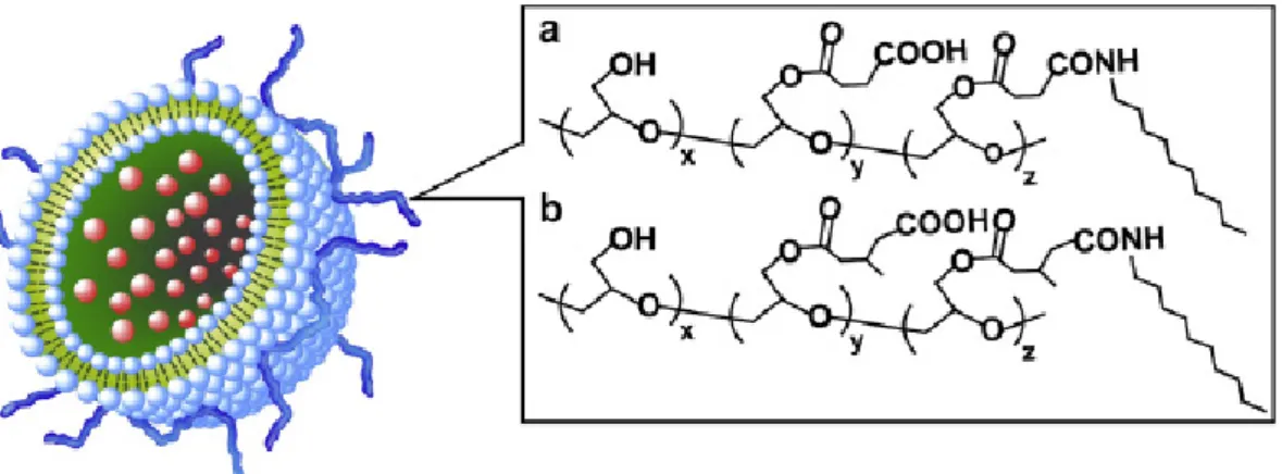
Figure 2-1. Structures of SucPG (a), MGluPG (b) and pH-sensitive polymer-modified
liposome.
advantages, such as noninvasive needle-free administration and induction of both mucosal and systemic immune responses [11–14]. Correlation of fusogenic properties of the liposomes with their ability to activate cellular immunity was described.
2. 2. Materials and Methods 2. 2. 1. Materials
EYPC and L-dioleoyl phosphatidylethanolamine (DOPE) were kindly donated by NOF Co. (Tokyo, Japan). N-(7-nitrobenz-2-oxa-1, 3-diazol-4-yl)dioleoyl phosphatidylethanolamine (NBD-PE) and lissamine rhodamine B-sulfonyl phosphatidylethanolamine (Rh-PE) were purchased from Avanti Polar Lipids (Birmingham, AL, USA). Monophosphoryl lipid A (MPL), Freund’s complete adjuvant (CFA) and OVA were purchased from Sigma (St. Louis, MO.). SucPG and MGluPG were prepared using poly(glycidol) with number average and weight average molecular weights of 1.6 × 10⁴ and 2.5 × 10⁴, respectively, as previously reported [10,15]. Molar percentages of glycidol/carboxylated glycidol/n-decylamine-attached unit in the resultant SucPG and MGluPG were determined to be 18/74/8 and 9/81/10, respectively, using 1HNMR.
16
DC2.4 cells, which were an immature murine DC line, were provided from Dr. K. L. Rock (Harvard Medical School, USA) and were grown in RPMI 1640 supplemented with 10% FBS (MP Biomedical, Inc.), 2 mM L-glutamine, 100 µM nonessential amino acid, 50 µM 2-mercaptoethanol (2-ME) and antibiotics at 37 °C [16]. EL4, a C57BL/6 mice-derived T lymphoma, was obtained from Tohoku University (Sendai, Japan). E.G7-OVA, which is a chicken egg OVA gene-transfected clone of EL4 and which presents OVA with MHC class I molecules, was obtained from the American Type Culture Collection (Manassas, VA) [17]. CD8-OVA1.3 cells, a T–T hybridoma against OVA257–264/H-2Kb complex, were kindly
provided by Dr. C.V. Harding [18], and were grown in Dulbecco’s modified Eagle’s medium supplemented with 10% FBS, 50 µM 2-ME, and antibiotics. OT4H.1D5 cells, a T–T hybridoma against OVA265–277/I-Ab complex, were kindly provided by Dr. J.A. Kapp [19],
and were cultured in RPMI 1640 medium supplemented with 10% FBS, 50 µM 2-ME, and antibiotics.
2. 2. 3. Animals
Female C57BL/6 mice (H-2b, 7 weeks old) were purchased from Oriental Yeast Co., Ltd. (Tokyo, Japan). The experiments were carried out in accordance with the guidelines for animal experimentation in Osaka Prefecture University.
2. 2. 4. Generation of Murine Bone Marrow-Derived DCs
Bone marrow-derived dendritic cells (BMDCs) were prepared according to the method of Lutz et al. with slight modification [20]. Briefly, bone marrow cells flushed from the femurs and tibias of C57BL/6 mice were seeded at 5 × 10⁶ cells per sterile 100-mm bacterial grade culture dish in 10 ml of RPMI 1640 containing 10% FBS, 10 ng/ml recombinant murine granulocyte/macrophage colony-stimulating factor (GM-SCF, PeproTech EC Ltd.), 50 µM 2-ME, and antibiotics. On day 5, another 10 ml of culture medium was
17
added to the dish for medium replenishment. Nonadherent cells were harvested on days 6–8 as immature BMDCs.
2. 2. 5. Preparation of Liposomes
Liposomes were prepared by two kinds of methods, namely vortex and extrusion. Liposomes were prepared using vortex as follows: a dry thin membrane of a mixture of EYPC, DOPE, and SucPG or MGluPG (EYPC/DOPE. 1/1, mol/mol; lipids/polymer. 7/3, w/w) was dispersed in PBS by vortex to afford liposomes. Also, liposomes were prepared by extrusion method as follows: a dry thin membrane of a mixture of lipid and polymer was suspended by a brief sonication using a bath-type sonicator and the obtained liposome suspension was extruded through a polycarbonate membrane of pore sizes of 100 nm [9,10,15]. For liposomes encapsulating OVA, PBS containing OVA (4 mg/ml) was used for hydration of the lipid/polymer membranes. Free OVA was removed by ultracentrifuge for vortex method and by gel filtration using a Sepharose4B column for extrusion method. Unmodified liposomes were also prepared according to the above procedure using dry membrane of a lipid mixture without polymer.
2. 2. 6. Dynamic Light Scattering and Zeta Potential
Diameters and zeta potentials of the liposomes were measured using a Nicomp380 ZLS dynamic light scattering instrument (Particle Sizing Systems, Santa Barbara, CA) equipped with a 35 mW laser (632.8 nm wavelength). Zeta potentials were measured by equipped an Avalanche photodiode detector, and were detected at an 18.9 angle treated with 9.75 mV. Data was obtained as an average of more than three measurements on different samples.
18
Fusion between plain EYPC liposomes and polymer-modified liposomes was detected by measuring resonance energy transfer between NBD-PE and Rh-PE [21,22]. Polymer-modified liposomes containing NBD-PE and Rh-PE were prepared according to the above procedure using the lipid/polymer membrane containing NBD-PE (0.6 mol%) and Rh-PE (0.6 mol%) and extrusion through a polycarbonate membrane with a pore size of 50 nm. Probe-free plain liposomes were also prepared according to the same procedure. The labeled liposomes (final concentration of lipid 0.125 mM) were mixed with fluorescent probe-free EYPC liposomes (final concentration of lipid 0.25 mM) in 25 mM MES and 125 mM NaCl solution of varying pH. Their fusion was followed by monitoring the fluorescence intensity ratio of NBD-PE to Rh-PE (R). The excitation wavelength of NBD-PE was 450 nm and monitoring wavelengths for NBD-PE and Rh-PE were 520 nm and 580 nm, respectively. Percentage increase in R was defined as:
Percentage increase in R = (Rt − R0)/(R100 − R0 ) × 100 (1)
where R0 and Rt represent the initial and intermediary R values. R100 is the R of the labeled
liposomes when the liposomes fused completely. The fluorescent lipid-labeled liposomes and the unlabeled liposomes were dissolved in methanol, dried by evaporation, and resuspended in MES buffer. The R value of the suspension was taken as R100 [21].
2. 2. 8. Cellular Uptake
The DC2.4 cells (1 × 10⁵ cells) cultured for 2 days in a 12-well plate were washed with Hank’s balanced salt solution (HBSS, Sigma) and then incubated in culture medium. The liposomes containing FITC-labeled OVA or the liposomes which lipids were substituted by Rh-PE (1 mol%) were added gently to the cells and incubated for 4 h at 37 °C. The cells were washed with HBSS three times, and then the detached cells using trypsin were applied to flow cytometry [23].
19
2. 2. 9. Microscopy
The DC2.4 cells (2 × 10⁵ cells) cultured for 2 days in 35-mm glass-bottom dishes were washed with HBSS, and then incubated in serum-free medium. The liposomes containing FITC-labeled OVA (50 µg), in which lipids were substituted by Rh-PE (1 mol%), were added gently to the cells and incubated for 4 h at 37 °C. After the incubation, the cells were washed with HBSS three times and then replaced by serum-free medium. LysoTracker Red DND-99 (Molecular Probes) was used by the staining of intracellular acidic compartments according to the manufacturer’s instructions. Briefly, LysoTracker Red was added to cells at the final concentration of 75 nM. After the 5 min-incubation, the cells were washed with HBSS three times. Confocal laser scanning microscopic (CLSM) analysis of these cells was performed using LSM 5 EXCITER (Carl Zeiss Co. Ltd.).
2. 2. 10. In Vitro Antigen Presentation Assay
The BMDCs were seeded in a 96-well culture plate at a density of 2 × 10⁴ cells/well and cultured for 12 h at 37 °C. Each well was washed twice with HBSS, and then the cells were treated with various liposomes containing OVA or OVA solution at various OVA concentrations. After 3 h incubation at 37 °C, the cells were washed three times with HBSS. Subsequently, the cells were co-cultured with 2 × 10⁴ CD8-OVA1.3 or OT4H.1D5 cells for 24 h at 37 °C. The response of stimulated CD8-OVA1.3 or OT4H.1D cells was assessed by determining the amount of IL-2 released into an aliquot of culture medium (100 µl) using a murine IL-2 ELISA KIT (PeproTech EC Ltd.).
2. 2.11. Immunization
Mice were nasally immunized with 10 µl aliquots of polymer-modified liposomes or unmodified liposomes containing 100 µg of OVA on days 0 and 14. Other group of mice was nasally immunized with OVA solution and another group of mice was subcutaneously
20
immunized with CFA/OVA emulsion.
2. 2. 12. CTL Assay
Splenocytes from immunized mice were suspended in RPMI 1640 medium supplemented with 10% FBS, 100 U/ml penicillin, 100 µg/ml streptomycin, 50 µM 2-ME, and 20 U/ml recombinant murine IL-2 (Peprotech, London, UK). Seven days after the second immunization, splenocytes were obtained from five mice, and the splenocytes were pooled and stimulated with mitomycin C-treated E.G7-OVA cells at a ratio of 10:1 for 5 days. The stimulated splenocytes were used as effector cells for the cytotoxicity assay. The CTL activity was evaluated at various ratios of effector cells to target cells (E.G7-OVA or EL4 cell), which were defined as E/T ratios, using a lactate dehydrogenase (LDH) cytotoxicity detection assay (Takara Biomedicals, Tokyo, Japan).
2. 3. Results and Discussion
2. 3. 1. Characterization of Liposomes
The author measured sizes and zeta potentials of prepared liposomes to characterize the liposomes. Table 2-1 presents particle sizes and zeta potentials of various liposomes prepared by vortex method or extrusion method. When the liposomes were prepared using the vortex method, unmodified liposomes were of about 1500 nm diameter. Although the SucPG-modified and MGluPG-modified liposomes were prepared according to the same method, they showed much smaller diameters of around 400–500 nm, probably because negatively charged polymers bound on the liposome surface enhanced hydration of themembrane surface and increased the colloidal stability of the liposomes. When prepared using the extrusion method, all liposomes showed similar particle sizes of around 110 nm, which corresponds to the pore size of the membrane used for their extrusion.
21
zero, indicating that their surface was electrically neutral. However, the liposomes modified with SucPG or MGluPG showed negative zeta potentials of around -11 mV because of carboxylate anions on the polymer chains. This result demonstrates that these polymer-modified liposomes have electrically similar surface characteristics, irrespective of the polymers attached on their surface.
2. 3. 2. Fusogenic Activities of Liposomes
In previous studies, it was showed that modification with SucPG and MGluPG provides pH-dependent fusion ability to stable EYPC liposomes [9,10]. In this study, the author used a mixture of EYPC and DOPE as liposomal lipids because inclusion of DOPE is shown to increase the fusion ability of liposomes [24]. The author examined the fusion abilities of these polymer-modified liposomes using resonance energy transfer between NBD-labeled and Rh-labeled lipids in the liposome membranes. The labeled liposomes were mixed withunlabeled plain EYPC liposomes and incubated for 1 h at various pH. Fusion between these liposomes was evaluated by monitoring the ratio of fluorescence intensity of NBD to that of Rh (R value).
Fig. 2-2 portrays the percent increase in the R value during 1 h incubation as a function of pH. The unmodified liposomes exhibit a very low extent of the increase in R throughout the experimental pH region, indicating the lack of fusion ability for unmodified liposomes. The SucPG-modified liposomes also showed a very low extent of the increase in R at neutral pH, but the R value increased below pH 6, suggesting that the SucPG-modified
22
Figure 2-2. pH-dependent fusogenic properties of liposomes. Percent increase in R values
for fluorescent lipid-labeled unmodified (diamonds), SucPG-modified (squares) and MGluPG-modified (triangles) EYPC/DOPE liposomes after 1 h incubation with the unlabeled plain EYPC liposomes at varying pH was shown. Measurements were performed in 25 mM MES and 125 mM NaCl at 37 °C. Each point is the mean ± SD (n = 3). Liposomes prepared by extrusion method were used.
liposomes generated fusion ability at weakly acidic pH. Although a similar pH-dependence of fusion was apparent for the MGluPG-modified liposomes, the extent of increase in the R value in the weakly acidic pH region was greater, indicating that the MGluPG-modified liposomes generated higher fusion ability than SucPG-modified liposomes. Because MGluPG has more hydrophobic side chains than SucPG, polymer chains of MGluPG might strongly disrupt the liposome membrane, resulting in more intensive fusion of the liposomes. These results indicate clearly that SucPG and MGluPG provided fusion ability to stable EYPC/DOPE liposomes and that the latter gave higher fusion ability than the former, which is consistent with our previous observation [10].
2. 3. 3. Association of Liposomes with DCs
23
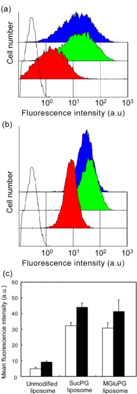
of antigenic proteins to cytoplasm of DC, the author examined the association of the liposomes with DCs after their incubation. The DC2.4 cells were incubated with various liposomes labeled with Rh-PE for 4 h; then fluorescence intensities of the cells were measured using a flow cytometer. As presented in Figs. 2-3a and 3b, cells treated with the labeled liposomes exhibited higher fluorescence intensities than intact cells, indicating the association of liposomes to the treated cells. In addition, the cells treated with SucPG-modified and MGluPG-modified liposomes displayed stronger fluorescence of Rh-PE, suggesting more efficient association of these polymer-modified liposomes to DCs. To evaluate their ability to associate DCs further, the average of cellular fluorescence intensities was calculated (Fig. 2-3c). Compared with the cellular association of the unmodified liposomes, on average, 6.6 and 6.2 times higher amounts of liposome association were observed, respectively, for SucPG-modified and MGluPG-modified liposomes. Actually, DCs are known to have scavenger receptors, which recognize the microorganisms or apoptotic cells with an anionic component [25]. As described in a previous report, these polymer-modified liposomes are likely to be taken up by DCs through interaction of their anionic surfaces with scavenger receptors [23].
Comparison of liposomes prepared by the extrusion method to those prepared by the vortex method shows that the former exhibited slightly higher cellular association than the latter, indicating that DCs engulf small liposomes more efficiently than large liposomes with diameters of 500–1500 nm, although the difference was slight.
2. 3. 4. Cytoplasmic Delivery of Antigenic Protein to DCs
The author examined the ability of the polymer-modified liposomes to deliver OVA, which was used as an antigenic protein into cytosol of DCs. To monitor the inracellular distribution of the liposomes, the encapsulated OVA and the liposome membrane were labeled, respectively, using FITC and Rh-PE. The labeled liposomes encapsulating
24
Figure 2-3. Evaluation of cellular association of various liposomes labeled with Rh-PE
using flow cytometry. DC2.4 cells were treated with SucPG-modified (green), MGluPG-modified (blue) and unmodified (red) liposomes prepared by vortex method (a) or extrusion method (b). Fluorescence intensity of untreated cells (black lines) is also shown. (c) Mean fluorescence intensity of the liposome-treated DC2.4 cells. Each point is the mean ± SD (n = 3). Cellular treatment was performed by incubating with liposomes prepared by vortex method (open) or extrusion method (closed) in serum-free medium at 37 °C for 4 h.
25
Figure 2-4. CLSM images of DC2.4 cells treated with various liposomes labeled with
Rh-PE and loaded with FITC-OVA. Liposomes prepared by extrusion method were used. DC2.4 cells were incubated with liposomes at 37 °C for 4 h in the serum-free medium, washed with HBSS and observed with CLSM. Scale bar represents 10 µm. Concentration of liposomal lipids for cellular treatment was 0.1 mM.
FITC-OVA were added to DC2.4 cells and incubated for 4 h. Then, the cells were washed with buffer and observed using CLSM (Fig. 2-4). When DC2.4 cells were treated with the unmodified liposomes, punctate fluorescence of FITC-OVA was observed in the cells. In
26
addition, fluorescence of Rh was observed at the same places that FITC-OVA fluorescence appeared. This result suggests that OVA molecules loaded in the unmodified liposomes were still trapped in the endosomes and/or the lysosomes. For cells treated with the SucPG liposomes, the fluorescence of FITC-OVA was also mostly punctate and overlapped with Rh-PE fluorescence. In contrast, for cells treated with MGluPG liposomes, diffuse fluorescence of FITC-OVA was observed in the cells, while fluorescence of Rh-PE remained punctate, indicating that the labeled OVA molecules were transferred into cytosol from endosome, where liposomal membrane remained trapped. Probably, the highly fusogenic MGluPG liposomes generate strong fusion ability in the weakly acidic environments of endosome and fuse with endosomal membrane, resulting in the release of entrapped OVA molecules into cytosol [9,10]. The SucPG liposomes only slightly induced the transfer of OVA into cytosol. Therefore, it is implied that their fusion ability is insufficient to induce fusion or destabilization of endosomal membrane for efficient transfer of OVA molecules into cytosol (Fig. 2-2).
2. 3. 5. Induction of Cellular Immune Responses
The author examined the ability of these polymer-modified liposomes to activate cellular immunity. Although immunization is generally conducted via subcutaneous injection of antigen, the author exploited the administration of antigen through nasal mucosa because antigen administration can be achieved using a noninvasive, needleless method in this immunization. It can also induce both mucosal and systemic immunities [11–13].
For this study, the C57BL/6 mice were immunized nasally twice with OVA-loaded liposomes containing MPL in the membrane as an adjuvant. One week later, splenocytes were collected from the immunized mice and stimulated with mitomycin C-treated E.G7-OVA cells. Then their toxicity toward E.G7-OVA cells, which are OVA-presenting recombinant cells derived from EL4 cells, was measured to estimate the induction of OVA-specific CTLs.
27
Figure 2-5. OVA-specific cytotoxic T cell responses in spleen at Day 21 after nasal
immunization with OVA solution (●), polymer-unmodified liposomes ( ◆ ), SucPG liposomes (■) and MGluPG liposomes (▲). Liposomes prepared by vortex method were used. Cytotoxic activity was measured by a LDH assay at indicated E/T ratios. E.G7-OVA cells (a) and EL4 cells (b) were used as target cells. T cell responses from mice without treatment (○) were also shown as a negative control.
Fig. 2-5a shows cellular toxicities of the stimulated splenocytes recovered from mice that had been immunized with various OVA-loaded liposomes or free OVA. When free OVA was used for the immunization, CTL activity was almost identical to that of the case of untreated mice, indicating that free OVA has no ability to induce cellular immunity under experimental conditions. In addition, administration of the OVA-loaded unmodified liposomes increased the CTL activity only slightly. In contrast, when the polymer-modified liposomes containing OVA were used for immunization, higher CTL activities were observed. Especially, MGluPG-modified liposomes more efficiently induced activation of CTLs than the SucPG-modified liposomes did. Indeed, the same stimulated splenocytes did not exhibit cellular toxicity toward EL4 cells (Fig. 2-5b). Therefore, immunization with these polymer-modified liposomes containing OVA induced activation of OVA-specific CTLs. Considering that these polymer-modified liposomes showed a similar degree of cellular association with DCs (Fig. 2-3), it is likely that the difference in fusion ability between these liposomes caused their different abilities for cellular immune activation.
28
2. 3. 6. Estimation of Antigen Presentation
The highly fusogenic MGluPG-modified liposomes induced cellular immune response efficiently. Because the cellular immunity is activated through antigen presentation on the MHC class I molecules of antigen presenting cells (APCs), such as DCs, the author expected that the liposome-mediated delivery of OVA molecules into cytosol of APCs caused the antigen presentation on the MHC class I molecules, engendering activation of cellular immunity. To confirm this mechanism, the author further examined whether the MGluPG-modified liposomes actually possess the capability of inducing antigen presentation on the MHC class I on DCs.
The OVA-loaded liposomes or free OVA were added to BMDCs; then BMDCs were co-cultured with CD8-OVA1.3 or OT4H.1D5 cells, which respectively recognize MHC class I/peptide complexes or MHC class II/peptide complexes. Subsequently, MHC class-restricted antigen presentation was evaluated by detection of IL-2 secretion in supernatants of the co-cultured medium. Fig. 2-6 shows IL-2 production levels of the co-cultured medium of BMDCs treated with the OVA-loaded liposomes and free OVA. As depicted in Fig. 2-6a, interaction with BMDCs and OVA-loaded MGluPG-modified liposomes strongly enhanced IL-2 release from CD8-OVA1.3 cells. The BMDCs treated with either OVA-loaded in the plain liposomes or free OVA also promoted IL-2 release from the CD8-OVA1.3 cells, but the promotion occurred to a much lesser degree than the case of the OVA-loaded MGluPG-modified liposomes. This result suggests that the OVA-loaded MGluPG-modified liposomes induced antigen presentation through the MHC class I molecules on BMDCs more efficiently than free or plain liposome-encapsulated OVA. In contrast, BMDCs treated with these OVA-loaded liposomes or free OVA enhanced IL-2 release from OT4H.1D5 cells only slightly, suggesting that the OVA-loaded MGluPG-modified liposomes have no ability to induce MHC class II-mediated antigen presentation. These results demonstrate that the MGluPG-modified liposomes can induce
29
antigen presentation through MHC class I molecules.
2. 3. 7. Influence of Liposome Size on Cellular Immunity Activation
Finally, the author examined the influence of liposome size on their ability to activate cellular immunity. First, MGluPG liposomes with diameters around 500 nm and 110 nm were prepared, respectively, using vortex and extrusion methods (Table 2-1). The author compared the respective efficiencies of the intracellular OVA delivery of these liposomes with different sizes. The amounts of encapsulated OVA per 1 nmol of liposomal lipid were estimated to be 0.77 µg for the large liposomes and 0.2 µg for the small liposomes. When the same amount of FITC-OVA was added to DC2.4 cells as encapsulated in the liposomes, approximately double the amount of FITC-OVA was taken up by the cells using the large liposomes than those using the small liposomes (Fig. 2-7a).
The author also examined the intracellular distribution of OVA molecules delivered
Figure 2-6. Presentation of OVA-derived epitope peptides via MHC molecules in BMDCs.
BMDCs were incubated with free OVA (diamonds), and OVA-loaded MGluPG-modified (triangles) and unmodified (squares) liposomes at varying OVA concentrations for 3 h. Liposomes prepared by extrusion method were used. Concentrations of IL-2 in the medium after co-culture of OVA-treated BMDCs with (a) CD8-OVA1.3 (specific for
OVA257–264/H-2Kb complex) and (b) OT4H.1D5 (specific for OVA265–277/I-Ab complex)
cells for 24 h as a function of OVA concentration during the BMDCs treatment were shown. Each point represents means ± SD.
30
by these MGluPG liposomes of different sizes. The DC2.4 cells were incubated with these MGluPG liposomes encapsulating FITC-OVA for 4 h and then observed using CLSM (Fig. 2-7b). Strong and punctate fluorescence of FITC-OVA was observed in cells treated with large MGluPG liposomes. Punctate fluorescence of FITC-OVA is mostly overlapped with fluorescence of LysoTracker Red. Therefore, most FITC-OVA molecules remained trapped in the endosomes and lysosomes. The punctate fluorescence of FITC-OVA was still observed in the liposome-treated cells even after 24 h incubation, indicating that FITC-OVA molecules were retained in endosome and lysosome for 24 h (data not shown). In contrast, cells treated with the small MGluPG liposomes displayed diffuse fluorescence of FITC-OVA, suggesting that these small liposomes delivered the antigenic protein efficiently into the cytosol.
The large MGluPG liposomes prepared by vortex showed less endosomal escape, compared to the small extruded MGluPG liposomes, although these liposomes were modified with the same fusogenic polymer MGluPG. Because the large liposome should have a multilamellar structure, only the outermost layer of the bilayer membranes with low curvature might contact with the target membrane, resulting in low efficiency of fusion. In fact, the author evaluated fusion ability of the large MGluPG liposome by the same method used for evaluation of the small MGluPG liposome fusion (Fig. 2-2) and observed only 10% increase in R value after 3.5 h incubation at pH4.0, indicating a low fusion ability of the large MGluPG liposome (data not shown).
These liposomes encapsulating OVA were administered nasally; then CTL responses were evaluated as described above (Fig. 2-7c). Splenocytes of the immunized mice exhibited high toxicity toward OVA-presenting E.G7-OVA cells but almost no toxicity toward their parent EL4 cells, indicating that both liposomes were able to induce OVA-specific CTLs, irrespective of their size difference. Moreover, the CTLs derived with these OVA-loaded liposomes showed an almost identical level of toxicity to E.G7-OVA cells within the error bars. The large MGluPG liposomes delivered more OVA molecules to cells than the small
31
liposomes. However, the latter introduced OVA molecules into the cytosol more efficiently than the former. Consequently, these liposomes might achieve CTL activation to a similar degree.
To evaluate the potential of MGluPG liposomes as an adjuvant, the author compared their induced CTLs activity with that of CFA, which is widely used for induction of immune responses [26]. The result is also presented in Fig. 2-7c. Apparently, CTL activity induced by CFA is of a comparable level to that of MGluPG liposomes.
2. 4. Conclusions
For this study, by modification of liposomes with carboxylated poly(glycidol) derivatives, such as SucPG and MGluPG, the author prepared pH-sensitive fusogenic liposomes that can deliver antigenic proteins into the cytosol of DCs. The author then investigated their ability to activate cellular immune response. Indeed, these polymer-modified liposomes were shown to have capabilities of delivering antigenic OVA into cytosol of DCs and inducing antigen presentation through the MHC class I molecules on DCs. Nasal administration of these liposomes encapsulating OVA activated the antigen-specific cellular immunity in mice. Especially, highly fusogenic MGluPG liposomes exhibited high ability for CTL activation, which is comparable to the widely used adjuvant CFA. Despite its high ability to activate immune response, CFA is known to induce inflammatory reactions at the site of administration. Consequently, CFA is not applicable to humans. In contrast, MGluPG liposomes comprise phospholipid and biocompatible poly(glycidol) derivatives [27]. For that reason, MGluPG liposomes and their relevant liposomes might be promising antigen carriers for establishment of cancer immunotherapy and mucosal vaccination.
32
Figure 2-7. (a) Effect of liposome size on delivery of FITC-OVA by MGluPG liposomes.
DC2.4 cells were treated with FITC-OVA-loaded liposomes prepared by extrusion or vortex method in the serum-free medium at 4 °C (open) and 37 °C (closed) for 4 h, and then cellular association of FITC-OVA was estimated using flow cytometry. Each point is the mean ± SD (n = 3). OVA concentration was 50 µg/ml. (b) CLSM images of DC2.4 cells treated with FITC-OVA-loaded liposomes prepared by vortex method or extrusion method. DC2.4 cells were incubated with MGluPG-modified liposomes encapsulating FITC-OVA (50 µg of OVA/ml) in the serum-free medium at 37 °C for 4 h. (c) Effect of liposome size on OVA-specific cytotoxic T cell response. Mice were nasally immunized with OVA-loaded MGluPG liposome prepared by vortex method (triangles) or by extrusion method (squares). Mice were also immunized with CFA/OVA emulsion subcutaneously (circles). Cytotoxic activity of splenocytes of the immunized mice was measured by a LDH assay at indicated E/T ratios. E.G7-OVA cells (closed symbols) and EL4 cells (open symbols) were used as target cells.
33
2. 5. References
1. J. Banchereau and RM. Steinman, Nature 1998, 392, 245–252. 2. I. Mellman and RM. Steinman, Cell 2001, 106, 255–258. 3. NC. Fernadez et al., Nat. Med. 1999, 5, 405–411.
4. T. Akagi et al., Biomaterials 2007, 28, 3427–3436.
5. T. Yoshikawa et al, Biochem. Biophys. Res. Commun. 2008, 366, 408–413. 6. YJ. Kwon et al., J. Control Release 2005, 105, 199–212.
7. J. Kunisawa et al., J. Immunol. 2001, 167, 1406–1412. 8. L. Bungener et al., Vaccine 2002, 20, 2287–2295.
9. K. Kono et al., Biochim. Biophys. Acta. 1997, 1325, 143–154. 10. N. Sakaguchi et al., Bioconjugate Chemistry 2008, 19, 1040–1048. 11. J. Mestecky et al., Behring Inst. Mitt. 1997, 98, 33–43.
12. FR. Brennan et al., J. Virol. 1999, 73, 930–938. 13. S. Sjolander et al., Vaccine 2001, 19, 4072–4080.
14. MT. De Magistris et al., Adv. Drug Del. Rev. 2006, 58, 52–67. 15. K. Kono et al., Biochim. Biophys. Acta 1994, 1193, 1–9. 16. Z. Shen et al., J. Immunol. 1997, 158, 2723–2730.
17. MW. Moore et al., Cell 1988, 54, 777.
18. CV. Harding et al., J. Immunol. 1994, 153, 4925–4933. 19. Y. Li et al., J. Leukoc. Biol. 1994, 56, 616–624.
20. MB. Lutz et al., J. Immunol. Methods 1999, 223, 77–92. 21. K. Kono et al., Gene Ther. 2001, 8, 5–12.
22. DK. Struck et al., Biochemistry 1981, 20, 4093–4099. 23. E. Yuba et al., J. Controlled Release 2008, 130, 77–83. 24. N. Bergstrand et al., Biophys. Chem. 2003, 104, 361–379. 25. ML. Albert et al., J. Exp. Med. 1998, 188, 1359–1368.
34
26. Y. Ke et al., Eur. J. Immunol. 1995, 25, 549–553.
35
Chapter 3
Carboxylated Hyperbranched Poly(glycidol)s
for Preparation of pH-Sensitive Liposomes
3. 1. Introduction
Cytoplasmic delivery of bioactive molecules such as proteins and nucleic acids, which are unable to permeate a cellular membrane themselves, is important to establish therapies––such as immunotherapy and gene therapy––based on these molecules. Although various systems have been attempted for application to cytoplasmic delivery, one of promising systems is pH-sensitive liposome, which induces destabilized and/or fusogenic activity under mildly acidic conditions. Various methods have been applied to produce pH-sensitive liposomes. For example, pH-sensitive amphiphiles, such as oleic acid and cholesteryl hemisuccinate, have been mixed with non-bilayer-forming phospholipid dioleoylphosphatidylethanolamine (DOPE) to yield pH-sensitive liposomes [1,2]. Another efficient method for pH-sensitization of liposome is modification of stable liposomes with pH-sensitive membrane active molecules such as fusion peptides derived from viral fusogenic proteins, or synthetic polymers with carboxyl groups such as poly(alkyl acrylic acid)s [3,4]. Earlier studies by the authors developed a series of carboxylated poly(glycidol) derivatives for pH-sensitization of liposomes [5–7]. These polymers have a linear backbone structure similar to that of poly(ethylene glycol) (PEG) and carboxyl groups on the side chains, which control interaction of the polymer backbone with lipid membranes in a pH-dependent manner. Earlier studies showed that these polymer-modified liposomes are stable at neutral pH, but that they exhibit considerable destabilization under mildly acidic conditions and deliver contents into cytosol by membrane fusion with endosome/lysosome membranes [5–7].
36
For example, enveloped viruses of various kinds have proteins that promote fusion of their envelope with cellular membranes to invade target cells. A very well studied viral fusion protein is influenza virus hemagglutinin (HA), which forms a fusion-active trimeric structure in the intracellular acidic compartment endosome and causes membrane fusion [8]. Considering these protein-mediated fusion processes, it might be important that fusogenic proteins having a bulky steric structure interact with a membrane for efficient membrane fusion because such interaction might generate a defective area and initiate membrane fusion.
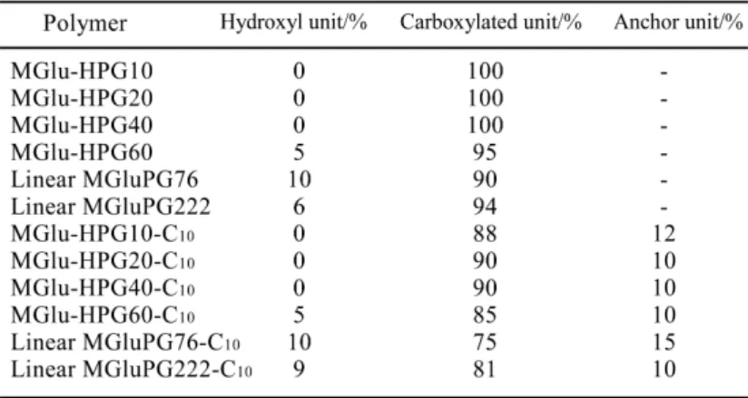
Synthetic polymers of various kinds reportedly interact with membranes and induce membrane fusion [4,5,7,9,10]. Considering that these synthetic polymers generally have a linear structure, it is presumed that their interaction with membranes might not be so effective to generate defective regions for initiation of membrane fusion as sterically bulky proteins do. To date, the influence of the backbone structure of fusogenic polymers on their membrane fusion activity remains unknown. Hyperbranched polymers tend to take on a three-dimensional and spherical structure, which differs from those of linear polymers taking on a random coil structure [11–15]. Recently, 3-methyl-glutarylated poly(glycidol) was synthesized by the authors. It destabilizes phospholipid membranes under a weakly acidic environment and causes membrane fusion [7]. For the present study, its analogous polymers were prepared using hyperbranched poly(glycidol)s (HPGs) with different degrees of polymerization (DP), 3-methyl-glutarylated HPGs (MGlu-HPGs). Results described herein demonstrate that the DP and backbone structure of the pH-sensitive polymers affected their pH-sensitive fusion properties and their performance as intracellular delivery vehicles.
3. 2. Materials and Methods 3. 2. 1. Materials
HPGs with DPs of 10, 20, 40 and 60, which are respectively designated as HPG10, HPG20, HPG40 and HPG60, were provided by Daicel Chemical Industries, Ltd. (Osaka,
37
Japan). Egg yolk phosphatidylcholine (EYPC) and L-dioleoyl phosphatidylethanolamine (DOPE) were kindly donated by NOF Co. (Tokyo, Japan). Pyrene, pyranine, 1-aminodecane and Triton X-100 were obtained from Tokyo Chemical Industries Ltd. (Tokyo, Japan).
p-Xylene-bis-pyridinium bromide (DPX) was from Molecular Probes (Oregon, USA). N-(7-Nitrobenz-2-oxa-1,3-diazol-4-yl)dioleoyl phosphatidylethanolamine (NBD-PE) and
lissamine rhodamine B-sulfonyl phosphatidylethanolamine (Rh-PE) were purchased from Avanti Polar Lipids (Birmingham, AL, USA). 3-Methylglutaric anhydride was obtained from Aldrich (Milwaukee, WI). 4-(4,6-Dimethoxy-1,3,5-triazin-2-yl)-4-methyl morpholinium chloride (DMT-MM) was from Wako Pure Chemical Industries Ltd. (Osaka, Japan). Ovalbumin (OVA) and fluorescein isothiocyanate (FITC) were purchased from Sigma (St. Louis, MO.). 3-Methylglutarylated linear poly(glycidol) (MGluPG) (Fig. 1) was prepared as previously reported using two kinds of poly(glycidol)s with different molecular weights: PG76 with number average molecular weight (Mn) of 5.6 × 10³ and weight average molecular weight (Mw) of 8.7 × 10³, and PG222 with Mn of 1.6 × 10⁴ and Mw of 2.5 × 10⁴, which were evaluated using gel permeation chromatography with Shodex KD-803 and KF805L columns (Showa Denko) and poly(ethylene glycol)s as the standard. Obtained polymers were designated as MGluPG76 and MGluPG222, respectively [7]. FITC-OVA was prepared by reacting OVA (10 mg) with FITC (11.8 mg) in 0.5 M NaHCO3 (4 mL, pH 9) at 4 °C for three
days and subsequent dialysis.
38
3. 2. 2. Synthesis of Hyperbranched Poly(glycidol) Derivatives
3-Methyl-glutarylated hyperbranched poly(glycidol) (MGlu-HPG) was prepared by reaction of HPG with varying DP with 3-methylglutaric anhydride. HPG10 (0.765 g) and LiCl (0.765 g) were dissolved in pyridine (18 mL) and 3.0 equiv. of 3-methylglutaric anhydride (3.98 g) was added to the solution. The mixed solution was kept at 115 °C for 24 h with stirring. Then, the reaction mixture was evaporated and dialyzed against water for 3 days. The product was recovered by freeze-drying. HPG20, HPG40 and HPG60 were also reacted with 3-methylglutaric anhydride by the same procedure. As anchor moieties for fixation of MGlu-HPG onto liposome membranes, 1-aminodecane was combined with carboxyl groups of MGlu-HPG. Each polymer was dissolved in water around pH 7.4, and 1-aminodecane (0.18 equiv. to carboxyl group of polymer) was reacted to carboxyl groups of the polymer using DMT-MM (0.18 equiv. to carboxyl groups of polymers) at room temperature for three days with stirring. The obtained polymers were purified by dialysis in water.
3. 2. 3. Cell Culture
DC2.4 cells, which were an immature murine DC line, were provided from Dr. K. L. Rock (Harvard Medical School, USA) and were grown in RPMI 1640 supplemented with 10% FBS (MP Biomedical, Inc.), 2 mM L-glutamine, 100 µM non-essential amino acids (Gibco, Inc.), 50 µM 2-mercaptoethanol (2-ME) and antibiotics at 37 °C [16].
3. 2. 4. Precipitation pH
Precipitation pH of polymers was determined by measuring the optical density of aqueous polymer solutions (0.25 mg/mL) at various pH. Polymers were dissolved in 30 mM sodium acetate and 120 mM NaCl aqueous solution of various pH. After 5 min incubation at 25 °C, optical densities of the polymer solutions at 500 nm were measured by using a spectrophotometer (Jasco V-520). Precipitation pH was determined using optical density-pH