CASE REPORT
Proximal Femoral Rotational Osteotomy for Symptomatic
Femoral Retroversion : A Case Report
Yasuaki Tamaki, M.D. ; Tomohiro Goto, M.D., Ph.D. ; Tomoya Takasago, M.D. ; Keizo Wada, M.D., Ph.D. ; Daisuke Hamada, M.D. Ph.D. ; and Koichi Sairyo, M.D., Ph.D.
Department of Orthopedic, Institute of Biomedical Sciences, Tokushima University Graduate School, Tokushima, Japan
Abstract : The Rotational osteotomy for femoral retroversion has been extremely rare despite the known as-sociation between femoral neck retroversion, hip pain, and osteoarthritis. Here, we describe a case of femoral neck retroversion for which proximal femoral rotation osteotomy. A 16-year-old boy with a past history of developmental dysplasia of the both hip treated conservatively presented with a complaint of pain in left hips. On physical examination, flexion of the left hip was limited to 90° with terminal pain. Internal rotation was also limited to 10°. Computed tomography (CT) showed -7.1° anteversion of the left femur. We performed rotational osteotomy to increase femoral anteversion because conservative treatment was not effective. The postopera-tive course was uneventful. At 12 postoperapostopera-tive months, his left hip pain was completely disappeared and fem-oral anteversion was 34° on CT scans. Retroversion of the femur is a distinct dynamic factor that should be con-sidered in the evaluation of mechanical causes of hip pain. Restoring the normal rotational alignment of the hip resulted in cure of the impingement due to femoral retroversion. J. Med. Invest. 67 : 214-216, February, 2020
Keywords : rotational osteotomy, retroversion, femoral neck retroversion, impingement
INTRODUCTION
Large variability in native femoral neck anteversion has been described in several reports to be in the range of -27.1° to 54.5° (1–3). Pierrepont et al. reported that 14% of patients requiring total hip replacement had extreme anteversion (< 0° or > 30°) and the incidence of femoral retroversion was 11% in males and 5% in females (3). Hartel et al. reported an incidence of retro-version of 7.8% in a general population (2). Because increased and decreased femoral anteversion are both associated with degeneration of the hip joint (4), corrective osteotomy for symp-tomatic patients is required. Femoral derotational osteotomy for excessive femoral anteversion has been well described (5–8). Al-though femoral neck retroversion can be a cause of hip pain and/ or osteoarthritis, rotational osteotomy for femoral retroversion has been extremely rare despite the known association between femoral neck retroversion, hip pain, and osteoarthritis (9, 10).
Here, we describe a case of femoral neck retroversion for which proximal femoral rotation osteotomy. The patient provided writ-ten informed consent for his data to be submitted for publication. The patient was treated in accordance with the Declaration of Helsinki.
CASE REPORT
A 16-year-old boy (height : 173 cm, body weight : 57 kg, body mass index : 19.0 kg/m2) with a past history of developmental
dysplasia of the both hip treated conservatively presented with a complaint of pain in left hip that started during a soccer match 3 years earlier. The pain was progressive and persisted during
daily activities, so he visited our hospital. On physical exam-ination, flexion of the left hip was limited to 90° with terminal pain and there was external rotation on hip flexion. Flexion of the right hip was limited to 90° without pain. Internal rotation was limited to 10° in both hips. Other range of motion values of the hip were below (extension : 20°/20° [right/left] ; abduc-tion : 50°/50° ; adducabduc-tion : 20°/20° ; external rotaabduc-tion : 90°/90°). The anterior impingement sign was positive in the left hip. Intraarticular injection of 1.0% xylocaine effected some relief temporarily. Japanese Orthopedic Association Score (JOA) score was 77 (range, 0 [worst] to 100 [best]).
Plain radiography and computed tomography (CT) showed -7.1° anteversion of the left femur and -8.0° anteversion of the right hip (Figure 1). Center edge (CE) angle was 23.6° for the left hip and 25.8° for the right hip. Both hips were considered to show mild acetabular dysplasia. On magnetic resonance imag-ing (MRI), no abnormal findimag-ings such as bone marrow lesion, osteoarthritis, or labral injury were detected. He was diagnosed as having femoroacetabular impingement due to femoral neck retroversion. Because conservative treatment was not effective, we planned a rotational osteotomy of the left femur to increase femoral neck anteversion.
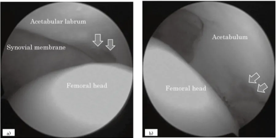
Intraoperative arthroscopic findings of the left hip revealed redness of the anterosuperior labrum and injury to the posterior acetabular cartilage of International Cartilage Repair Society (ICRS) grade 4 (Figure 2). After the arthroscopy, we performed rotational osteotomy to increase femoral anteversion. First, we performed subtrochanteric osteotomy and then internal rotation of the distal fragment by about 40° from -7° to 30° of anteversion using reference wires, which were inserted in the proximal and distal femur. Second, we performed osteosynthesis using an in-tramedullary nail (Gamma long nail 120°, 10×320 mm ; Stryker, Kalamazoo, MI). Partial weight bearing was permitted for 6 weeks postoperatively.
The postoperative course was uneventful. At 12 postoperative months, femoral anteversion was 34° on CT scans (Figure 3). Bone union at the osteotomy site was confirmed on plain radiog-raphy. Range of flexion and internal rotation of the left hip were
The Journal of Medical Investigation Vol. 67 2020
Received for publication July 29, 2019 ; accepted January 6, 2020. Address correspondence and reprint requests to Yasuaki Tamaki, MD, Department of Orthopedic, Institute of Biomedical Sciences, Tokushima University Graduate School, 3-18-15 Kuramoto, Tokushi-ma 770-8503, Japan and Fax : +81-88-633-0178.
215
The Journal of Medical Investigation Vol. 67 February 2020
Figure 3. Findings at 12 months postoperatively. a), b) Plain radiography shows bone union at osteotomy site. c), d) CT shows 35° femoral anteversion. Dotted line, posterior condylar axis. Solid line, femoral neck axis.
Figure 2. Intraoperative arthroscopy. a) Antero-lateral acetabular labrum shows slight redness but no detachment. b) Posterior acetabular cartilage with localized ICRS grade 4 defect.
Figure 1. Preoperative findings. a) A-P view of plain radiography revealed mild dysplasia of the both hip. b) c) Computed tomography of the right femur d) e) Computed tomography of the left femur. Dotted line: posterior condylar axis. Solid line: femoral neck axis.
Femoral head Acetabular labrum Synovial membrane Acetabulum Femoral head a) b)
216
Y. Tamaki, et al. Proximal femoral rotational osteotomy
improved to 115° and 50°, respectively, and JOA score was 98.The left hip pain were completely improved.
DISCUSSION
Femoroacetabular impingement is a condition involving abnor-mal contact that may arise as a result of abnorabnor-mal morphologic features involving the proximal femur and/or the acetabulum (11). Repetitive abutment between the femur and the acetabular rim was reported to occur during hip motion (12). As one cause, femoral rotational malalignment was associated with pain and functional disability and may be a contributing factor to hip im-pingement as well as instability (13). Retroversion of the femur may engage the rim much sooner, resulting in significant pain and loss of internal rotation during activities daily of living (14). Retroversion of the femur is a distinct dynamic factor that should be considered in the evaluation of mechanical causes of hip pain. We detected impingement between the anterolateral aspect of the femoral neck and the anterosuperior acetabular rim using three-dimensional (3D) simulation based on preoperative CT data. In the simulation, bony impingement was found to occur on 90° flexion and 23° internal rotation of the hip. This impinge-ment point was matched to the intraoperative findings of redness of the acetabular labrum. Based on these results, we hypothesize the pathology in this case as follows. First, the impingement was attributable to abnormal hip congruency due to femoral retroversion. Second, a shearing force that was amplified by the principle of the leverage pushed on the femoral head in the posterior acetabulum. Third, the posterior acetabular cartilage defect was the result of repetitive abutment between the femur and the acetabular rim.
The standard treatment for femoroacetabular impingement is to modified abnormal alignment such as surgical hip dislocation, open osteochondroplasty, arthroscopic surgery and correction osteotomy. Rotational femoral osteotomy has, to our knowledge, been described in only two reports in the literature. Wagner et al. described proximal femoral rotational and distal femoral varus osteotomies for femoral retroversion and genu valgum (15), and Tonnis et al. reported that corrective procedures for decreased femoral anteversion should be considered in order to correct the anatomical deformity, decrease pain, and avoid the development of osteoarthritis (10). They also stated that the goal is to achieve 15° to 20° of anteversion. Our preoperative plan was for a total amount of correction of about 40°, from-8° to 30° anteversion, which is somewhat large compared with normal femoral anteversion and was decided on in order to avoid undercorrection of postoperative femoral anteversion and to reduce stress distribution on the acetabular joint surface of the posterior aspect. The 3D simulation based on postoperative CT scans showed bony impingement with 90° flexion and 52° internal rotation of the hip. The range of internal rotation was significantly improved postoperatively. In this case, the right femur had a similar morphology of 8° retroversion compared with the left hip, although there was no right hip pain. If the patient develops right hip pain in the future, we may consider a similar treatment option.
In conclusion, we performed rotational proximal femoral oste-otomy to correct femoral retroversion. Restoring the normal ro-tational alignment of the hip resulted in cure of the impingement due to femoral retroversion.
CONFLICT OF INTEREST
The authors have no conflict of interest to declare.
REFERENCES
1. Bargar WL, Jamali AA, Nejad AH : Femoral anteversion in THA and its lack of correlation with native acetabular anteversion. Clin Orthop Relat Res 468 (2) : 527–32, 2010 2. Hartel MJ, Petersik A, Schmidt A, Kendoff D, Nuchtern J,
Rueger JM, Lehmann W, Grossterlinden LG : Determina-tion of Femoral Neck Angle and Torsion Angle Utilizing a Novel Three-Dimensional Modeling and Analytical Tech-nology Based on CT Datasets. PLoS One 11 (3) : e0149480, 2016
3. Pierrepont JW, Marel E, Bare JV, Walter LR, Stambouzou CZ, Solomon MI, McMahon S, Shimmin AJ : Variation in femoral anteversion in patients requiring total hip replace-ment. Hip Int : 1120700019848088, 2019
4. Reikerås O, Høiseth A : Femoral neck angles in osteoarthri-tis of the hip. Acta Orthop Scand 53 (5) : 781–4, 1982 5. Iobst CA, Ansari A : Femoral derotational osteotomy using
a modified intramedullary nail technique. Tech Orthop 33 (4) : 267–70, 2018
6. Matuszewski PE, Herzenberg JE : Percutaneous femoral derotational osteotomy for excessive femoral torsion. Ortho-pedics 37 (10) : 653–4, 2014
7. Mei-Dan O, McConkey MO, Bravman JT, Young DA, Pascual-Garrido C : Percutaneous femoral derotational osteotomy for excessive femoral torsion. Orthopedics 37 (4) : 243–9, 2014
8. Nelitz M : Femoral derotational osteotomies. Curr Rev Mus-culoskelet Med 11 (2) : 272–9, 2018
9. Tönnis D, Heinecke A : Diminished femoral antetorsion syn-drome : a cause of pain and osteoarthritis. J Pediatr Orthop 11 (4) : 419–31, 1991
10. Tönnis D, Heinecke A : Acetabular and femoral antever-sion : relationship with osteoarthritis of the hip. J Bone Joint Surg Am 81 (12) : 1747–70, 1999
11. Ganz R, Parvizi J, Beck M, Leunig M, Nötzli H, Siebenrock KA : Femoroacetabular impingement : a cause for osteoar-thritis of the hip. Clin Orthop Relat Res 417 : 112–20, 2003 12. Genovese E, Spiga S, Vinci V, Aliprandi A, Di Pietto F,
Coppolino F, Scialpi M, Giganti M : Femoroacetabular im-pingement : role of imaging. Musculoskelet Surg 97 (Suppl 2) : S117–26, 2013
13. Waisbrod G, Schiebel F, Beck M : Abnormal femoral an-tetorsion-a subtrochanteric deformity. J Hip Preserv Surg 4 (2) : 153–8, 2017
14. Bedi A, Dolan M, Leunig M, Kelly BT : Static and dynamic mechanical causes of hip pain. Arthroscopy 27 (2) : 235–51, 2011
15. Wagner R, Barcak EA : Simultaneous proximal femoral rotational and distal femoral varus osteotomies for femoral retroversion and genu valgum. Am J Orthop 41 (4) : 175–8, 2012