One-, two - dimensional silver(I) coordination polymer with poly(benzylsulfanyl)lbenzene and
photo-induced property
Yusaku Suenaga,*a,b Hisashi Konaka,a Takeshi Sugimoto,a Emi Miyako,a Takayoshi Kuroda-Sowa,a Masahiko Maekawab and Megumu Munakataa
a Department of Chemistry , Kinki University, Kowakae, Higashi-Osaka, Osaka 577-8502, Japan E-mail: [email protected]
b Research Institute for Science and Technology , Kinki University, Kowakae, Higashi-Osaka, Osaka 577-8502, Japan
(Received, November, 15, 2002)
Abstract
Three silver(I) complexes [Ag2(3bsb)2(C104)2] (1) (3bsb; 1,3,5-tris(benzylsulfanyl)benzene), [Ag(4bsb)](C104) (2) (4bsb; 1,2,4,5-tetrakis(benzylsulfanyl)benzene) and [Ag2(6bsb)(C2F5C00)](C2F5C00)(toluene)2 (3) (6bsb;
hexakis(benzylsulfanyl)benzene) have been prepared and their molecular structures were determined by X-ray crystallography. In 1, the silver ion prefers a tetrahedral coordination geometry and produces a two-dimensional sheet structure. Irradiation of this complex I with UV light changes its color from white to red; Heat makes the irradiated sample revert back to white. However, only 3bsb has no characteristic like this photochromism. Complex 2 formed linear chain structure consist of Ag-S in the silver coordination polymer. On the other hand complex 3 have zigzag chain structure consist of silver ions with trigonal coordination geometry. However, the latter two complexes have no indicated photochromism such as complex 1.
Key words: Silver(I) complexes, Poly(benzylsulfanyl)benzene, Photo-induced property, Crystal structures
1. Introduction
The study of the self-assembling process of metal complexes continues to be a subject of considerable current interest in the context of developing new solid-state polymeric materials with specific
architectural and functional features.' Some of the complexes exhibit interesting behavior due to external stimuli and have been the subject of a number of physical investigations.2
Metal complexes with chromotropic property in the solid state have been reported. (1) Charge-transfer mechanism; for example [Ni(1,2-dione- dioximate)2]3. (2) Hydrogen bonding cleavage mechanism; for example [Cu(N,N' -di ethyl-
ethylene diamine)2](C104)24. (3) Rearrangement of
coordination geometry; for example [Ni(2,3- buthanediamine)2]Br25. (4) Mix valence mechanism; for example like Fe(II)-Fe(III) complexes.' Also, M. Munakata7 reported copper(I) coordination polymer with dithienylethene undergoes reversible color changes, turning orange and red under the effect of UV irradiation.
The silver(I) ions are regarded as extremely soft acids favoring coordination to soft bases, such as ligands containing S and unsaturated N. Silver(I) complexes with these soft ligands give rise to an interesting array of stereo chemistries and geometric configurations, with the coordination numbers of two to six all occurring. Therefore, silver(I) is often used to construct network structures.' Recently, we have been exploring ways
to modify the substituents to tailor the polymer structures with silver(I)/poly(phenysulfanyl)- substituted aromatic systems, which are well known for their host-guest chemistry in the solid state,' as to the mode of polymerization, dimensionality and framework.10
In this paper, we report three silver(I) coordination polymer with poly(benzylsulfanyl)benzene, which are different the number and position of the substituted groups. Irradiation of complex 1 with light changes its color from white to red. The crystal structure and the photo-induced property of these complexes are discussed.
2. Experimental
General
Preparations were performed using the usual Schlenk techniques. All solvents were dried and distilled by standard methods before use. The standard chemicals were obtained from Wako Chemical Co., Japan, and used without further purification. The IR spectra were measured as KBr discs on a JASCO FT-8000 spectrometer. The 1H NMR spectra were obtained with a Varian MERCURY-300 FT spectrometer at 23 °C.
Tetramethylsilane was used as the internal reference. ESR spectra were obtained on a JEOL JES-TE200 ESR spectrometer. Diffuse reflection spectra were obtained with a HITACHI U-4000 in the 350-800 nm ranges.
Syntheses
1,3,5-Tris(benzylsulfanyl)benzene (3bsb): In a three-necked flask equipped with a magnetic stirring bar, septum cap and argon gas inlet tube was placed 250 mg (1.44 mmol) of 1,3,5-
benzenetrithiol (Nippon Fine Chemical Co., Ltd.) and 50 cm3 of dry dimethylformamide (Tokyo Chemical Industry Co., Ltd.). To this solution was added with stirring 107 mg (4.46 mmol) of sodium hydride. Pale yellow precipitates were obtained after 2 h. To this stirred suspension was added bis(triphenylphosphine)palladium(II) dichloride as the catalyst and benzylchloride (1.09 g, 8.61 mmol). After 20 h at 65 °C, the TLC (silica gel, 3:1 hexane-diethylether) showed only one spot. At this point the reaction mixture was added to 100 cm3 of cool water and a yellow precipitate was filtered off and copiously washed with water and then sparingly with cold methanol to yield 460 mg (72 %) of a pale yellow powder. Recrystallization from toluene-n-pentane yielded yellow plate-like crystals (281 mg, 44 %), mp 60-61 °C. IR (KBr disc): v/cm 1 w, 3054w, 3028w, 2925w, 1553s, 1492m, 1451m, 1408m, 1241m, 1107m, 1071m, 922m, 834m, 795m, 782m, 763m, 748m, 718m and 704s. 1H NMR (300 MHz, CDC13): bH/ppm
3.979 (6 H, s), 6.970 (3 H, s), 7.207-7.322 (15 H, m). EI Mass spectrum: m/z 444 (100%, M).
(Found: C, 73.10; H, 5.59. Calc. for C27H24S3: C, 72.92; H, 5.45 %).
1,2,4,5-Tetrakis(benzylsulfanyl)benzene
(4bsb)": In a three-necked flask equipped with a magnetic stirring bar, septum cap and argon gas inlet tube was placed 250 mg (1.21 mmol) of 1,2,4,5-benzenetetrathiol (Nippon Fine Chemical Co., Ltd.) and 50 cm3 of dry dimethylformamide (Tokyo Chemical Industry Co., Ltd.). To this solution was added with stirring 125 mg (5.21 mmol) of sodium hydride, pale yellow precipitates were obtained after 2 h. To this suspension was added bis(triphenylphosphine)palladium(II) dichloride as catalyst and benzylchloride (1.23 g, 9.72 mmol), stirring. After 4 h at 65 °C, the TLC (silica gel, 3:1 hexane-diethylether) showed only one spot. At this point the reaction mixture was added to 100 cm3 of cool water and a yellow precipitate was filtered off and copiously washed with water and sparingly with cold methanol.
Recrystallization from toluene-n-pentane yielded yellow columns (610 mg, 89%), mp 148-149 °C.
IR (KBr disc): v/cm-1 3060m, 3028m, 2920m, 1601m, 1493m, 1454m, 1425s, 1306m, 1253m, 1120m, 1065m, 859m, 775m, 761m, 693s, 659m and 499m. 1H NMR (300 MHz, CDC13): 6H/ppm 3.886 (8 H,^), 6.903 (2 H, s), 7.200-7.317 (20 H, m). EI Mass spectrum: m/z 566 (100%, M).
(Found: C, 72.05; H, 5.37. Calc. for C34H30S4: C, 72.03; H, 5.35%).
Hexakis(benzylsulfanyl)benzene (6bsb): In a three-necked flask equipped with a magnetic stirring bar, septum cap and argon gas inlet tube was placed 5.0 g (mmol) of sodium hydride (Aldrich Chemical Co., Inc.) and 80 cm3 of dry dimethylformamide (Tokyo Chemical Industry Co., Ltd.). To this suspension was added with stirring 14.1 ml (120 mmol) of toluenethiol under cooling bath. After stirring during 30 min, 2.85 g (10 mmol) of hexachlorobenzene via a syringe through the septum cap. The addition was at a relatively fast rate, resulting in a mildly exothermic reaction.
After 2 h at room temperature, the TLC (silica gel, 3:1 hexane-diethylether) showed only one spot. At
nn
this point the reaction mixture was added to 400 cm3 of cold methanol and pale yellow needles was filtered off and copiously washed with water and sparingly with ethanol to yield of pale yellow needles. Recrystallization from THF-n-pentane yielded yellow needles (2.19 g, 27%), mp 129 °C.
IR (KBr disc): v/cm-1 3058m, 3026m, 2928m, 1599m, 1491s, 1451m, 1428m, 1271m, 1066m, 1026m, 772s and 701s. 1H NMR (300 MHz, DMF): 8H/ppm 4.112 (12 H, s), 7.252 (30 H, m).
EI Mass spectrum: m/z 810 (100%, M). (Found: C, 70.55; H, 5.20. Calc. for C48H42S6: C, 71.07; H, 5.22%).
[Ag2(3bsb)2(C1O4)2] 1: Single crystals suitable for X-ray analysis were obtained by the reaction of AgC1O4 (4.1 mg, 2 mM) dissolved in 5 cm3 of acetone and an acetone solution (5 cm3) containing 3bsb (4.3 mg, 0.01 mM). The mixture was stirred for 1 h and the yellow filtrate was then transferred to a glass tube and layered with 2 cm3 of n-pentane as the diffusion solvent. After standing for 14 d at 5 °C, the colorless plate crystals of complex 1 were isolated. Yield: 61%
based on silver. IR (KBr disc): v/cm-1 w, 3054w, 3029w, 2925w, 1553s, 1493m, 1452m, 1408m,
1241m, 1144s, 1114s, 1089s, 923m, 835m, 795m, 782m, 762m, 748m, 718m, 704s, 628m and 462m.
[Ag(4bsb)](C104) 2: Crystals suitable for X- ray analysis were obtained by procedures similar to complex 1. AgC1O4 (6.4 mg, 5 mM) was dissolved in 1 cm3 of acetone transferred to a glass tube and layered with a THF solution (1 cm3) containing 4bsb (18.2 mg, 2 mM). After standing for 7 d at ambient temperature colorless plate crystals of complex 2 were isolated. Yield: 63%
based on silver. IR (KBr disc): v/cm-1 m, 3059w, 3028w, 2924w, 1493m, 1454m, 1427m, 1089s, 771m, 695m, 627m and 467m.
[Ag2(6bsb)(C2F5COO)](C2F5COO)(tolue ne)2 3: Crystals suitable for X-ray analysis were obtained by procedures similar to complex 1.
AgC2F5COO (13.5 mg, 10 mM) was dissolved in 5 cm3 of toluene and to this solution was added a toluene solution (5 cm3) containing 6bsb (20.3 mg,
5 mM) and layered with 2 cm3 of n-pentane as the diffusion solvent. After standing for 7 d at ambient temperature yellow brick crystals of complex 3 were isolated. Yield: 54% based on silver. IR (KBr disc): v/cm 1 3061w, 3028w, 2965w, 1663s, 1493m, 1454m, 1325m, 1210s, 1160s, 1029s, 731s and 698s. (Found: C, 46.71; H, 3.26. Calc. for C54H42Ag2F 1004S6: C, 47.94; H, 3.13%).
Crystallography
Crystal data for complexes 1, 2 and 3 are given in Table 1. The structures were solved by a direct method12 and refined by full-matrix least-squares
analysis on F2. Data collection for these complexes were performed on an MSC Mercury CCD area detector coupled with a Rigaku AFC-7R diffractometer. All the full-occupancy non- hydrogen atoms were anisotropically refined. The positions of all the hydrogen atoms were determined from difference electron density maps and included, but not refined. Atomic scattering factors and anomalous dispersion terms were taken from the usual sources.13 Computations were carried out using TEXSAN.14 The selected bond lengths and bond angles for 1, 2 and 3 are listed in Table 2.
Table 1.
1
Crystallographic data for complexes
2 3
Chemical formula Formula weight Crystal system Space group
Unit Cell Parameters a/ A
b/ A c/ A a/ ° /3/°
Unit Cell Volume V/ A3
Z
µ(Mo-Ka) / c m- 1 No. reflections measured No. observed [I >2.006(1 )]
R1 wR2 77K
reflections
C5 4H4 8A g2 C 12 O8 S6 1303.97
Triclinic PI (no . 2)
12.390(1) 15.297(2) 15.576(2) 1 1 1.798(6) 90.357(3) 93.627(5) 2734.3(6) 2 10.93 32372 10664 0.033 0.070 150.0
C3 4H3 0AgC1O4 S4 774.17
Orthorhombic Pca2,(no. 29)
18.0568(4) 9.1575(5) 19.5834(4)
3238.2(2) 4
9.99 53561 6658 0.020 0.045 150.0
C6 8H5 8Ag2 F 1 0O4 S6 1537.28
Monoclinic P2 ,/n(no. 14)
15.480(1) 14.755(1) 28.506(2)
88.861(4)
6509.6(8) 4 8.69 39344
11901 0.052 0.116 120.0 RI=E11Fol - (Fc1 1 /E[Fol, wR2 =[1w(Fo2 - Fc2 )2/ Eco(Fo2 )2 ~ 1 / 2
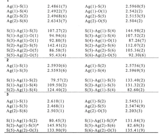
Table 2. Selected bond length(A) and bond angle(°) for complexes
1
Ag(1)-S(1) Ag(1)-S(4) Ag(2)-S(2) Ag(2)-S(6) S(1)-Ag(1)-S(3) S(1)-Ag(1)-O(1) S(3)-Ag(1)-O(1) S(2)-Ag(2)-S(5) S(2)-Ag(2)-O(5) S(5)-Ag(2)-O(5)
2.4861(7) 2.4922(7) 2.4968(6) 2.6343(7) 107.27(2)
94.94(6) 92.80(5) 142.41(2) 86.58(5) 95.43(4)
Ag(1)-S(3) Ag(1)-O(1) Ag(2)-S(5) Ag(2)-O(5) S(1)-Ag(1)-S(4) S(3)-Ag(1)-S(4) S(4)-Ag(1)-O(1) S(2)-Ag(2)-S(6) S(5)-Ag(2)-S(6) S(6)-Ag(2)-O(5)
2 2 2 2
.5960(5) .542(2) .5153(5) .504(2)
144 107 87 112 105 92
.98(2) .52(2) .26(7) .07(2) .36(2) .30(6) 2
Ag(1)-S(1) Ag(1)-S(3) S (1)-Ag(1)-S(2) S(1)-Ag(1)-S(4) S(2)-Ag(1)-S(4)
3 Ag(1)-S(1) Ag(1)-S(3) Ag(2)-S(6) S(1)-Ag(1)-S(2) S(2)-Ag(1)-S(3)*
S(51-Aa(21-O(31
2.5930(6) 2.5593(6)
79.57(2) 109.50(2) 124.40(2)
2.610(1) 2.468(1) 2.503(1)
80.45(3) 145.95(3) 133.90(9)
Ag(1)-S(2) Ag(1)-S(4) S(1)-Ag(1)-S(3) S(2)-Ag(1)-S(3) S(3)-Ag(1)-S(4)
Ag(1)-S(2) Ag(2)-S(5) Ag(2)-O(3) S(1)-Ag(1)-S(3)*
S(5)-Ag(2)-S(6) S(61-Aa(21-O(3 )
2.
2.
5756(5) 5969(5)
133 131 82
2 2 2
.40(2) .32(2) .60(2)
.545(1) .5474(9) .203(3)
131.
82.
135.
84(3) 69(3) 41(9)
3. Results and discussion
X-ray crystal structure
The molecular structure together with the atomic numbering scheme is given in Fig. 1. The solid- state structure of complex 1 is quite unusual in that it contains two slightly different structure motifs (Figs. 1(a) and (b)) in the same crystal. The silver(I) ion is coordinated to three S atoms from three different 3bsb donors and the 0 atom from the counter anion in a distorted tetrahedral coordination sphere. The Ag-S distances are in the range of 2.4861-2.6343 A, and the Ag-0 distances are in the range of 2.504-2.542 A. The shortest S•••S distances are 4.09 A. The distances between the two core benzene rings are 3.37 A in the dimeric molecule. The Ag•••Ag contact for adj acent metal ions in Fig. 1(a) is 8.15 A while in Fig. 1(b), it is 8.17 A. The crystal packing structure is shown in Fig. 2. Each dimeric species acts as a bridge to produce a two dimensional sheet having a
cavity structure. The cavity size is 7.86 A X 11.38 A, and occupied by two counter anions. The inter-sheet interaction, the shortest S•••S distances, are 17.31 A, indicative of negligible interactions of S•••S contacts due to the long separations (far greater than twice the van der Waals radius of 3.70 A ).
coordinated by two 4bsb donors in a tetrahedral coordination sphere (Fig. 3). The ligand acts as a bridge between pairs of Ag atoms to produce a linear chain polymer. Four benzyl groups are perpendicular to the core benzene ring plane in complex 2.
(a)
Fig.1 Ortep views of the dimeric structure (a) and (b) in complex 1, showing 50% thermal ellipsoids.
Fig.3 Linear chain structure, showing 50% thermal ellipsoids and schematic view of complex 2.
Fig.2 Two-dimensional schematic view of complex omitted for clarity).
sheet I (side
structure and benzyl groups
On the other hand a single-crystal structure determination reveals that coordination compound 2 contains an infinite chain structure. In the cation, all 4bsb units are attached to the two silver centers on the S atoms. The Ag-S distances are in the range of 2.5593(6)-2.5969(5) A. Each silver ion is
[Ag2(6bsb)(C2F5COO)](C2F5COO)(toluene)2 3 were obtained by using 6bsb in place of 4bsb as the ligand. The ligand acts as a bridge between pairs of Ag atoms to produce a chain polymer, only five out of a possible six sulfur atoms are coordinated. This is probably due to the steric constrains that would be imposed by complexation. The un-coordinated legs play an important role in deteunining the one- dimensional polymer structure of the complex. The silver(I) coordination geometry is different from complex 2. Ag(1) is coordinated by three sulfur atoms of 6bsb donors. Also Ag(2) is coordinated by two sulfur atoms of 6bsb donors and one oxygen atom from the counter anion. As a result silver ion has distorted trigonal geometry. The three Ag-S distances are in the range of 2.468(1)-2.610(1) A.
The Ag(2)-O(3) distance is 2.230(3) A. The crystal structure of complex 3 is shown in Fig. 4. In contrast linear chain structure of complex 2, complex 3 consist of a zigzag chain structure. As the coordination sphere of many metal ions is flexible, a large number of possibilities exists for the network for a given ligand topicity. In the complex I of 3bsb which is tritopic ligand, the observed networks are 3-connected. The silver(I) is
coordinated to three sulfurs of 3bsb and an oxygen of perchlorate anion. Therefore, the structure is characterized by a two-dimensional net.
to lu en e
photochromism is due to a solid-state transformation and that coloration-decoloration is not the result of a surface oxidation reaction.
Furthermore, the process must be a relatively low- energy transformation since it requires only temperatures in the range of 50-100 °C to effect bleaching.
O2CFSC2 02CF5C2
Aa
p2CF5C2O2CF5C2
Fig.4 (a) Ortep view showing 50% thermal ellipsoids. (b) One-dimensional zigzag chain structure (side benzyl groups omitted for clarity).
(c) Schematic view of complex 3.
Photo-induced property
Complex I is a white, crystalline solid which quickly turns red when exposed to light or solar irradiation. It can be shown, as follows, that this change in color is due to a solid process: The infrared spectrum of a KBr pellet before and after coloration is unchanged (Fig. 5). Heating of a colored sample pellet returns to white color.
Exposure of a sample to ultraviolet or visible light under strictly anaerobic conditions has no deleterious effect on the photochromism. The above observations indicate that the
3000 2000 1500 1000 400 wave number (cm-1)
Fig.5 Infrared spectra of complex 1. (a) before irradiation (b) after irradiation.
In order to gain more insight into the mechanism responsible for the observed effect, the ESR and powder reflectance spectra were measured.
A red sample whose color was generated by exposure to light showed a ESR signal, but white sample whose de-color was generated by heating showed no signal (Fig. 6). The two closely spaced lines, one at g = 2.008 and another at g = 2.003 with approximate line widths of 14 and 10 G, respectively, were observed at room temperature.
These two g values could correspond to unpaired electrons centered on the sulfur and carbon, respectively, and the fact that the line width does not change with temperature implies that the electrons are localized on these atoms. Heating this sample above its bleaching temperature caused the two sharp signals to disappear (Fig. 6(b)). This photo-induced color change behavior is similar to 4bsb." However, interestingly, this reversible
chromic phenomenon has been realized by the Ag- 3bsb complex for the first time, but not by 3bsb.
solubility for solvents. This is change.
irreversible color
Fig.6 ESR of complex 1 in the solid state. (a) after irradiation (b) heating of (a).
As shown in Fig. 7, the powder reflectance spectra of before and after the coloration are clearly different. The spectra of complex 1 (white) and after coloration of 1 (red) have Amax values of 347 nm and 341, 425, 609 nm, respectively. The bands are tentatively assigned as follows by comparison with the spectra of 4bsb. The bands observed at 341 and 425 nm for 1 (red) are ascribed to the metal-to- ligand and metal-to-ligand(radical) charge transfer, respectively. A strong, broad band at 609nm in assigned to thyil radicals.
For complex 2 there is no visible color change upon any reasonable period of irradiation. Complex 3 showed color change after irradiation in the single-crystalline state. The powder reflectance spectra of complex 3 (yellow) and after coloration of 3 (green) have Xmax values of 477 nm and 466, 627 nm, respectively. However, decomposition of complex structure is occurred resulting increased
wavelength (nm)
Fig.7 Powder reflectance spectra of complex 1. (a) after irradiation (b) before irradiation.
In conclusion, we have prepared three Ag(I) complexes with poly(benzylsulfanyl) substituted benzene. The different Ag(I) coordination polymer were obtained depend on position and the number of the benzylsulfanyl substituents on the benzene ring. We also discovered a new photochromic silver(I) coordination polymer, which is constructed of a two-dimensional sheet structure, and appears to be unique. This behavior can explain the separation of the benzyl radicals or thyil radicals from results of the ESR signals. The Ag-S distance is shorter by 0.08 — 0.1 A than the other complexes. Activation by Ag ions is easy to cause homolytic cleavage of the benzyl-sulfur bonds to produce radical species.
4. Acknowledgements
This work was partially supported by a Grant-in-Aid for Science Research (c) (no. 12640547) from the Japan Society for the Promotion of Science. The authors thank Professor S. Ito and Dr. M. Iwasaki (Kinki University) for the powder reflectance spectra measurements.
References
[1] M. Munakata, L. P. Wu and T. Kuroda-Sowa, Adv. Inorg. Chem., 1999, 46, 173; J. -M. Lehn, Angew.
Chem., Mt. Ed. Engl., 1988, 27, 89; A. Muller, H. Reuter and S. Dillinger, Angew. Chem., Int. Ed. Engl., 1995, 34, 2328; V. Balzani, Terahedron, 1992, 48, 10443; E. C. Constable, Adv. Inorg. Chem., 1989, 34, 1; M. Munakata, T. Kuroda-Sowa, M. Maekawa , A. Hirota and S. Kitagawa, Inorg. Chem., 1995, 34, 2705.
[2] S. L. Gilat, S. H. Kawai and J.-M, Lehn, Chem. Eur. J., 1995, 1, 275; S. H. Kawai, S. L. Gilat, R.
Ponsinet and J.-M. Lehn, Chem. Eur. J., 1995, 1, 285; M. Irie, In Photoreactive Materials for Ultrahigh Density Optical Memory, M. Irie, Ed., Elsevier, Amsterdam , 1994.
[3] I. Shirotani, A. Kawamura, K. Suzuki, W. Utsumi and T. Yagi, Bull. Chem. Soc. Jpn., 1991 64 1607; I.
Shirotani, K. Suzuki, T. Suzuki, T. Yagi and M. Tanaka, Bull. Chem. Soc. Jpn., 1992 65 1078.
[4] I. Grenthe, P. Paoletti, M. Sandstrom and S. Glikberg, Inorg. Cem., 1979, 18, 2687; L. Fabbrizzi, M.
Micheloni and P. Paoletti, Inorg. Chem., 1974, 13, 3019 .
[5] Y. Ihara, Y. Fukuda and K. Sone, Inorg. Chem., 1987, 26, 3745; K. L. Bray and H. G. Drickamer, J.
Phys. Chem., 1989, 93, 7604; K. L. Bray and H. G. Drickamer, J . Phys. Chem., 1990, 94, 2154; K.
Nakajima, M. Kojima, S. Azuma, R. Kasahara, M. Tsuchimoto, Y. Kubozono , H. Maeda, S. Kashino, S.
Ohba, Y. Yoshikawa and J. Fujita, Bull. Chem. Soc. Jpn ., 1996, 69, 3207.
[6] S. E. H. Etaiw and A. M. A. Ibrahim, J. Organomet. Chem., 1996, 522, 77.
[7] M. Munakata, L. P. Wu, T. Kuroda-Sowa, M. Maekawa, Y. Suenaga and K. Furuichi, J. Am. Chem.
Soc., 1996, 118, 3305.
[8] Y. Suenaga, M. Maekawa, T. Kuroda-Sowa and M. Munakata, J. Chem. Soc., Dalton. Trans., 2000, 3620 and references therein.
[9] D. D. MacNicol and D. R. Wilson, J. Chem. Soc., Chem. Commun., 1976, 494; A. D. U. Hardy, D. D.
MacNicol and D. R. Wilson, J. Chem. Soc., Perkin II, 1979, 1011.
[10] Y. Suenaga, M. Maekawa, T. Kuroda-Sowa and M. Munakata, J. Chem. Soc., Dalton Trans., 2000, 3620; Y. Suenaga, K. Kitamura, T. Kuroda-Sowa, M. Maekawa and M . Munakata, Inorg. Chim. Acta,
2002, 328, 105.
[11] S. D. Cox, C. W. Dirk, F. Moraes, D. E. Wellman, F. Wudl, M. Soltis and C. Strouse, J. Am. Chem. Soc., 1984, 106, 7131; F. Maiolo, L. Testaferri, M. Tiecco and M. Tingoli , J. Org. Chem., 1981, 46, 3070; P.
A. Odorisio, S. D. Pastor, J. D. Spivack and R. K. Radebaugh , Phosphorus Sulfur, 1982, 13, 309.
[12] DIRDIF: Direct methods for Difference structures - an automatic procedure for phase extension and refinement of difference structure factors. P. T. Beurskens, Technical Report 1984/1, Crystallographic
Laboratory, Toenooived, Nijimegen, 1984.
[13] International Tables for X-Ray Crystallography, Kynoch Press, Birmingham, 1974, vol 4.
[14] TEXSAN-IEXRAY, Structure Analysis Package, Molecular Structure Corporation, The Woodlands, TX, 1985.