筋活動による骨格筋損傷とその治癒過程における一酸化窒素の関与
2013 年
櫻井 智野風
目 次
第1章 研究の背景と目的
1.1. はじめに・・・・・・・・・・・・・・・・・・・・・・・・・・・・・・・・・・・・・・・・・・・・・・3 1.2. 先行研究
1.2.1. 一酸化窒素 (Nitric Oxide. NO) とは ・・・・・・・・5 1.2.2. 骨格筋活動とメカニカルストレス ・・・・・・・・・・・・7 1.2.3. 骨格筋損傷と炎症 ・・・・・・・・・・・・・・・・・・・・・・・9 1.2.4. 骨格筋損傷の再生機序 ・・・・・・・・・・・・・・・・・10 1.2.5. 骨格筋損傷と遅発性筋痛 ・・・・・・・・・・・・・・・・13 1.3. 研究の目的と研究課題 ・・・・・・・・・・・・・・・・・・・・・・・・・・・・・・・・15 第2章 筋活動時の NO 生成 2.1. 走運動負荷 ・・・・・・・・・・・・・・・・・・・・・・・・・・・・・・・・・・・・・・・・・・・・・・・・19 2.2. 過重力負荷 ・・・・・・・・・・・・・・・・・・・・・・・・・・・・・・・・・・・・・・・・・・・・・・・・34 2.3. 外力負荷 ・・・・・・・・・・・・・・・・・・・・・・・・・・・・・・・・・・・・・・・・・・・・・・・・・・54 第3章 外力負荷時の NO 生成と筋損傷治癒 ・・・・・・・・・・・・・・・・・・・・73
第4章 外力負荷時の NO 生成と遅発性筋痛 ・・・・・・・・・・・・・・・・・・100
第5章 まとめ ・・・・・・・・・・・・・・・・・・・・・・・・・・・・・・・・・・・・・・・・・・・・・・116
引用文献 ・・・・・・・・・・・・・・・・・・・・・・・・・・・・・・・・・・・・・・・・・・・・・・・・・・・・122 謝 辞 ・・・・・・・・・・・・・・・・・・・・・・・・・・・・・・・・・・・・・・・・・・・・・・・・・・・・140
第 1 章 研究の背景
1.1. はじめに
われわれの生体において運動刺激は生理反応に変調をきたすストレス要因であり, 身体の諸器官に影響を及ぼす.そのストレスは,時として骨格筋の組織に損傷を引き 起こす.運動刺激時の骨格筋内では,酸素消費量の増大,血流の増加などの代謝的 ストレス,あるいは炎症反応を伴う伸張性筋収縮などのメカニカルストレスの増加により フリーラジカル生成が亢進するが,この生成が過剰となると,脂質の過酸化やタンパク 質酸化,細胞膜障害,血管透過性抗進などにより組織の損傷を惹起する(Best et al., 1999: Tatsumi et al., 2002).また,損傷の回復時にはこのフリーラジカルの減少が伴う ことも明らかになっている(Tatsumi, 2010) .生体内に発生するガスである一酸化窒素(Nitric Oxide, NO)は,生態現象の多くの 場面で様々な現象を引き起こすメディエーターとして知られている(Anderson et al., 2000; Kobzik et al.,1995; Moncada et al.,1991; Moncada et al.,1993).NO はフリーラジ カルの一種であり,生体内において抗酸化酵素の活性を減弱させ酸化ストレスを増大 させる一方で,その殺菌力により炎症を縮小しアポトーシスを抑制するという善悪両面 の作用を持つことが知られている(Alderton et al.,2001; Soneja et al.,2005; Sakurai et al.,2005; Sriwijitkamol et al.,2006; Filippin et al.,20111; Filippin et al.,20112).また,
NO は細胞内情報伝達物質として,炎症性メディエーターであるサイトカイン・ケモカイ ンの分泌を調節することが知られている(Rigamonti et al., 2013). これらメディエーターの分泌調節は,骨格筋損傷後に発生する遅発性筋痛の発症 に関与すると考えられる.このように,NO は骨格筋損傷と治癒,炎症や発痛作用に関 与していることが考えられるが,その詳細は不明である. 本研究では動物モデルを対象に, 伸張性収縮モデル,高強度走運動,過重力負 荷といった様々な筋活動時のメカニカルストレスと骨格筋内における NO 生成の関連 を検討し,メカニカルストレスにより作成した骨格筋損傷とその治癒に及ぼす NO の役 割について明らかにすることを目的とする.
1.2. 先行研究
1.2.1. 一酸化窒素
Furchgott と Zawadzki(1980)により内皮由来血管弛緩因子(EDRF)が発見され,後 にそれが NO であることが明らかにされたことが契機となり,生体内で生成される新しい 細胞間情報伝達物質としての NO に関する研究が世界中で盛んに行われるようになっ た.NO は血管内皮細胞や白血球(好中球,マクロファージ)で NO シンターゼによって アルギニンから生成される.NO はグアニル酸シクラーゼの活性化により細胞内サイクリ ック GMP(cGMP,環状グアノシンーリン酸)を増加させ代謝を亢進させるとともに,細 胞内 Ca2+濃度を低下させることで血管弛緩作用も併せ持つ.しかし一方で NO はフリ ーラジカルでもあるため,炎症や異物の除去の目的で白血球などが高濃度に NO を 放出する状況下では,O2-と NO が反応し,ラジカル ONOO-(ベルオキシニトライト peroxynitrite)が発生する.O2-,NO,ONOO-などのラジカルは周辺の血管や組織を破 壊して炎症を引き起こす(Crow et al.,1996). NO により血管平滑筋が弛緩し血管拡張が引き起こされると,血圧が低下し血流は 増加する.この作用は動脈だけでなく静脈やリンパ管にも及び,腎臓では血管平滑筋 だけでなく腎臓交換神経中枢に作用して血管を弛緩し血圧を低下させている.また, 循環系だけでなく,神経系,免疫系の各分野において内因性 NO が重要な生理機能に関与すると共に,過剰に生成された場合には種々の病態に関与することが分かり NO のもつ二面性が各々の分野で明らかになった. 生体内で NO を生成する NO 合成酵素(NOS)には,細胞内カルシウム濃度の上昇 により活性化される構成型 NO 合成酵素(cNOS)と,病的刺激で誘導される誘導型 NO 合成酵素(iNOS)の2種類があり,cNOS には主として血管内皮細胞にある eNOS と神経細胞にある nNOS がある.一般に,NO は臓器血流の維持,血小板凝集の抑制, 抗動脈硬化作用,シナプス可塑性,神経伝達,記憶・学習などの生体保護作用を有 している (Elrod et al., 2008).これらは cNOS により生成される少量の NO により維持さ れるが,エンドトキシンショック,神経細胞死,細胞毒性,炎症などの生体障害作用は iNOS により生成される大量の NO により生じることが示唆されている (Thompson et al., 1996).したがって,NO が関与する病態は,生理作用に関与する cNOS 由来の NO の 機能低下と iNOS で生成される過剰の NO による細胞障害に大別できる.
1.2.2. 骨格筋活動とメカニカルストレス
骨格筋はその長さを収縮することにより力を発揮する.その収縮形態には 3 つのタ イ プ が存 在 す る.筋 がその 長 さを 短 くし ながら 力 を発 揮 す る収 縮を 短 縮 性 収 縮 (concentric contraction:CON),引き延ばされながら収縮しようと力を発揮するのを伸 張性収縮(eccentric contraction:ECC)と称し,これら短縮性と伸張性を合わせて動的 収縮(等張性収縮:isotonic contraction)と呼ぶ.短縮性の中でも収縮速度が一定の場 合を等速性収縮(isokinetic contraction)という. また,筋がその長さを変えないで力を 発揮する形態を等尺性収縮(isometric contraction:ISO)と呼ぶ.これらの収縮形態に より筋の発揮張力も異なる.収縮速度が速いほど発揮張力は低くなり,CON の発揮力 は,ISO と比べて低い.また ECC は ISO と比べて高い張力を発揮する.これらの骨格 筋収縮により筋や靭帯が引き伸ばされ,関節内では軟骨や骨がひずむという現象が 生じる.こうして骨格筋組織や骨格筋内の血管はメカニカルストレスを受容する. 生体内で働くメカニカルストレスとしては,ずり応力,静水圧,引張力,振動・衝撃力 が挙げられる.ずり応力とは,血流により血管内皮細胞に負荷されるストレスであり,静 水圧は,血圧によって血管内皮や血管平滑筋に加わる圧ストレスを指す.また引張力 は,血圧や筋の緊張/弛緩に伴って血管平滑筋細胞や骨格筋細胞に働くストレスで あり,振動・衝撃力とは,骨や聴覚器官に加わるストレスを指す.メカニカルストレスは,受容する組織・細胞において,それぞれ恒常性の維持,病気の発症,感覚,そして器 官の発生や分化に関与していることが分かっている.このストレスによって骨格筋機能 が改善され,あるいは骨や軟骨のマトリクス生成が亢進する.in vitro の実験において, 物理的な刺激は細胞の分化や組織構築が影響を及ぼすこともわかっており,メカニカ ルストレスは生体組織の構築,細胞分化に重要な役割を果たしていることが示唆され ている(Akimoto et al., 2005). 骨格筋活動に伴う筋損傷は,大きな張力を発揮する ECC 時に顕著に引き起こされ ることが知られている(Armstrong, 1984; Smith, 1991; Nosaka et al., 1996).しかし, ECC によって骨格筋損傷が引き起こされるメカニズムの詳細は,現在のところ明らかに なっておらず,以下の二説が有力と考えられている.筋が収縮方向と反対方向に伸展 される際,筋節長の不均一が生じ弱い筋節が大きく引き伸ばされるために引き起こさ れるという説がある(Morgan et al., 2004).一方,ECC 時のメカニカルストレスに伴い筋 細胞内膜系を含む興奮収縮連関のいずれかに損傷が生じ,筋細胞内カルシウムイオ ンの恒常性が破綻するという説(Peake et al., 2005) が存在するが,解明には至ってい ない.
1.2.3. 骨格筋損傷と炎症
骨格筋損傷時には,損傷部位を除去し新たな組織を再生する防御機構が動員され る.すなわち損傷した骨格筋中には炎症が引き起こされるが,そこには特徴的な白血 球の活動が観察できる.炎症免疫反応において重要な役割を担う白血球は, その核 の形態によって, 多形核白血球と単核球に分類されている. このうち多形核には, 染 色色素の染まる形によって好酸球, 好塩基球, 好中球に分類され, 単核球は, リンパ 球と単球に分類されている.白血球のなかで多数を占める好中球は生体の防御機能 を担っている.好中球は,損傷後早期に,スーパーオキサイドに依存したメカニズムに より筋膜の溶解やミエロペルオキシダーゼのリリースに寄与する (Fielding et al., 1993; Nguyen et al., 2005).ミエロペルオキシダーゼにより生成されるマクロファージは,侵入 してきた病原微生物などの異物を貪食し,活性酸素を生成して殺菌するとともに,筋 原性前駆細胞の融合を維持し活性化させる(Tidball et al., 2007; Brunelli et al., 2008; Tidball et al.,2010).マクロファージは,組織に浸潤する初期段階において腫瘍壊死因 子α (Tumor Necrosis Factor α, TNFα) や単球走化性タンパク質-1 (Monocyte Chemoattractant Protein-1, MCP-1) を含むサイトカインやケモカインを生成する.これ らのサイトカイン・ケモカインは血管内皮細胞に作用し,細胞接着分子であるセレクチ ンの発現により血中に流れる白血球の速度を低下させるとともに,白血球の血管内皮 細胞表面上への接着を促す.また白血球もケモカインの影響を受けて血管内皮を通りぬけ損傷部位へと浸潤していき損傷部位の貪食を早める.このような炎症性サイトカイ ンの誘導による炎症反応増強は,自己を構成する正常な組織までも攻撃しより深刻な 組織損傷を起こすことがある.こうした過剰な反応を抑制するため,炎症の後期段階で は,骨格筋由来の IL-6 が,インスリン様成長因子 1 (IGF-1) および IL-10 といった抗 炎症性のサイトカインを分泌し反応を沈静化させることにより,筋線維の再構成を維持 することが示唆されている (Pedersen, 2011).
1.2.4. 骨格筋損傷の再生機序
身体活動を担う運動器である骨格筋は,多核細胞である筋線維によって構成され, 他の細胞と異なった形態的,機能的特性を示す.その機能特性の一つとして筋収縮 によって誘発される様々なストレスに対して高い可塑性を有することがあげられる.筋 収縮は,メカニカルストレスの他に,低酸素,代謝ストレス等を含んだ複合的な刺激を 誘発するストレスである.筋収縮によるメカニカルな刺激は,一次的損傷である筋線維 微 細 構 造 の 崩 壊 を 生 じ さ せ る . さ ら に , 筋 細 胞 膜 の 崩 壊 や 機 械 受 容 チ ャ ネ ル (stretch-activated channel:SAC)の開孔を誘発し,Ca2+が筋細胞内へ流入することでカ ルパイン(Calpain) などのタンパク質分解酵素の活性を惹起し,筋タンパクの分解が 促進する.細胞内 Ca2+濃度が上昇することで筋線推の浮腫,貧食細胞の浸潤などの炎症反応の過程で二次的損傷を引き起こすが,その後,損傷した筋線維は再生過程 に向かう. 再生医学の進歩により,様々な組織に幹細胞(stem cells)が存在することが明らか にされ,多分化能として機能する可能性が示唆されている.成熟した骨格筋にも衛星 細胞(satellite cell)と呼ばれる幹細胞が存在し,筋の成長,損傷後の筋の再生に関与 している(Hawke et al., 2001).衛星細胞は自己増殖が可能であり,骨格筋損傷により 生 じ た 各 種 ス ト レ ス に 応 じ て 増 殖 し , myogenic factor 5 (Myf5) や Myogenic differentiation 1 (MyoD) などの筋特異的転写調節因子の発現によって筋形成前駆 体となる.その後,myogenin, Paired box 3 (Pax3),Ladybird homeobox 1 (Lbx1) など の遺伝子発現を経て筋芽細胞となる.一つの筋細胞は複数の体節に由来する筋芽細 胞から形成される.筋芽細胞は増殖した後,一方向に配列しながら細胞融合をして筋 管細胞という多核の筋細胞を経て,増殖・融合を繰り返して骨格筋細胞へ分化する (Zammit et al., 2006) (Fig.1).骨格筋損傷の再生時には,未分化な衛星細胞が,中 心的な役割を果たす.すなわち骨格筋損傷などにより,衛星細胞が活動状態になり分 裂・増加し,筋菅細胞を経て骨格筋を再生する.しかし,骨格筋の再生過程における 筋分化誘導能を持つ MyoD が,筋細胞のアポトーシスを積極的に誘導する作用も有 することなども報告されている.(Sakuma et al.,1999).このように,骨格筋損傷の再生 過程における見解は未だ明らかにされるに至っていない.
Fig.1 Schematic of satellite cell myogenesis and markers typical of each stage. Satellite cells are quiescent in normal adult muscle and can be activated by, for example, muscle damage. Once activated, satellite cells divide to produce satellite cell-derived myoblasts that further proliferate, before committing to differentiation and fusing to form myotubes, which then mature into myofibers (for clarity, satellite cell selfrenewal is not included). CD34, Pax7, and Myf5/b-gal are expressed in quiescent satellite cells. Satellite cell activation is marked by the rapid onset of MyoD expression, whereas myogenin later marks the commitment to differentiation. The temporal expression pattern of MLC3F-tg is typical of many structural muscle genes such as skeletal muscle actin and MyHC, which mark sarcomeric assembly in the later stages of differentiation. (Zammit et al., 2006)
1.2. 5. 骨格筋損傷と遅発性筋痛
身体運動より骨格筋損傷や筋肉痛が引き起こされることは古くから認識されており, 現在に至るまで多くの研究が報告されている.1970 年代からはじまった動物を対象に した骨格筋損傷モデルの作成により,筋が伸ばされながら力を発揮する伸張性筋活 動は筋の微細損傷を引き起こしやすいことがわかってきた(Lieber, 1992).しかしその メカニズムは完全には解明されておらず,ヒトの身体運動の結果,どの程度の骨格筋 損傷が生じるかについての知見や,損傷の治癒・回復過程における細胞構造の再生 に関する知見も未だ乏しい.遅発性筋痛に関しては,損傷説(Hough, 1902)と温度上 昇説(Armstrong et al., 1984)が有力とされているが,現在のところ,損傷・炎症が遅発 性筋痛のトリガーとなるという損傷説が支持されている(Cleak et al., 1992)が詳細な解 明には至っていない.骨格筋損傷や炎症反応を反映すると考えられる CPK や白血球 浸潤等の間接的指標と遅発性筋痛との間には,「経時的な変化」や「損傷度と痛みの 大きさの関係」においても一致をみない(Nosaka et al., 1996).また,筋ジストロフィー などの筋疾患においては,骨格筋損傷に伴う組織炎症時にも痛みは発生しないという 報告もある(Henriksson et al., 1988).このことから,遅発性筋痛の原因が骨格筋損傷 そのものであることには疑問が残る.著者は骨格筋損傷後数日遅れて組織内に増加 する一酸化窒素(NO)が,その治癒・炎症反応の制御においても関与している可能性 を示した(Sakurai et al., 2005).フリーラジカルと遅発性筋痛の関連を示唆する報告(Close et al., 2005)もあることから,損傷の治癒をコントロールする NO を含む活性酸素 種が,一方で筋痛の発生に関与していることも考えられる.しかし現在のところ遅発性 筋痛と骨格筋損傷治癒との関連は国内外を問わず不明である.
1.3. 研究の目的と研究課題
筋活動時の伸張性収縮の繰り返しにより骨格筋損傷が生じるが,その詳細なメカニ ズムや治癒・回復過程に関する知見は未だ乏しい.近年, NO は組織の炎症との関 連性が報告されている.その役割は酸化ストレスを増大させる一方で,その殺菌力に より炎症を縮小するという善悪両面の作用を持つことが知られている.骨格筋損傷と治 癒には NO の動態が関与していることが考えられるが,その詳細は不明である. そこで,本研究では動物モデルを用い,筋活動に伴い筋組織中における NO の生 成と,損傷および治癒との関連を明らかにすることを目的とする.実際の身体運動によ り骨格筋内に生成される NO 動態を検証するために,ラットに一過性の高強度走運動 を負荷したモデルを作成した.次に,身体運動の遂行は骨格筋に過重力状態を作り 出すことから,過重力環境そのものが骨格筋に及ぼす影響を検証するために,ラット に過重力を負荷したモデルを作成し検討する.また,骨格筋損傷における NO 動態を 探るために,下肢筋を対象に電気刺激とモーター駆動を用いた骨格筋損傷モデルの 作成を試みる.これらのモデルにより,下肢骨格筋に対し総括的なメカニカルストレス を想定したデータを収集する(表 1).NO が持つ特性から,メカニカルストレスにより引 き起こされた骨格筋損傷による炎症状態の産物にとどまらず,損傷治癒との関連性も 予想される.そこで,骨格筋損傷度の作成・確認が容易であるラットの損傷モデルを対象に,骨 格筋損傷の治癒・回復および同時に進行する遅発性筋痛の発症と NO 生成の関連性 についても検討する.以上の課題解明のために,以下の 5 つの研究課題を遂行する. 表 1. 本研究における骨格筋損傷モデル作成 筋活動 メカニカルストレス 振動・衝撃力 ずり応力・静水圧 引張力 外的刺激 血流・血圧 筋の緊張/弛緩 研究 1 走運動負荷 ◎ ○ ○ 研究 2 過重力負荷 ○ ◎ ○ 研究 3 外力負荷 ○ ○ ◎ ◎:特に影響があると考える ○:影響があると考える
研究課題 1. 走運動負荷による NO 生成 実際の筋運動における筋内 NO 生成を調べるために,短時間の急性高強度ラン ニングを負荷したラットの下肢骨格筋における NO と誘導性 NO 合成酵素(inducible Nitric oxide synthase:iNOS)の動態を観察する.
研究課題 2. 過重力負荷による NO 生成 普段の筋運動と異なるメカニカルストレスである過重力が,骨格筋に及ぼす影響 について検討する. 研究課題 3. 外的負荷による NO 生成 ラビットの脚部にメカニカルな伸張性筋損傷モデルを作成し,筋組織内に生成さ れる NO と iNOS の動態を観察する.これにより,損傷骨格筋に生成される NO の生成 機序について検討する. 研究課題4. 外力負荷時の NO 生成と筋損傷治癒 メカニカルな骨格筋損傷モデル作成後,筋タンパク分解を促進するプロテアーゼ である Calpain や骨格筋損傷部位の再構築に働く酵素である Myo-D および Myogenin と,骨格筋内 NO 生成との関連について検討する.
研究課題5. 外力負荷時の NO 生成と遅発性筋痛 メカニカルな骨格筋損傷モデル作成後,骨格筋内における発痛物質やその受容 体の変化を調べ,NO が遅発性筋痛の発生に関与している可能性について検討す る. これらの検討により,高強度走運動モデル,過重力負荷モデル,伸張性収縮モデ ルといった骨格筋活動と NO 生成との関係について明らかにするとともに,骨格筋損 傷治癒や筋痛の発生への NO の関与ついても検討する.
第 2 章 筋活動時の NO 生成
2.1. 走運動負荷
(論文Ⅰ)
The effects of high-intensity running on inducible nitric oxide synthase expression in rat skeletal muscle.
要約 電気刺激とモーター駆動による損傷モデルと,実際の走運動による骨格筋損傷と骨 格筋内 NO 生成の違いを明らかにするために, Wistar 系雄性ラットに高強度(毎分 23m)および低強度(毎分 10m)の短時間のトレッドミル走運動を負荷した.ランニング は伸張性筋収縮による骨格筋損傷が増大される下り坂走行ではなく,通常の上り坂走 行で行い純粋なランニングの効果を探ることを試みた.走行直後, 3,24 時間後に屠 殺・解剖し速やかに腓腹筋(Gas),長指伸筋(EDL),前脛骨筋(TA)を摘出した.iNOS タンパク質は,低強度走行ではいずれの骨格筋組織についても観察されなかったが,
高強度の走行後に Gas と TA の両方で増加した.高強度走行後の iNOS mRNA 発現 は Gas と TA でみられるものの EDL においては観察されなかった.高強度走行後のラ ット下肢骨格筋では, iNOSmRNA が,筋腱接合部,筋腹の順に発現し近位部には 観察されなかった.これらの結果より,NO の合成酵素である iNOS の mRNA 発現は高 強度走行後にみられるが,骨格筋の部位によりその発現は異なることが考えられた. 各骨格筋は筋線維組成が異なることより,酸素消費の多少と NO 生成の関連や骨格 筋内の NO 発生分布に関連があることが示唆された.
(1) INTRODUCTION
NO is an important cardiovascular signaling molecule that has been reported to play an integral role in promoting exercise hyperemia in animals (Hirai et al., 1994; King et al., 1994) and humans (Schrage et al., 2004), and furthermore it is a key signaling molecule involved in adult skeletal muscle homeostasis, including its regeneration after injury (De Palma et al., 2012; Filippin et al., 2009; Stamler etal., 2001; Nisoli et al., 2007). In particular, NO promotes myogenic precursor cell activation and fusion and chronically damaged muscles (Buono et al., 2012). NO is synthesized from L-arginine
by NOS enzymes. In healthy subjects, skeletal muscle expresses the constitutive nNOS (Silvagno et al., 1996) and the eNOS isoforms. nNOS and eNOS are mostly calcium/calmodulin -dependent and usually expressed constitutively (Green et al., 1996). On the other hand, iNOS is independent of intracellular calcium/calmodulin concentration and typically is expressed when cells are stimulated by immunological stimuli. iNOS has been suggested to be responsible for the profound and long-lasting production of NO. The specific contribution of iNOS to muscle healing has not been investigated yet. Because iNOS is expressed by specific macrophage populations, understanding its function may shed light on the mechanisms and role of inflammation in muscle repair. The regeneration effects of iNOS-derived NO, particularly after dynamic exercise, remain controversial. For example, our laboratory reported recently that iNOS and NO levels are elevated in response to a controlled stretch injury (Sakurai et al., 2005).
The fibre-type and contraction-intensity dependent vascular effects of NO may account for the unchanged skeletal muscle blood flow following NOS inhibition during low speed treadmill running (Copp et al., 2010b) which recruits primarily oxidative slow- and fast twitch muscle (Gollnick et al., 1974; Laughlin & Armstrong, 1982). It is possible that high speed treadmill running which additively recruits highly glycolytic
fast-twitch muscle may unmask obligatory NO control during running exercise.
The purpose of this study was to investigate the changes in iNOS expression at 0, 3 and 24 hours following high-intensity running exercise in rat skeletal muscle.
(2) METHODS
1) Animal care and acute exercise program
35 untrained male Wistar rats (13 weeks of age) with initial body weight of 200-250 g were used for this study. Rats were housed individually and fed food and water ad libitum in a temperature-controlled room at 23 °C with a 12:12 hour light-dark cycle. Some of the rats were subjected to exercise on a treadmill set at a 5° incline for 60 min at 17 m/min. The sedentary control rats were not subjected to running on a treadmill. In the exercise groups, the rats were scarified to be dislocated at the cervical vertebra immediately after exercise (0 h group), 3 h after exercise (3 h group), or 24 h after exercise (24 h group), and the skeletal muscle were removed. After completion of exercise, the rats were scarified at the indicated time. All experiments conducted in this study were approved by the Animal Care Committee of the Tokyo Metropolitan University Graduate School of Science and Animal Care Committee of Tokyo University of Agriculture.
2) Tissue Harvest
Rats were euthanized immediately after running exercise, the right extensor digitorum longus (EDL), tibialis anterior (TA) and gastrocnemius (Gas) muscles were
excised at 0 hr (n=5), 3 hr (n=5) and 24 hr (n=5) post exercise for each exercise intensity, frozen in isopentane precooled to its freezing point by liquid nitrogen, and stored at -80°C. In addition to the five rats were used to estimate baseline (rest) iNOS levels.
3) iNOS Protein Content
SDS polyacrylamide gel electrophoresis (SDS-PAGE) in combination with an enhanced chemiluminescence plus (Amersham, Arlington Heights, IL) detection system was used to determine iNOS protein content in skeletal muscle. Tissue samples were homogenized in 50 mM Tris HCl, 1 mM EDTA, and 0.1% phenylmethyl- sulfonylfluoride (PMSF). Relative amount of iNOS was determined utilizing an anti-iNOS (IgGl) antibody (Transduction Laboratories, San Diego, CA). Autoradiographic signals were assessed using a BioRad scanning densitometer (BioRad, Hercules, CA). Results are expressed in arbitrary optical density units.
4) RT-PCR Analysis for expression of iNOS.
Tissue samples were collected in RNAlater solution (Qiagen). Total RNA was extracted from the skeletal muscles using ISOGEN solution (Nippon Gene, Tokyo,
Japan). To determine the expression levels of genes, extracted RNA was reverse-transcribed and amplified with PCR using specific primers. The genes analyzed were for inducible nitric oxide synthase (iNOS). The following primer sets were used for the respective genes:
iNOS 5’-GGAAGAGTTCCCATCATTGC 5’-TCTGCAGGATGTCTTGAACG GAPDH 5’-CCAAAAGGGTCATCATCTCC
5’-GGAGTTGCTGTTGAAGTCAC
Specific GAPDH primers were used for the internal control to normalize the sample cDNA amounts. Band intensities of the RT-PCR products were quantified using Image J application (NIH).
(3) RESULTS
1) iNOS protein content
Fig. 1 demonstrates the expression of the three iNOS protein content rat hindlimb muscle. Fig. 2 shows the density ratio of iNOS protein. iNOS protein was not observed in any of the tissues at rest or following low intensity exercise. iNOS protein was increased following high intensity exercise in both Gas and TA at 0 and 3 hrs.
2) RT-PCR Analysis for expression of iNOS mRNA.
Expression of iNOS mRNA (Fig. 3) and densitometric analysis (Fig. 4) demonstrated in rat hindlimb muscle. iNOS mRNA in Gas was not detectable at rest and was higher after 3 hr than 0 and 24 hr (6.02±1.33vs.1.76±0.91 and 0.38±0.07, p<0.05). iNOS mRNA in TA was only observed 3 hr after H exercise, and its level was lower compared to that in Gas. iNOS mRNA was undetectable in EDL after either H or L exercise. In Gas 3 hours after H exercise, iNOS mRNA expression was highest at the musculotendinous junction (MTJ) and lowest in the proximal muscle (Fig 5).
(4) DISCUSSION
The present study demonstrated that the changes in iNOS expression at 0, 3 and 24 hours following high-intensity running exercise in rat skeletal muscle. This is evidenced by the following experimental results. For the first time we demonstrated that (a) generation of iNOS protein is dependent on the intensity of running exercise; (b) iNOS protein is observed in TA and Gas muscle after 3 hr high intensity exercise; and (c) iNOS mRNA expression was highest at the musculotendinous junction (MTJ) and lowest in the proximal muscle.
In the normal rat skeletal muscle, iNOS was almost undetectable and was overexpressed only after damage. This suggests the causal involvement of the NO system in the process of muscle damage. The increased iNOS protein in leg skeletal muscle after running exercise that was found in our study might have a physiological role since NO modulates vascular diameter and therefore causes vasodilatation and increased perfusion, which is essential for remodeling the damaged muscle after high intensity running exercise.
A major known function of NO is the regulation of blood vessel tone and therefore blood flow (Rådegran et al., 1999; Shen et al., 1995). We have shown that iNOS protein was not observed in any of the tissues at rest or following low intensity exercise and
was increased following high intensity exercise in both Gas and TA at 0 and 3 hrs. The blood flow in the leg skeletal muscles may be estimated by exercise intensity.
Another function of NO is to regulate the contractile force of skeletal muscles (Reid, 2001; Corona et al., 2013). The expression of iNOS mRNA in high intensity muscle was significantly higher compared with low intensity muscle, at all-time points that were measured. 3 h after the high intensity exercise muscle, the density ratio of iNOS in Gas was 185% higher than that of low intensity exercise muscle. These data are compatible with the known property of NO to regulate contractile force in skeletal muscle (Reid, 2001; Corona et al., 2013), therefore supporting our speculation of the involvement of muscle fiber type in the pathogenesis of damage.
iNOS mRNA was extensive in response to running exercise and was largely restricted to areas of high MTJ concentration and high mechanical force near the Achilles tendon. It suggests that running exercise induced muscle damage selectively increases transcription of the iNOS gene and promotes a rapid change in the iNOS at the MTJ. And NO may play a role in MTJ conductance of contractile and stretching forces.
These findings suggest that iNOS expression increases after high intensity exercise in rat skeletal muscle and the level of expression varies with muscle fiber type.
EDL
Gas
TA
L
exercise
(10 m/min)H
exercise
(23 m/min)Fig.2. High intensity exercise (23m/min running) change in iNOS protein expression in rat hindlimb muscle. Data were quantified by scanning densitometry and pooled (n=5). Low intensity exercise showed no detectable iNOS expression.
Fig.3. Running exercise specifically change iNOS mRNA expression in rat hindlimb muscle. Gas:Gastrocnemius, TA:Tibialis Anterior, EDL: Extensor Digitorum Longus. Skeletal muscles were taken from control and exercise rat immediately after treadmill running at times indicated.
Fig.4. Relative iNOS mRNA abundance normalized with GAPDH expression. Data are Mean ± S.E. (N=5)
Fig.5. After 3 hour high Intensity exercise in increase iNOS mRNA expression in rat Gas. Expression was highest at the musculotendinous junction (MTJ) and lowest in the proximal muscle.
2.2. 過重力負荷
(論文Ⅱ) 短時間の過重力負荷が骨格筋内 NO 生成に及ぼす影響 (1) 緒言 微小重力への曝露は人間や実験動物に対して,生理学的に重要な影響を及ぼす. 例えば,ラットやマウスを宇宙飛行させた研究では,主にヒラメ筋をはじめとする抗重力 筋に,萎縮や変性などの大きな変化が観察されている (Allen et al.,1996; Ohira et al.,2002; 後藤ら,2007).これらの現象は,地上での微小重力模擬実験でも確認されて いる (Ohira et al.,1999; Goto et al.,2003).しかしながら,宇宙飛行を利用した研究に おいては,宇宙滞在中の微小重力への曝露以外に,打ち上げならびに帰還時に,急 激に増加する重力(過重力)が宇宙船に加わっている.実際,スペースシャトルの場合, 打ち上げ時には 3.2-G ,帰還時には 1.4-G の過重力が発生している (Stowa et al.,2008).したがって,宇宙飛行を利用した研究により得られた結果には,宇宙滞在中 の微小重力による影響に加え,この過重力の影響が含まれている可能性がある.しか しながら,これまで宇宙船の打ち上げや帰還時を想定した数分間の過重力負荷が骨 格筋に及ぼす影響については,あまり明らかにされていない.通常の地球 1G 環境下において,数分間の高強度運動を負荷した際には骨格筋 に損傷が生じる.一方,過重力環境下における数十秒間のスクワット・トレーニングは, その筋への負荷量が1G 環境下での高強度トレーニングに相当することが報告されて いることから (Yang et al.,2007),過重力環境そのものが骨格筋に影響を与えることが 予想される.また,1G 環境下での数分間の高強度運動による骨格筋への過負荷は, 骨格筋損傷の指標物質であるβ-グルクロニダーゼ (Koskinen et al.,2001) や,炎症性 サイトカイン(TNFα, インターロイキン 6(IL-6) )(Kawamura et al.,2002; Ostrowski et al.,1998) や NO の合成酵素 (Rosendal et al.,2005; Tidball et al.,1998) の生成を誘導 することが報告されている.
そこで本研究では,宇宙船の打ち上げ時を想定した短時間の過重力負荷は,1G 環境下における高強度運動等の過負荷と同様に,骨格筋の組織的損傷を引き起こし, β-グルクロニダーゼ, TNFα,IL-6,iNOS および NO 生成を増加させるという仮説を立 て検討した.
(2) 実験方法 1) 実験動物および飼育条件 実験動物には,Wistar 雄性ラット 14 匹(コントロール群,n=7,過重力負荷群,n=7) を用いた.体重は 393.8g±15.2(平均±標準偏差)であった.ラットは個別ゲージ内で餌 および水は自由摂取とし,明暗サイクル(12 時間:12 時間)と室温の制御した環境にて 飼育した.すべての実験プロトコルは,東京農業大学動物倫理委員会規定ならびに 日本大学実験動物規定に従い行った. 2) 過重力負荷実験 過重力負荷には日本大学医学部所有の動物用遠心過重力装置 (Kita et al.,2006) を用いた.本装置はモーターに直結した 2 本の水平なアーム(アーム長:0.7 m)の先 に 4 つのゴンドラが設置され,回転により重力を負荷するものである.過重力負荷とし てラット 7 匹(過重力負荷群)に対し,宇宙船の地上からの打ち上げ時を想定した 3.0-G の過重力負荷を 8 分間実施した (Stowa et al.,2008) .各ゴンドラには 1 匹ずつ ラットを乗せ,回転数 51 rpm にて過重力を負荷した. 3) 骨格筋標本の抽出
ラットは過重力負荷終了直後に,ペントバルビタール(0.5 mg/kg 腹腔内投与)にて 深麻酔後,右後肢の膝関節下部の皮膚を切開し,ヒラメ筋,腓腹筋,前脛骨筋および 長指伸筋を摘出した.筋サンプルは摘出直後,重量を測定し液体窒素で冷却したイソ ペンタン内で急速冷凍した後,-80℃にて保存した. 尚,mRNA 測定のための筋サン プルは RNAlater 溶液(Qiagen, Hilden, Germany)中に保存した.筋サンプルは,十分 に冷やしたホモジネイトバッファー(0.1 M K2HPO4-KH2PO4 buffer (pH 7.4; wt:vol
1:10))内でモーター駆動のポッター・グラス・ホモジナイザーにて均質化し,β-グルクロ ニダーゼ活性,TNFα 濃度,IL-6 濃度および NO 含有量の測定に用いた . 4) 組織化学染色 すべての骨格筋サンプルにおいて凍結切片をヘマトキシリンとエオジン(HE)により 染色し,過重力負荷が骨格筋に与える組織学的な影響を光学顕微鏡(BX-10, Olympus,Tokyo, Japan)を用いて観察した(Fig.1). 5) β-グルクロニダーゼ活性測定 β-グルクロニダーゼ活性は Koskinen ら (2001) の方法に従い,測定した.骨格筋サ ンプルをホモジネイト後に 3,000 × g (4 °C) で 10 分間の遠心を行い,バッファーの 上澄液 50 µl に 450 µl の 0.1 M acetate buffer (pH 4.2) を加え 37 °C の恒温槽にて
5 分 間 イ ン キ ュ ベ ー ト し た . 次 に , サ ブ ス ト レ イ ト 250 µl ( 5 mM p-nitrophenyl-β-D-glucuronide; Sigma-Aldrich, St. Louis, MO, USA) を加え,37 °C の恒温槽にて 18 時間反応させた. 培養後,氷冷したグリシンバッファー(0.1M,1.5ml, pH 10)を加えた.吸光度は 420nm の波長で測定した. 結果は可溶性タンパク質量と 反応時間単位で規定し計算した. サンプルのタンパク質濃度は Lowry 法(DC プロテ インアッセイキット:BioRad 社 Richmond, CA, USA)により測定した.
6) TNFα 濃度の測定
骨 格 筋内 の TNFα 濃度の測定には,ラット TNFα免疫測定キット(BioSource International Camarillo, CA, USA)を使用した.筋サンプルはビオチン標識された抗ラ ットTNFα 抗体とともに室温で 1 時間反応させた.各サンプルは,Streptavidin-ホースラ ディシュ・ペルオキシダーゼ(HRP;Horseradish Peroxidase)溶液と共に室温で 45 分間 反応させた後,30 分間安定させた. 停止液の添加の後,分光光度計を用い波長 450nm で測定した.
7) IL-6 濃度の測定
IL-6 濃度は ELISA 法により測定した.測定にはマウス IL-6 測定キット (Endogen, Woburn, MA, USA) を使用した.筋サンプルはビオチン標識された抗マウス IL-6 抗
体とともに 20℃で 1 時間反応させた.各サンプルは,Streptavidin-HRP 溶液と共に 20℃で 30 分間反応させた後,20℃にて 30 分間安定させた. 停止液の添加の後,分 光光度計を用い波長 450nm で測定した.検出限界は 1 pg/ml であった.
8) iNOS mRNA 分析
RNAlater 溶液中に保存した筋サンプルより,ISOGEN (Nippon Gene, Tokyo, Japan) により総リボ核酸を抽出し,Oligo dT プライマー逆転写ポリメラーゼ連鎖反応(RT-PCR) 法により iNOS 遺伝子の解析を行った. PCR の条件は,Taq polymerase 活性化のた め 94℃で 7 分間温めたのちに,94℃で 1 分,55℃で 2 分,72℃で 3 分間を 1 サイク ルとして 30 サイクル行った.使用したプライマーはそれぞれ以下に示すとおりであり, インターナルコントロールとして GAPDH を用いた. iNOS 5'-GGAAGAGTTCCCATCATTGC 5'-TCTGCAGGATGTCTTGAACG GAPDH 5'-CCAAAAGGGTCATCATCTCC 5'-GGAGTTGCTGTTGAAGTCAC RT-PCR のイメージ解析には,NIH イメージプログラム(Ver.1.62)を使用し定量化し た.
9) NO 含有量の測定
筋サンプルを 15,000 × g (4 °C) で 15 分間の遠心の後,ろ過した上澄液を 5,000 × g (4 °C) で 60 分間遠心し,上清を測定に供した.測定には NO2/NO3 Assay Kit-CII
(Dojindo, Kumamoto, Japan)を使用した.
10) 統計処理
すべてのデータは平均±標準誤差で表した. コントロール群と過重力負荷群の間 における有意差の検定には Mann-Whitney U-tests を用い,有意水準は 5%未満 (P<0.05)とした.
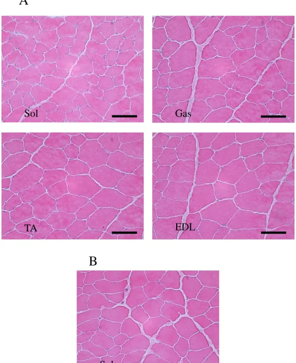
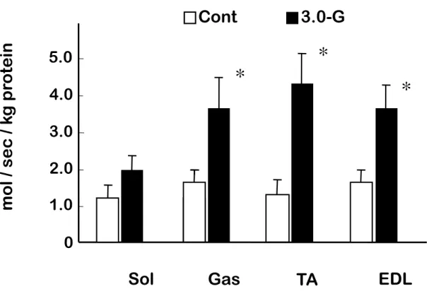
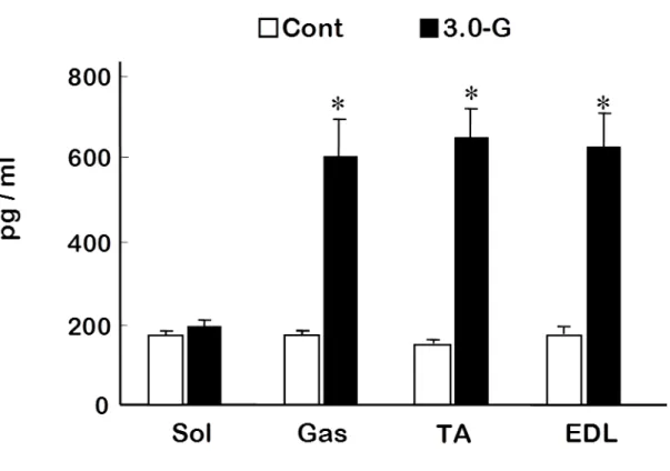
(3) 結果 1) HE 染色による観察 Fig.1 は,コントロール群のヒラメ筋と,過重力負荷群における腓腹筋,ヒラメ筋,前脛 骨筋および長指伸筋の筋組織画像である.各筋ともに過重力負荷群とコントロール群 の間に光学顕微鏡による観察上で差はみられなかった. 2) β-グルクロニダーゼ活性 骨格筋損傷の指標である β-グルクロニダーゼ活性は,腓腹筋,前脛骨筋および長 指伸筋においてコントロール群に比べて,過重力負荷群のほうが有意に高い値を示し た(P<0.05).しかし,ヒラメ筋では両群の間に差を認めなかった(Fig.2). 3) TNFα 濃度 TNFα 濃度は,腓腹筋,前脛骨筋および長指伸筋において,コントロール群に比べ て,過重力負荷群のほうが有意に高い値を示した(P<0.05)(Fig.3).しかしながら,ヒラ メ筋では両群の間に有意な差を認めなかった. 4) IL-6 濃度
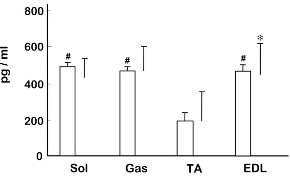
IL-6 濃度は,過重力負荷群とコントロール群の両群において前脛骨筋が他の筋より も低い傾向を示した.しかし,いずれの筋においても過重力負荷群とコントロール群の 間に有意な差を認めなかった(Fig.4). 5) iNOS mRNA iNOS mRNA はコントロール群では検出されなかったが,過重力負荷群の腓腹筋, 前脛骨筋および長指伸筋において検出された(Fig.5). 6) NO 含有量 NO は腓腹筋,前脛骨筋および長指伸筋において,コントロール群に比べて,過重 力負荷群のほうが有意に高い値を示した(P<0.05)(Fig.6).しかしながら,ヒラメ筋では 両群の間に有意な差を認めなかった.
(4) 考察 本研究において,8 分間という短時間の過重力負荷により下肢骨格筋の腓腹筋,前 脛骨筋および長指伸筋において骨格筋損傷の指標であるβ-グルクロニダーゼ,TNFα, iNOS および NO が増加することをはじめて明らかにした.しかしながら,骨格筋損傷を 引き起こす 1G 環境下での高強度運動等の過負荷とは異なり,IL-6 にはいずれの筋に おいても変化を認めなかった.また,ヒラメ筋においては他の筋とは異なりすべての指 標において,過重力負荷による影響を認めなかった. 宇宙空間における微小重力への曝露は,生体の主要なシステムに影響を及ぼす. なかでも,骨格筋は最も強く影響を受けるシステムの 1 つであり,その変化は形態 (Allen et al.,1996; Riley et al.,1996),収縮機能 (Caiozzo et al.,1996; Widrick et al.,1999),遺伝子発現 (Caiozzo et al.,1996) に及ぶことが,宇宙飛行を利用した研究 により,解明されている.特にヒラメ筋などにおいて顕著な変化が認められており (Allen et al.,1996; 後藤ら, 2007; Ohira et al.,2002),抗重力筋は微小重力曝露の影響 を受けやすいと考えられる.一方,宇宙船の打ち上げや帰還時には過重力の影響を 受けるが,これら数分間の過重力負荷が骨格筋の組織的損傷やその指標物質等に 及ぼす影響については,あまり明らかにされていない.
本研究における 8 分間 3.0G の過重力負荷により,骨格筋損傷の指標であるβ-グル クロニダーゼ活性には増加が認められたが,骨格筋損傷特有の組織学的な変化は観
察されなかった.通常の 1G 環境下おいて骨格筋損傷の指標であるβ-グルクロニダー ゼ活性は,レジスタンス運動による骨格筋損傷後数日で有意に増加する 19).さらに β-グルクロニダーゼ活性の総計は骨格筋損傷を反映だけでなく (Salminen et al.,1985), β-グルクロニダーゼ活性と病的組織変化との関連が示されている (Salminen et al.,1987).この組織学的変化に関する先行研究との結果の違いについては,プロトコ ル上の時間的な違いによる可能性が考えられる. 本研究で用いたラットは,8 分間の過重力負荷終了直後に下肢筋群を摘出した.通 常,骨格筋における生化学的な変化は運動等による過負荷直後に引き起こされるも のもあるが,組織学的な変化は過負荷の数時間後に観察される(Brickson et al.,2001; Sakurai et al.,2005).したがって,過重力負荷に対する生化学的な変化と組織学的変 化との解離については,今後,β-グルクロニダーゼおよび骨格筋内の損傷状態を反映 する他のリソソーム酵素(Salminen et al.,1985) の経時的な観察を行うことにより,説明 することが可能となるかもしれない. IL-6,TNFα,iNOS および NO などの様々なサイトカインは,組織に過負荷が加わっ た時に炎症の進行や治癒において重要な役割を担い,損傷骨格筋内にて増加する (Langberg et al.,2002; Rosendal et al.,2005).また,NO やその合成酵素である NOS は 筋の収縮や 1.0-G 環境下での過負荷となる高強度運動においても増加することがわ かっている(Alonso et al.,2006; Gomez-Cabrera et al.,2005; Sakurai et al., 2005; Tidball
et al.,1998).特に,誘導型の NO 合成酵素である iNOS は,マクロファージ,血管内皮 細胞,血管平滑筋細胞等により発現され,活性化されると炎症過程を刺激するのに十 分な NO を生成することが知られている (Beck et al.,1999; Torres et al.,2004). 本研 究では,3.0-G の過重力負荷により,TNFα 発現は腓腹筋,前脛骨筋および長指伸筋 において増加し,その程度は,一過性の高強度運動で引き起こされる骨格筋損傷 (Kawamura et al.,2002) とほぼ同レベルであった.さらに,3.0-G 過重力負荷後には iNOS も過剰発現し,NO も増加していた.しかしながら,高強度運動時には即座に応 答し増加するサイトカインである IL-6 (Heinrich et al.,1990; Rosendal et al.,2005) は, 本研究における過重力負荷では増加を示さなかった.また,IL-6 は前脛骨筋が他の 筋群に比べ低いことが示された.速筋である前脛骨筋は筋活動維持のためのグルコ ース取込を維持する目的で,骨格筋内より IL-6 を放出していると考えられ (Febbraio et al.,2002),この傾向は過重力負荷時においても変化しなかったものと考えられる.す なわち,短時間の過重力負荷により,TNFα は他の過負荷や炎症性反応と類似の変 化を示すものの,IL-6 においては他の過負荷に対する反応とは異なっていた.このこ とより,短時間の過重力負荷は高強度運動に代表されるような過負荷や炎症性反応と は異なる過程によって骨格筋に変化を及ぼしている可能性が考えられた.さらに 8 分 間という短時間の過重力負荷中に,炎症性反応と同様な生化学的変化の過程を経る ことが可能かどうかは,時間的観点からも疑問が残る.
本研究の結果では,8 分間の 3.0-G 過重力負荷がヒラメ筋には明らかな影響を及 ぼさなかった.先行研究において,慢性の過重力はさまざまな器官やシステムに影響 することが報告されており(Kita et al.,2003; Pecaut et al.,2004; Shibata et al.,2004; Kita et al.,2006),なかでも,ヒラメ筋筋線維の発達もしくは異化は,慢性の過重力負荷と密 接に関連していることが示されている (Bozzo et al.,2004; Chi et al.,1998; Pecaut et al.,2004; Picquet et al.,2005).一方,急性の過重力負荷(2G)曝露に対しても,ヒラメ筋 は 1G 環境下またはそれ以上の筋活動を示すことが報告されている (Wang et al.,2006).これらのことより,本研究においてヒラメ筋に明らかな生化学的変化を観察し なかったことは,ヒラメ筋の抗重力筋としての活動量の相違ではないと考えられる.同 様に,ヒラメ筋以外の筋群で観察された TNFα 増加による炎症性反応は,異なる筋線 維タイプ特性による応答性の違いよるものと示唆される.さらに速筋である前脛骨筋に おいては通常の状態でも他の筋に比べ IL-6 の発現量が少ないことから,筋線維タイ プによる特異性が伺える. 本研究においては iNOS の mRNA を測定した.骨格筋の収縮能や血管の緊張度, 血流の調節などに重要な役割を果たしている NO (Anderson et al.,2000; Kobzik et al.,1995; Moncada et al.,1995) の合成酵素には,iNOS の他に,血管内皮性および神 経性の NOS が存在する (Nathan et al.,1994).この 3 種類の酵素ともに L-アルギニン から NO を合成することから,本研究における過重力負荷により増加した NO について
は,血管内皮性もしくは神経性の NOS も関係していた可能性を完全には否定できな いと考えられる.
A
B
Fig. 1. Representative cross-sections of rat skeletal muscle stained with hematoxylin and eosin. A, 3.0-G hypergravity; B, control.; Sol, soleus muscle; Gas, gastrocnemius muscle; TA, tibialis anterior muscle; EDL, extensor digitorum longus muscle. Bars, 50 μm.
Sol
Gas
TA
EDL
Fig. 2. Changes in β-glucuronidase activity in rat skeletal muscle. Cont, Control group, open column; 3.0-G, hypergravity group, closed column; Sol, soleus muscle; Gas, gastrocnemius muscle; TA, tibialis anterior muscle; EDL, extensor digitorum longus muscle. Values represent mean ±SEM. *P<0.05 for 3.0-G vs. control.
0
2.0
4.0
Gas
TA
EDL
Sol
*
*
*
□
Cont
■
3.0
-
G
3.0
1.0
5.0
m
o
l /
se
c
/ k
g
pr
o
te
in
Fig. 3. Changes in tumor necrosis factor-α (TNFα) concentration in rat skeletal muscles. Cont, Control group, open column; 3.0-G, hypergravity group, closed column; Sol, soleus muscle; Gas, gastrocnemius muscle; TA, tibialis anterior muscle; EDL, extensor digitorum longus muscle. Values represent mean ±SEM. *P<0.05 for 3.0-G vs. control.
Fig. 4. Changes in Interleukin-6 (IL-6) concentration in rat skeletal muscle. *#P<0.05 vs. TA.
0
400
800
Gas
TA
EDL
Sol
pg /
m
l
*
□
Cont
■
3.0-G
600
200
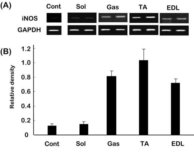
# # #Fig. 5. Expression of inducible nitric oxide synthase (iNOS) mRNA ( A ) and densitometric analysis data ( B ) in rat skeletal muscle following 3.0-G hypergravity. The value is related to the optical density of GAPDH.
GAPDH
iNOS
EDL
Gas
TA
Sol
0
0.2
0.4
0.6
0.8
1.0
1.2
Gas
TA
EDL
Sol
(A)
(B)
R e la ti ve d e ns it yCont
Cont
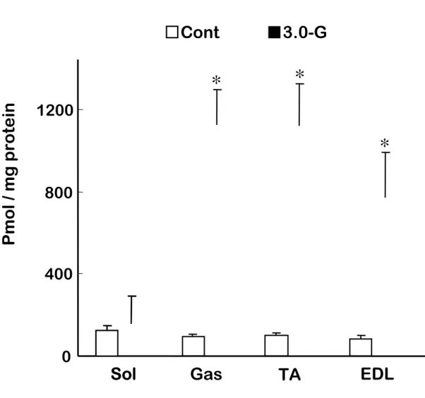
Fig. 6. Nitric oxide (NO) content in rat skeletal muscles. *P<0.05 for 3.0-G vs. control.
0
400
800
1200
Gas
TA
EDL
Sol
Sol
P
m
o
l/
m
g
pr
o
te
in
*
*
*
□
Cont
■
3.0-G
2.3. 外力負荷
(論文Ⅲ)
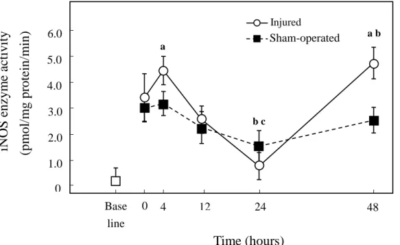
Changes in nitric oxide and inducible nitric oxide synthase following stretch induced injury to the tibialis anterior muscle of rabbit
要約
麻酔下の New Zealand White Rabbits の下肢前脛骨筋遠位腱を露出させ,モータ ー軸にクリッピングした.モーター駆動により前脛骨筋にメカニカルな伸張性筋損傷を 引き起こさせ骨格筋損傷モデルを作成し,NO 生成と骨格筋損傷の関連を検討した. 伸張性筋損傷処置直後・4・12・24・48 時間後のラット骨格筋損傷モデルを作成しその 変化を検討した.損傷筋における NO 生成は損傷直後より 4 時間後まで有意に上昇し その後 24 時間まで低下するが,48 時間後に再び有意な上昇を示した.また損傷筋で は 4 時間及び 48 時間後に iNOS タンパク発現がみられ,iNOS 活性も 4 時間及び 48 時間後に二峰性の有意な上昇を示した.この上昇は非損傷筋に比べ有意に大きいも のであり,観察期間全般においても損傷筋と非損傷筋の間には有意な差が示された. 以上の結果より,伸張性筋損傷後に引き起こされる NO 生成は,骨格筋肉細胞の中に
生成される iNOS およびその活性増加によって増大することが示唆されたが,損傷後 24 時間以降に増加する NO 生成の役割は明確にされるには至らなかった.
(1) INTRODUCTION
Nitric oxide (NO) generation occurs through the conversion of L-arginine to NO and L-Citrulline, which is controlled by NO synthases (NOS) (Moncada et al.,1991). There are three forms of NOS that catalyze NO production (Nathan et al., 1994). NO is a ubiquitous molecule involved in a variety of physiological functions including vascular regulation, neurotransmission, immune regulation and cell signaling (Reid et al.,1998; Reid et al.,2001; Tidball et al., 1998). NO is also known to have profound effects on skeletal muscle function, including contractility, blood vessel tone, blood flow, and satellite cell activation (Anderson et al., 2000; Kobzik et al.,1995; Moncada et al.,1991; Moncada et al.,1993). Resting skeletal muscle contains two constitutive types, neural NOS (nNOS), and to a lesser extent, endothelial NOS (eNOS) (Frandsen et al.,1996; Kobzik et al.,1994; Kobzik et al.,1995). The third or inducible form (iNOS) is expressed mainly in leukocytes and macrophages in response to inflammation. In contrast to eNOS and nNOS, iNOS can produce large amounts of NO over a prolonged period of time (Nathan et al.,1997; Xie et al.,1994), and skeletal muscle has been shown to induce iNOS mRNA and protein following endotoxin administration (Thompson et al.,1996). Because NO has both pro-inflammatory and anti-inflammatory properties, several possible roles for NO in muscle injury have been postulated. For example,
leukocyte concentrations within muscle tissue following unloading and reloading can be decreased by NOS inhibitors (Joshin et al.,1999). Moreover, a recent study reported that rat skeletal muscle produced sustained increases in both iNOS activity and protein levels in response to crush injury (Rubinstein et al.,1998). In contrast to these effects of NO on inflammation, NO can also have direct effects on muscle function, growth, and repair (Tidball, 1995). However, the pathogenesis of acute muscle stretch injury and the factors that regulate inflammation and repair are not well understood. In order to clarify whether NO modulates either inflammation or regeneration/repair, or both, following muscle trauma, it would be important to simultaneously examine the time course for NO levels and nitric oxide synthase following muscle injury.
Stretch injury to skeletal muscle typically occurs following repeated stretch-contraction cycles of the joint and associated muscles and is a common problem for clinicians and patients. In this communication, we measured the time course for NO levels, iNOS protein content and iNOS activity in stretch-injured skeletal muscle, using an anesthetized rabbit model. We employed an isokinetic injury apparatus that quantifies tibialis anterior (TA) muscle-tendon shortening, while allowing us to generate a reproducible stretch injury similar to clinical muscle-tendon stretch injury in humans (Best et al.,1999).
(2) METHODS 1) Animal care
Thirty-three male New Zealand white rabbits (weight 2.7- 3.0 kg) were housed individually and fed food and water ad libitum in a temperature-controlled room on a 12:12 hour light-dark cycle. The University of Wisconsin-Madison Research Animals and Resource Center Review Committee approved the animal use protocol.
2) Injury Model and Protocol
Our model of stretch-induced injury to skeletal muscle has been described (Best et al.,1999). Briefly, all animals were anesthetized with an intramuscular preparation of ketamine (100 mg/kg), xylazine (2.5 mg/kg), and acepromazine (3 mg/kg). A 6-mm incision was made over the dorsum of the right foot just distal to the ankle joint to isolate the tendinous portion of the TA. The peroneal nerve was isolated through a 4 mm skin incision at the knee. The animal was placed supine in the test apparatus and the muscle-tendon unit shortened 1.3 cm (Best et al.,1998) . With the muscle-tendon unit in the shortened position, the TA muscle was stimulated to tetany (Best et al.,19981). Once muscle tetany was achieved, the ankle was plantarflexed through a 90° arc at 450°/sec. The torque-angular displacement-time behavior was recorded. A sham operation of skin
incisions was performed in the left foot and knee. The 4-h animals were maintained under anesthesia with 1 cc of i.m. Ketamine every 45 min until tissue harvest.
For the remaining four groups (12, 24 and 48 h), all skin incisions were closed with 4-0 Ethilon suture, and the animals were returned to their cages after recovery from anesthesia with unrestricted activity. Evaluation 7 days after single stretch-induced injury in this protocol demonstrated a functional deficit (percent ankle isometric torque; injured leg versus uninjured leg) of 47.5% +/- 5.4% (mean +/- SD) (Best et al.,19982).
3) Tissue Sampling
The entire TA muscle-tendon unit was surgically removed at its proximal attachment to the tibia and distal insertion into the foot. A one cm2 block (approximately 2 g) of muscle tissue was removed from the myotendinous junction region of both the stretch-injured and sham-operated controls. This sample always contained the area of visible hematoma and maximum injury. The tissue block was cut and one section (1.8 g) was snap-frozen in liquid nitrogen. Animals were sacrificed with Beusthanasia (0.4 cc/kg), i.v., placed into the marginal ear vein immediately following muscle harvest. In addition to the thirty animals evaluated following injury, three rabbits were used to estimate baseline NO levels and iNOS activity. Measurements were made in both legs
of the three animals not exposed to stretch injury or skin incisions to ascertain the contribution of the surgical procedures to overall NO levels and iNOS activity.
4) NO Content
Samples were homogenized at 0 - 4°C in ice-cold 0.1 M K2HPO4-KH2PO4 (pH 7.4)
buffer (wt:vol of 1:10) with a motor-driven Potter-Elvehjem teflon glass homogenizer. Quantification of NO content was performed spectrophotometrically (Nims et al.,1996). A range of standard solutions (concentration: 0-30 µM NO) was prepared using KNO3
as a standard. Because the Griess reagent measures only nitrite, and biological systems contain both nitrite and nitrate, addition of 0.01 U nitrate reductase (Sigma, St. Louis, MO) was added to each well, followed by addition of NADH (0.02 mM). Samples were read at 540 nm using a microplate reader (Molecular Devices, Sunnyvale, CA) and the results were expressed as pmol/mg protein. Pilot assays established inter/intraassay variability of less than 3% (data not shown).
5) Inducible Nitric Oxide Synthase (iNOS) Protein Content
SDS polyacrylamide gel electrophoresis (SDS-PAGE) in combination with an enhanced chemiluminescence plus (Amersham, Arlington Heights, IL) detection system
was used to determine iNOS protein content in skeletal muscle. Tissue samples were homogenized in 50 mM Tris HCl, 1 mM EDTA, and 0.1% phenylmethyl– sulfonylfluoride (PMSF). Relative amount of iNOS was determined utilizing an anti-iNOS (IgGl) antibody (Transduction Laboratories, San Diego, CA). Autoradiographic signals were assessed using a BioRad scanning densitometer (BioRad, Hercules, CA). Results are expressed in arbitrary optical density units.
6) Inducible Nitric Oxide Synthase (iNOS) Activity
Measurement of iNOS activity was performed by examination of the conversion of [3H]-arginine to [3H]-citrulline using the method of Thompson (1996). The reaction buffer contained 3 mM n-2-hydroxyethyl-piperazine-N’protein-ethanesulfonic acid (HEPES), 4 mM NADPH, 20 mM tetrahydrobiopterin, 20 µM flavin adenine dinucleotide (FAD), 20 µM flavin mononucleotide (FMN), 1 mM dithiothreitol (DTT), 4 µM L-arginine, 4 mM ethylene glycol-bis (b-aminoethyl ether)-N, N, N’, N’-tetraacetic acid (EGTA), and 2.0 mCi/ml L-[3H]-arginine (Amersham, Arlington Heights, IL). The reaction was incubated at 37°C for 1 hour, and then terminated by addition of buffer containing 40 mM HEPES, pH 5.2, 2 mM EDTA, and 2 mM EGTA. Samples were then applied to a 10 ml Dowex AG50WX-8 (Na+) column (BioRad,
Hercules, CA), and eluted with 40 mM HEPES. Effluent was collected in scintillation vials and quantified by liquid scintillation spectroscopy using a Wallac LKB 1211 Rack Beta counter (Wallac, Helsinki, Finland). 1 mM N-nitro-L-arginine methyl ester (L-NAME), a nonselective NOS inhibitor, served as a negative control to assess background activity and non-NOS-dependent conversion of L-[3H] arginine to L-[3H] citrulline. Results are expressed as picomoles per minute per microgram protein (pm/min/mg protein). Pilot experiments demonstrated inter/intra variability of less than 4%.
7) Statistical Analysis
Means ± SEM values were caECCulated for all data sets. Data were analyzed with a 2-way analysis of variance with repeated measures to evaluate the two main treatment effects, injury and time, and their interaction (StatView). The Fisher’s Least Significant Difference post hoc test was used to locate the differences between means when the observed F ratio was statistically significant (p < 0.05).
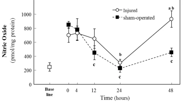
(3) RESULTS 1) NO Content
Baseline NO levels were approximately one third of sham-operated or injured tissue values (Fig. 1). And 0 h values were significantly higher than baseline (p<0.05). Sham-operated muscles had decreased NO content at 12, 24, and 48 h after surgery, as compared to 0 h (Fig. 1; p<0.05, for all three time points). Stretch injured muscles showed decreased NO content at 24 h, as compared to 0 h injured muscles (Fig. 1; p<0.05). At 48 h, sham-operated muscle continued to have lower NO than at 0 h, whereas stretch-injured muscles recovered NO to resting levels and showed a 146% higher NO content compared to sham-operated muscles (Fig. 1; p<0.05).
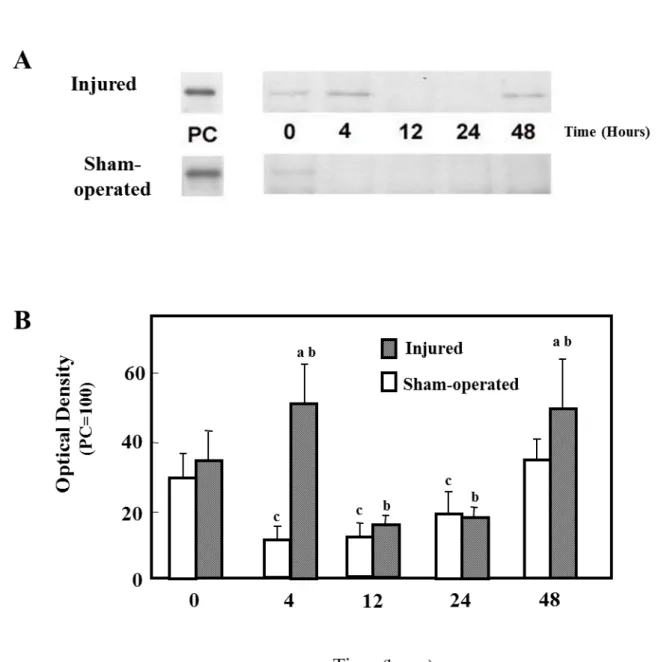
2) iNOS Content
Figure 2A shows the immunoreactive levels of iNOS protein in sham-operated and injured muscles. Figure 2B shows densitometric quantification of iNOS protein. iNOS protein expression in injured muscles was not different from sham-operated muscles, at 0, 12, and 24 h post injury. However, iNOS protein in injured muscles at 4 and 48 h was significantly higher than their sham-operated counterparts (P<0.05).
Stretch injured muscles showed increased iNOS protein expression at 4 and 48 h, as compared to 0 h injured muscles (Fig. 2B; p<0.05).
3) iNOS Activity
Similar to iNOS protein content, iNOS enzyme activity was higher at both 4h (p<0.05) and 48h (p<0.01) in injured vs. sham-operated muscles (Fig. 3). The time course of iNOS enzyme activity in injured muscles displayed two peaks, with higher levels at 4 and 48 h after injury, as compared with 0 h (p<0.05) (Fig. 3). At 24 h, injured and sham-operated muscles had decreased iNOS activity, as compared to 0 h (p<0.05) (Fig. 3). iNOS activity was fully inhibited by the addition of L-NAME.
(4) DISCUSSION
The major finding in the current study was that stretch-injured muscles showed an increase in NO levels 48 h after injury compared to sham-operated tissues. During the initial 24 h after injury, similar changes in NO levels were found in sham-operated muscles and injured muscles. No significant increase in NO levels was observed in sham-operated muscles 48 h after surgery, whereas a significant increase in NO levels was noted in injured muscles 48 h after injury. This finding may be of significance for muscle healing for several reasons. First, adequate levels of intracellular NO may be an important prerequisite for muscle healing after injury as it has been postulated that NO plays a central role in satellite cell activation and muscle regeneration (Moncada et al.,1991; Nguyen et al.,2003). Consistent with this role, various NO donors have been shown to stimulate myoblast proliferation in vitro (Ulibarri et al.,1999). Second, NO may provide some control on the extent of muscle inflammation and repair through its influence on leukocyte infiltration. In addition to influencing myofiber regeneration, NO has been shown to affect collagen synthesis, as NO generators can have both proliferative and inhibitory effects on the mitotic activity of fibroblasts (Evans et al.,1996). Finally, NO is a well-known vasodilative agent and conceivably increases blood flow to recovering myocytes following stretch injury (Thompson et al.,1996).