INTRODUCTION
Radiotherapy is a standard treatment for various malignant tumors. Ionizing radiation (IR) strongly induces growth arrest and/or apoptosis in a variety of cells.1, 2) However, responses to IR differ markedly among cell types. Importantly, most malignant cells do not show distinct apop- tosis immediately after IR exposure. In these cells, even 48 hours after IR exposure, little if any apoptosis is detectable, and thereafter weak apoptosis may occur following multiple cell divi-
sions.3!5) Thus, radiotherapy appears to be inef- fective for these radioresistant carcinoma cells.
To date, many attempts have been made to explore the roles of radiotherapy in the manage- ment of locally advanced tumors. To understand molecular radiobiology, strategies for enhancing tumor radiosensitivity have begun to focus on targeting the molecules regulating cellular re- sponses after irradiation. Chemotherapeutic agents that enhance the effectiveness of radia- tion treatment are termed radiosensitizers, Yubin Zhang1) , 2), Masaaki Adachi 1), Rina Kawamura1), Yaohua Liu1), 2),
Kohzoh Imai1), Masato Hareyama3), Yasuhisa Shinomura1)
1)
First Department of Internal Medicine, Sapporo Medical University School of Medicine, S
!1, W
!16, Chuo
!ku, Sapporo 060
!8543, Japan,
2)Department of Neurosurgery, First Clinical
College of Harbin Medical University, No. 23, Youzheng St. Nangang Dist. Harbin, Heilongjiang, China, and
3)Department of Radiology, Sapporo Medical University School
of Medicine, S
!1, W
!16, Chuo
!ku, Sapporo 060
!8543, Japan
ABSTRACT Radiation therapy is one of the main anti-
cancer therapies. However, there are consider- able numbers of radioresistant carcinoma cells and severe adverse effects occasionally occur af- ter radiation therapy. Therefore, there have been many attempts to identify radiosensitizers.
Histone hyperacetylation has begun to receive considerable attention as a potential target for
radiosensitization. Inhibitors of histone deacety- lase (HDAC) activity are becoming a major tool for modifying histone acetylation status. This ar- ticle reviews the classification of HDAC inhibi- tors and their potential as radiosensitizers. We also discuss possible molecular mechanisms by which the HDAC inhibitors radiosensitize can- cer cells.
Key words :Histone deacetylase (HDAC) inhibitors, Histone acetylation, JNK, Radiosensitivity, Apoptosis
HDAC inhibitors as radiosensitizers
<Review>
Tumor Res.41,15−21(2006) 15
which make cancer cells more susceptible to IR
!induced death than surrounding normal cells, and several such compounds are now available for the treatment of solid tumors6).
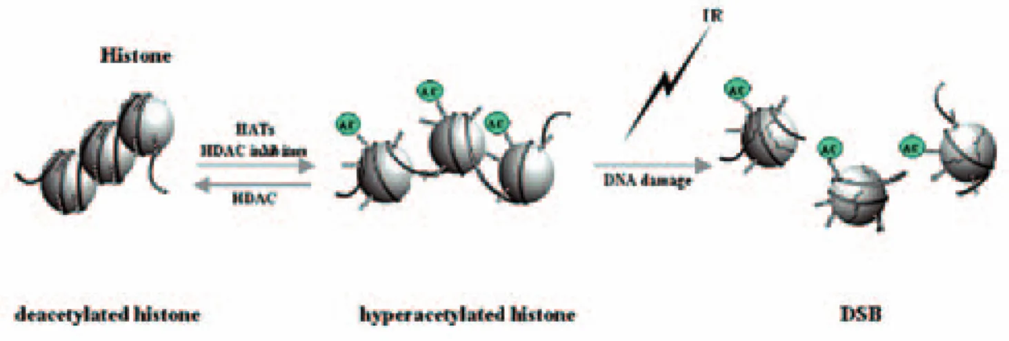
Modification of histone acetylation is one of the potential targets to enhance radiosensitivity, since histone hyperacetylation leads to loose chromatin structure which may affect suscepti- bility to DNA damage (Fig. 1)7, 8). Since histone acetylation levels are determined by the balance of histone acetylases (HATs) and histone deace- tylases (HDACs) activities, inhibition of HDAC activity may enhance radiosensitivity. In this context, considerable efforts have been put into the development of novel HDAC inhibitors, which often strongly induce apoptosis in tumor cells by themselves. Although extensive pre- clinical studies have already revealed that many HDAC inhibitors have antitumor activity and promising anticancer effects9), a limited numbers of recent studies discovered that some HDAC inhibitors can significantly enhance radiosensi- tivity. Importantly, a structurally diverse set of HDAC inhibitors has similar radiosensitizing ef- fects in a variety of cancer cell types. Moreover, their radiosensitizing effects actually increased the anticancer effects of radiotherapy in several human tumor xenografts. Therefore, HDAC in- hibitors are not only promising anticancer agents as single modalities or in combination with chemotherapeutic agents, but also promis- ing radiosensitizers. We here review biological
activities of HDAC inhibitors as radiosensitizers.
CLASSIFICATIONS OF HDAC INHIBITORS There are ranges of structurally diverse HDAC inhibitors which are either products iso- lated from natural sources or synthetically pro- duced compounds. These compounds can be di- vided into six groups based on their structure (Table 1). HDAC inhibitors include the short chain fatty acids, sodium butyrate (NaB), phenyl butyrate and valproic acid (VPA)10), the hydrox- amic acids, suberoylanilide hydroxamic acid (SAHA), trichostatin A (TSA)11), LAQ824, LBH 529 and PXD101; the tetrapeptides, FK228, Apicidine and Trapoxin; the benzamides, MS! 275 and CI!994 (N!acetyldinaline); and Ketones, Trifluoro!methyl ketone.
Although these HDAC inhibitors have their own structures, their inhibitory action is quite similar. The identified mechanism of the hy- droxamic acid groups is to interact with the catalytic site of HDAC, and thereby block bind- ing of substrates to active sites of HDACs11). Synthetic benzamides have a polar end to bind zinc ion within the HDAC catalytic pocket structure and inhibit their activity. Other HDAC inhibitors also bind to HDAC active site powerfully and inhibit HDAC activity.
Short!chain fatty acids are important, since they are abundantly found in feces and thus play major roles in anticancer activity of diet12). However, the action of these compounds is non!
Fig.1 Histone hyperacetylation induces relaxation of chromatin structure
16 Y.ZHANG et al.
specific and weak. It is thus hard to achieve ef- fective inhibiting concentrations in vivo. Among the members, VPA is a unique HDAC inhibitor.
VPA is currently marketed as an anti!epilepsy agent and anti!migraine headaches. It has re- cently been discovered to inhibit HDAC activity at clinically applicable concentrations. Interest- ingly, VPA inhibits the class I HDAC activity better than class II HDAC11), but its clinical trial shows that VPA alone has only a weak antican- cer effect in patients with acute myeloblastic leukemia. When VPA was combined with the demethylating agent, 5!aza!2'deoxycitabine, a significant number of refractory leukemia pa-
tients achieved a complete remission13). More precise studies are required for evaluation of its clinical efficacy.
MOLECULAR MECHANISMS OF RADIOSEN- SITIZING EFFECT OF HDAC INHIBITORS
Some HDAC inhibitors have been shown to radiosensitize a broad array of cancer cells14!17). HDAC inhibitors are unique anticancer drugs, since they induce histone hyperacetylation and activate some transcription factors, but do not affect DNA itself17!19), while many anticancer agents are genotoxic and interfere with DNA synthesis. This suggests that HDAC inhibitors Table 1
Group Compounds
Short!chain fatty acids
Hydroxamic acid!derived compounds
Cyclic tetrapeptides
Synthetic pyridyl carbamate derivative
Synthetic benzamides Ketones
Valproic acid (VA) Phenyl butyrate (PB) Phenyl acetate (PA) Sodium butyrate (SB) Trichostatin (TSA)
Suberoylanilide hydroxamic acid (SAHA)
M!carboxycinnamic acid bis!hydroxamide (CBHA) Azelaic bis!hydroxamic acid (ABHA)
NVP!LAQ824 LBH589 Oxamflatin PXD101 Scriptaid Pyroxamide
Depsipeptide (FK228, FR901228) Apicidine
Trapoxin HC!toxin Chlamydocin Depudesin CHAPS
AN!9 (Pivanex)
MS!275, CI!994 (N!acetyldinaline) Trifluoromethyl ketone
a!ketomides
41(2006) HDAC inhibitors as radiosensitizers 17
may not increase the occurrence of secondary malignancies after radiation therapy. In this con- text, development of HDAC inhibitors!mediated enhanced radiation therapy is important. To es- tablish a combination therapy, the precise mo- lecular mechanisms by which HDAC inhibitors enhance anticancer activity of radiation therapy must be discovered. Although the precise mechanisms remain uncertain, several possible explanations have been suggested as described below (Fig. 2).
First, K. Camphausen and et al18) investi- gated the effects of MS!275 on the radiosensitiv- ity of two human tumor cell lines, and found that MS!275!mediated enhanced radiosensitiv- ity is correlated well with histone hyperacetyla- tion and prolonged phosphorylation of histone H
2AX (H2AX) and its foci. This suggests a de- crease in the repair of radiation!induced DNA double!strand breaks (DSBs). In addition, A.
Munshi, et al19) reported that NaB and TSA ra- diosensitized two melanoma cell lines, A375 and MeWo, and the radiosensitization correlated with functional impairment of DNA repair as determined by the host cell reactivation assay.
Moreover, NaB significantly reduced the expres- sion of the repair!related genes Ku70 and Ku86 and DNA!dependent protein kinase catalytic subunit in melanoma cells at the protein and mRNA levels. They also found thatγ!H2AX foci persisted longer after ionizing exposure in the NaB!treated cells than in untreated cells. These accumulating data strongly suggest that HDAC inhibitors can inhibit DNA repair activity and
Fig2Possible mechanisms of HDAC inhibitor!mediated sensitization to radiation
18 Y.ZHANG et al.
enhance the anticancer effect of radiation.
Second, eukaryocytic DNAs are compacted with histones and other accessory proteins, while histone hyperacetylation may loosen com- pact DNA, allowing DNA to become more sus- ceptible to damage7, 8). Consistent with this data, CBHA strongly increased γ!H2AX and its foci formation immediately after IR exposure20). An- other HDAC inhibitor, FK228, similarly en- hanced IR!induced γ!H2AX within 1 hour after IR. Importantly, the enhanced γ!H2AX was in- itially diminished and thereafter increased at 12 hours after IR. These data suggest that HDAC inhibitors may enhance IR!induced DNA dam- age itself.
Third, it is well known that histone hyper- acetylation alters chromatin structure and in- creases the transcription activity of selected genes. To date, many apoptosis!related genes have been identified as target genes of HDAC inhibitor!mediated signals. Cyclin!dependent kinase inhibitor p21WAF1 and gelsolin are clearly induced by HDAC inhibitors and their induction is suggested to be important for antiprolifera- tive action or inhibition of tumor progression21!23). Interestingly, HDAC inhibitors can also activate the proapoptotic activity of BAX by acetylation of Ku70, which can abolish their physical inter- action and allow mitochondrial targeting of BAX24). We and others found that several HDAC inhibitors increase expression of a pro!apoptotic BH3!only protein Bim14, 25) and its related mole- cule Bmf26), and suggestted their crucial role in their proapoptotic activity. Importantly, knock- down of Bmf expression strongly inhibits IR!in- duced apoptosis27). These accumulating data suggest that an increase in these BH3!only pro- teins may be one of the central mechanisms of IR!induced apoptosis.
CONCLUSION
We strongly suggest that HDAC inhibitors are promising radiosensitizers. The agents are not genotoxic and thus might not enhance sec- ondary transformation after IR therapy. In addi- tion, HDAC inhibitors cooperatively increase IR
!induced DSB and induce apoptosis!related genes, especially Bim and Bmf. Our knowledge is obviously limited and the efficacy of combina- tion therapy has been demonstrated only in mice models. However, these accumulating in- formation may help us to develop a more potent HDAC inhibitors!combined IR therapy.
REFERENCES
1.Hartwell LH, Weinert TA. Checkpoints:
controls that ensure the order of cell cycle events. Science 1989; 246: 629!634.
2.Clarke AR, Purdie CA, Harrison DJ, Morris RG, Bird CC, Hooper ML, Wyllie AH. Thy- mocyte apoptosis induced by p53!depend- ent and independent pathways. Nature 1993;362: 849!852.
3.Harms!Ringdahl M, Nicotera P, Radford IR.
Radiation induced apoptosis. Mutat Res 1996;366:171!179.
4.Muschel RJ, Soto DE, McKenna WG, Bern- hard EJ. Radiosensitization and apoptosis.
Oncogene 1998;17:3359!3363.
5.McKenna WG, Bernhard EJ, Markiewicz DA, Rudoltz MS, Maity A, Muschel RJ.
Regulation of radiation!induced apoptosis in oncogene!transfected fibroblasts: influence of H!ras on the G2 delay. Oncogene 1996;12:
237!245.
6.Lawrence TS. Radiation sensitizers and targeted therapies.Oncology (Williston Park) 2003;17:23!28.
7.Hebbes TR., Clayton AL, Thorne AW, Crane!Robinson C. Core histone hyperace- tylation co!maps with generalized DNase I sensitivity in the chicken beta!globin chro- mosomal domain. EMBO J 1994;13:1823! 1830.
8.Martinez!Lopez W, Folle GA., Obe G, Jeppesen P. Chromosome regions enriched in hyperacetylated histone H4 are preferred sites for endonuclease! and radiation!in- duced breakpoints. Chromosome Res 2001;
9:69!75.
9.Hess!Stumpp H. Histone deacetylase inhibi- tors and cancer: from cell biology to the
41(2006) HDAC inhibitors as radiosensitizers 19
clinic. Eur J Cell Biol 2005;84:109!121.
10.Phiel CJ, Zhang F, Huang EY, Guenther MG, Lazar MA, Klein PS. Histone deacety- lase is a direct target of valproic acid, a po- tent anticonvulsant, mood stabilizer, and teratogen. J Biol Chem 2001;276:36734! 36741.
11.Finnin MS, Donigian JR, Cohen A, Richon VM, Rifkind RA, Marks PA, Breslow R, Pavletich NP. Structures of a histone deac- stylase homologue bounc to TSA and SAHA. Nature 1999; 401:188!193.
12.Myzak MC, Dashwood RH. Histone deace- tylases as targets for dietary cancer pre- ventive agents: lessons learned with bu- tyrate, diallyl disulfide, and sulforaphane.
Curr Drug Targets 2006;7:443!452.
13.Garcia!Manero G, Kantarjian HM, Sanchez! Gonzalez B, Yang H, Rosner G, Verstovsek S, Rytting M, Wierda WG, Ravandi F, Koller C, Xiao L, Faderl S, Estrov Z, Cortes J, O'Brien S, Estey E, Bueso!Ramos C, Fioren- tino J, Jabbour E, and Issa JP. Phase 1/2 study of the combination of 5!aza!2'!deoxy- cytidine with valproic acid in patients with leukemia.Blood. 2006;108:3271!3279.
14.Zhang Y, Adachi M, Zhao X, Kawamura R, Imai K. Histone deacetylase inhibitors FK 228, N!(2!aminophenyl)!4![N!(pyridin!3!yl! methoxy carbonyl) amino!methyl] ben- zamide and m!carboxycinnamic acid bis! hydroxamide augment radiation!induced cell death in gastrointestinal adenocarci- noma cells. Int J Cancer 2004;110:301!308.
15.Camphausen K, Scott T, Sproull M, Tofilon PJ. Enhancement of xenograft tumor radio- sensitivity by the histone deacetylase inhibi- tor MS!275 and correlation with histone hy- peracetylation. Clin Cancer Res 2004;10:6066
!6071.
16.Kim JH, Shin JH, Kim IH. Susceptibility and radiosensitization of human glioblastoma cells to trichostatin A, a histone deacetylase inhibitor. Int J Radiat Oncol Biol Phys 2004;
59:1174!1180.
17.Zhang Y, Jung M, Dritschilo A, Jung M. En-
hancement of radiation sensitivity of human squamous carcinoma cells by histone deace- tylase inhibitors. Radiat Res 2004;161:667! 674.
18.Camphausen K, Burgan W, Cerra M, Oswald KA, Trepel JB, Lee MJ, Tofilon PJ.
Enhanced radiation!induced cell killing and prolongation of gammaH2AX foci expres- sion by the histone deacetylase inhibitor MS!275. Cancer Res 2004;64:316!321.
19.Munshi A, Kurland JF, Nishikawa T, Tanaka T, Hobbs ML, Tucker SL, Ismail S, Stevens C, Meyn RE. Histone deacetylase inhibitors radiosensitize human melanoma cells by suppressing DNA repair activity.
Clin Cancer Res 2005;11:4912!4922.
20.Zhang Y, Adachi M, Zou H, Histone deace- tylase inhibitors enhance phosphorylation of histone H2AX after ionizing radiation Int J Radiat Oncol Biol Phys 2006 ;65:859!866.
21.Richon VM, Sandhoff TW, Rifkind RA, Marks PA. Histone deacetylase inhibitor se- lectively induces p21WAF1 expression and gene!associated histone acetylation. Proc Natl Acad Sci USA 2000;97:10014!10019 22.Han JW, Ahn SH, Park SH, Wang SY, Bae
GU, Seo DW, Kwon HK, Hong S, Lee HY, Lee YW, Lee HW. Apicidin, a histone deace- tylase inhibitor, inhibits proliferation of tu- mor cells via induction of p21WAF1/Cip1 and gelsolin. Cancer Res 2000;60:6068!6074.
23.Kim JH, Choi YK, Kwon HJ, Yang HK, Choi JH, Kim DY. Downregulation of gelsolin and retinoic acid receptor beta expression in gastric cancer tissues through histone deacetylase 1. J Gastroenterol Hepatol 2004;
19:218!224.
24.Cohen HY, Lavu S, Bitterman KJ, Hekking B, Imahiyerobo TA, Miller C, Frye R, Ploegh H, Kessler BM, Sinclair DA. Acetyla- tion of the C terminus of Ku70 by CBP and PCAF controls Bax!mediated apoptosis.
Mol Cell 2004;3:627!638.
25.Zhang XD, Gillespie SK, Borrow JM, Hersey P. The histone deacetylase inhibitor suberic bishydroxamate regulates the expression of
20 Y.ZHANG et al.
multiple apoptotic mediators and induces mitochondria!dependent apoptosis of mela- noma cells. Mol Cancer Ther 2004;3:425!435.
26.Zhang Y, Adachi M, Kawamura R, Imai K.
Bmf is a possible mediator in histone deace- tylase inhibitors FK228 and CBHA!induced apoptosis. Cell Death Differ 2006;13:129!140.
27.Zhang Y, Adachi M, Kawamura R, Zou HC, Imai K Hareyama M, Shinomura Y. Bmf contributes to histone deacetylase inhibitor! mediated enhancing effects on apoptosis af- ter ionizing radiation. Apoptosis 2006;11:
1349!1357.
(Accepted for publication, Jan. 10, 2007)
41(2006) HDAC inhibitors as radiosensitizers 21