55:483
はじめに
海綿静脈洞血栓症(cavernous sinus thrombosis; CST)は, さまざまな原因で海綿静脈洞内に血栓が形成され,神経症候 を呈する疾患である.その原因は感染性と非感染性に大別さ れるが,海綿静脈洞はその解剖学的性質上,脳や顔面,口腔 からの血管が集中する部位であり,副鼻腔炎や静脈洞炎,齲 歯にともなう感染性の血栓症が原因としてはもっとも多い. とくに蝶形骨洞炎・篩骨洞炎などの感染症が原因としては もっとも多いとされる1).近年では画像検査の進歩や,抗菌 薬の普及により,早期診断,治療が可能となったが,依然致 死率の高い疾患でもある1)2).今回われわれは未治療の齲歯が 原因で感染性海綿静脈洞血栓症を呈した症例を経験した.特 異な画像所見が診断に有用で,すみやかな治療により良好な 経過をたどった貴重な症例と考えられここに報告する. 症 例 患者:54 歳 女性 既往歴:高血圧.無症候性 HTLV-1 キャリアー.第 1 子流産. 家族歴:娘 2 人が全身性エリテマトーデス. 生活歴:飲酒:ビール 1 日 2 缶程度,喫煙なし. 現病歴:2012 年から齲歯に対して歯科通院中であったが, 2013年 5 月頃から歯科への通院を一時中断していた.2013 年 11月初旬から両側の下歯の痛みを自覚していたが,未治療で 経過をみていた.2013 年 11 月中旬に頭痛をともなう 38~ 39°Cの発熱が出現し,市販の解熱鎮痛剤を服用するも症状の 改善はなかった.入院 4 日前の明け方から右眼窩部に鈍痛を ともなう腫脹が出現した.その後も右眼窩部の疼痛と腫脹は 悪化,入院 2 日前には左眼窩部の腫脹も出現したため当科に 精査加療目的に入院した. 現症:身長 153.5 cm,体重 55 kg,脈拍 56/ 分・不整,血圧 149/76 mmHg.一般身体所見では,眼脂と両側でとくに右に 強く眼瞼浮腫・眼球充血がみられた(Fig. 1).右眼の自発開 眼は不可であった.聴診上,眼窩部に血管雑音はみられなかっ た.両側耳下腺に圧痛をともなわない可動性良好な弾性軟な 腫脹が触知されたが,明らかなリンパ節腫脹はみられなかっ た.口腔内や扁桃に明らかな異常は指摘できなかった.心音, 呼吸音に異常はみとめなかった.腹部は平坦・軟で正常腸蠕 動音を聴取し,両側下腿に浮腫はなく,全身皮膚には紅斑な どの異常はみられなかった. 神経学的所見で,意識は清明.項部硬直や Kernig 徴候は陰 性で,眼球運動制限は右眼(上転 : -2 下転 -3: 内転 : -3 外転 : -2),左眼(上転 : -1 下転 0: 内転 : 0 外転 : -2)と両側にみら れた.その他の脳神経に異常はなく,四肢に運動・感覚系の 異常をみとめなかった.四肢腱反射はやや活発で,Babinski 徴候は陰性.協調運動障害もみとめなかった. 検査所見において,血算は白血球が 1.4×10⁴/μl(好中球
症例報告
齲歯が原因で生じた感染性海綿静脈洞血栓症と
Lemierre
症候群の合併例
西田 明弘
1)緒方 利安
1)工藤 仁隆
1)福原 康介
1)深江 治郎
1)坪井 義夫
1)*
要旨: 症例は 54 歳女性.入院 1 年前に齲歯の治療中断歴があり,今回頭痛と発熱,眼窩部の腫脹疼痛を主訴に 当科入院.両眼瞼浮腫と眼球運動障害以外に神経学的異常をみとめなかった.血液検査では炎症反応,凝固線溶系 マーカーが上昇し,MRI 拡散強調画像で右上眼静脈から海綿静脈洞に高信号がみられ,造影 CT で同部位に造影不 良域をみとめたことから,感染性海綿静脈洞血栓症と診断した.血液培養の結果から齲歯による敗血症を原因とす る海綿静脈洞血栓症(cavernous sinus thrombosis; CST)と診断した.抗菌薬と抗凝固療法で治療を開始し,症 状は軽快,画像上海綿静脈洞内の血栓も縮小した.本症例は MRI と造影 CT の画像所見から CST を早期診断しえた 貴重な症例である.(臨床神経 2015;55:483-489)
Key words: 海綿静脈洞血栓症,Lemierre 症候群,齲歯
*Corresponding author: 福岡大学医学部神経内科学教室〔〒 814-0180 福岡市城南区七隈 7-45-1〕
1)福岡大学医学部神経内科学教室
(Received October 20, 2014; Accepted March 17, 2015; Published online in J-STAGE on June 4, 2015) doi: 10.5692/clinicalneurol.cn-000676
88.2%)と上昇し,赤血球数 356 × 104/μl,ヘモグロビン値 10.8 g/dl,ヘマトクリット 31%と軽度貧血の所見がみられた.血
小板数は 23.7 × 10⁴/μlと正常だった.生化学所見では C reactive
protein(CRP)が 22.2 mg/dl と著明に上昇し,Na 133 mmol/l,
K 2.7 mmol/l,Cl 92 mmol/l と電解質の異常,および alkaline
phosphatase(ALP)1,080 U/l,γ glutamyl transpeptidase(γGTP)
631 U/lと胆道系酵素の上昇をみとめたが,肝酵素の上昇はな
く,腎機能も正常であった.空腹時血糖 121 mg/dl,HbA1C: 5.5%と糖尿病の所見はなかった.凝固マーカーでは fibrinogen
degradation products 13 μg/ml(≦ 5 μg/ml),D-dimer 3.5 μg/ml
(≦ 1.0 μg/ml)と線溶系の上昇がみられた.免疫血清学的検 査ではプロカルシトニンが 0.70 ng/ml(≦ 0.05 ng/ml)と上昇 していたが,βD グルカンは陰性で抗 HIV 抗体もみとめなかっ た.リウマチ因子 6 IU/ml(≦ 15 IU/ml),抗核抗体 7.5(≦ 40), 抗double stranded DNA定量2.7 IU/ml(≦12 IU/ml),PR3-ANCA < 1.0(U/ml),MPO-ANCA < 1.0(U/ml)と膠原病や血管炎 を示唆する所見はなく,抗カルジオリピン β 抗体も陰性だっ た.腫瘍マーカーはいずれも陰性.HTLV-1 抗体は既知の通 り陽性だった. 経 過 入院時に施行した頭部 MRI 拡散強調像では左右の上眼静 脈が紐状に高信号を呈して,同部位は ADC 値が低下してい た.とくに右の上眼静脈が著明に拡大しており,静脈内血栓 と考えられた(Fig. 2).両側眼窩内には上眼静脈の血流を妨 げる腫瘤性変化はみられず,さらに拡散強調像では上眼静脈 から両側の海綿静脈洞にかけて高信号域連続性にみとめられ ており,海綿静脈洞内の血栓が示唆された.頭部の造影 CT では左右の上眼静脈と両側海綿静脈洞に増強効果が欠損した 領域をみとめ,海綿静脈洞内の血栓の存在を示唆した(Fig. 3). 明らかな副鼻腔炎の所見はなかった.臨床症状も眼球突出は みとめるが拍動,血管雑音は聴取されず,内頸動脈-海綿静 脈洞瘻を示唆する造影剤の漏出はなかった.以上の所見から 感染性 CST が診断としてもっとも考えられた.入院第 2 病日 に施行した入院時の歯科の診察では,右下 2 番部および左下 7番部に排膿している所見があり,化膿性根尖性歯周炎で あった.入院時に施行した血液培養検査で,口腔内常在菌で ある Streptococcus constellatus が陽性としてみとめられた.原 因としては膠原病や経口避妊薬の服用はなく,流産歴がある も抗リン脂質抗体症候群は陰性で,全身性エリテマトーデス Fig. 1 Clinical course in a 54-year-old woman diagnosed with cavernous sinus thrombosis.
Antibiotic (topmost panels) and anticoagulant (middle panel) treatment were administered over a 26-day period. The serum CRP (solid line) and D-dimer (dashed line) concentrations peaked early during the clinical course and then declined, and the APTT (dotted line) gradually increased. The blephaloptosis (bottom panel) decreased and resolved after 26 days of treatment. SBT/APC; Sulbactam/Ampicillin, PIPC/TAZ; Piperacillin/Tazobactam, APTT; activated partial thromboplastin time, CRP; C-reactive protein.
敗血症による海綿静脈洞血栓症の 1 例 55:485 の家族歴が濃厚であるも患者自身はそれを示唆する理学的所 見や抗体の異常はなかった.もともと齲歯の治療中断歴があ り,今回の発熱,眼瞼腫脹,眼痛の症状が出現する以前に未 治療部位の歯の疼痛があったことも合わせて,化膿性根尖性 歯周炎から敗血症にいたり,Streptococcus constellatus が海綿静 脈洞に大量に流入したことで海綿静脈洞内に血栓が形成され たことで CST を発症したと考えられた. 抗菌薬は入院当初よりセフトリアキソンを投与していた が,血液培養の感受性結果を確認の後,メロペネム 6 g/ 日に 変更した(Fig. 1).抗凝固療法としてヘパリンを開始し,活 性型トロンボプラスチン時間で 40 秒から 50 秒を目標にしな がら持続静注をおこなった.第 10 病日に施行した胸部 CT で は,複数の孤発性の異常陰影をみとめた.クリプトコッカス・ ネオフォルマンス抗原,ムコールはいずれも陰性で,ツベル クリン反応は弱~中等度陽性で結核感染を示唆する所見もみ られなかった.入院時の造影 CT にて左内頸静脈にも造影不 良域をみとめ,同部位の静脈血栓に合併が考えられ,肺病変 は同部位もしくは海綿静脈洞内血栓の一部が,血行性に肺に 到達し,肺塞栓症をきたした可能性が考えられた.動脈血液 ガスは PH 7.506,PCO2 38.8 mmHg,PO2 65.1 mmHg,HCO3- Fig. 2 MRI findings on presentation.
(a) Diffusion-weighted MRI. (b) Apparent diffusion coefficient. The bilateral superior ophthal-mic veins showed increased enhancement (arrows).
Fig. 3 Contrast-enhanced CT findings on presentation.
The bilateral superior ophthalmic veins were enlarged (arrows), and a right cavernous sinus thrombosis was evident (arrowhead).
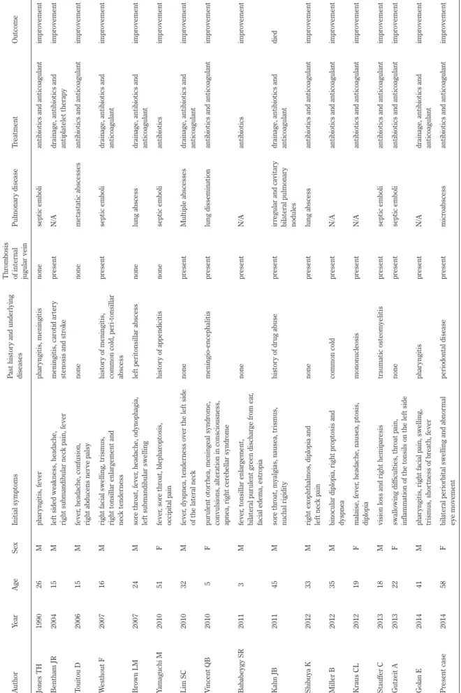
Table
1
R
epor
tedcases of patients with both L
emier
re syndr
ome and caver
nous sinus thr ombosis. A uthor Year Age Sex Initial symptoms Past histor y and underlying diseases Thr ombosis of inter nal jugular vein Pulmonar y disease Tr eatment Outcome Jones TH 1990 26 M phar yngitis, fever phar yngitis, meningitis none septic emboli
antibiotics and anticoagulant
impr ovement Bentham JR 2004 15 M lef
t sided weakness, headache,
right submandibular neck pain, fever
meningitis, car otid ar ter y stenosis and str ok e pr esent N/A
drainage, antibiotics and antiplatelet therapy
impr ovement Touitou D 2006 15 M fever , headache, confusion,
right abducens ner
ve palsy
none
none
metastatic abscesses
antibiotics and anticoagulant
impr ovement W esthout F 2007 16 M
right facial swelling, trismus, right tonsillar enlar
gement and
neck tender
ness
histor
y of meningitis,
common cold, peri-tonsillar abscess
pr
esent
septic emboli
drainage, antibiotics and anticoagulant
impr ovement Br own LM 2007 24 M sor e thr oat, fever , headache, odynophagia, lef t submandibular swelling lef t peritonsillar abscess none lung abscess
drainage, antibiotics and anticoagulant
impr ovement Yamaguchi M 2010 51 F fever , sor e thr oat, blephar optosis, occipital pain histor y of appendicitis none septic emboli antibiotics impr ovement Lim SC 2010 32 M fever , dyspnea, tender
ness over the lef
t side
of the lateral neck
none
pr
esent
Multiple abscesses
drainage, antibiotics and anticoagulant
impr ovement V incent QB 2010 5 F pur ulent otor
rhea, meningeal syndr
ome,
convulsions, alteration in consciousness, apnea, right cer
ebellar syndr ome meningio-encephalitis pr esent lung dissemination
antibiotics and anticoagulant
impr ovement Bababeygy SR 2011 3 M fever , tonsillar enlar gement, bilateral pur ulent gr een dischar ge fr om ear ,
facial edema, estr
opia none pr esent N/A antibiotics impr ovement Kahn JB 2011 45 M sor e thr
oat, myalgias, nausea, trismus,
nuchal rigidity histor y of dr ug abuse pr esent ir
regular and cavitar
y
bilateral pulmonar
y
nodules
drainage, antibiotics and anticoagulant
died
Shibuya K
2012
33
M
right exophthalmos, diplopia and left neck pain
none
pr
esent
lung abscess
antibiotics and anticoagulant
impr ovement Miller B 2012 35 M
binocular diplopia, right pr
optosis and dyspnea common cold pr esent N/A
antibiotics and anticoagulant
impr ovement Kraus CL 2012 19 F malaise, fever
, headache, nausea, ptosis,
diplopia
mononucleosis
pr
esent
N/A
antibiotics and anticoagulant
impr ovement Stauffer C 2013 18 M
vision loss and right hemipar
esis
traumatic osteomyelitis
pr
esent
septic emboli
antibiotics and anticoagulant
impr ovement Gutzeit A 2013 22 F swallowing difficulties, thr oat pain,
inflammation of the tonsils on the lef
t side
none
pr
esent
septic emboli
antibiotics and anticoagulant
impr ovement Golan E 2014 41 M phar
yngitis, right facial pain, swelling,
trismus, shor tness of br eath, fever phar yngitis pr esent N/A
drainage, antibiotics and anticoagulant
impr ovement Pr esent case 2014 58 F
bilateral periorbital swelling and abnor
mal eye movement periodontal disease pr esent micr oabscess
antibiotics and anticoagulant
impr
ovement
N/A, not available; M, male; F
敗血症による海綿静脈洞血栓症の 1 例 55:487 30.5 mmol/l,BE 7.1 mmol/l,と代謝性アルカローシスの所見 であり明らかな肺塞栓症を示唆する所見はみとめられなかっ たが画像所見と症状から本症例は Lemierre 症候群の合併と 診断した. 炎症所見は徐々に改善傾向で第 8 病日には左眼の自発開眼 も可能になり,眼瞼腫脹の改善と共に眼球運動制限も改善し てきた.第 11 病日にメロペネムを中止し,細菌の感受性がよ り狭域のスルバクタム・アンピシリンに変更した.その後も 眼瞼腫脹と眼球運動制限などの神経所見は徐々に改善した. 歯科では口腔内ブラッシングと外皮用殺菌消毒剤で洗浄し口 腔内衛生管理をおこないながら経過を観察し,最終的に第 21 病日に抜歯をおこない,第 25 病日には右眼の自発開眼が可能 なまで回復した.拡散強調像で指摘された紐状の高信号は症 状の改善とともに消退していった.海綿静脈洞内の血栓もみ とめられなくなった.また胸部 CT 上の異常陰影も治療経過 とともに消失した. 炎症の改善とともに電解質や胆道系の酵素も正常化した. 抗生剤は内服薬に変更し,敗血症の再燃,海綿静脈洞の血栓 の再形成の所見がないことを確認して第 40 病日に退院,その 後外来で抗生剤を中止したが症状の再発をみとめていない. 考 察 CSTの症状はその頻度や重症度において多岐にわたる.静 脈洞炎や副鼻腔炎,あるいは本症例のように齲歯からの波及 による炎症では,頭痛や発熱が早期から出現し,その後生じ る眼静脈の灌流障害にともない眼球突出,結膜浮腫,眼瞼周 囲の浮腫にともなう膨張,ひいては外眼筋の圧迫で外眼筋麻 痺をおこしやすい1)2).感染の波及による直接浸潤で髄膜炎, 脳炎や脳血管障害,下垂体壊死を合併したばあいには重篤化 あるいは致死性となる.致死率はおよそ 30%で,50%以上が 脳神経麻痺などの後遺症を残す予後不良の疾患と報告されて いる1).しかし近年の画像的診断の進歩により,症状出現後, 比較的早期に治療を開始することが可能となった. 歯肉の drainage vein である歯槽静脈は,翼突筋静脈叢に流 入し,翼突筋静脈叢は卵円孔を介して海綿静脈洞との連絡が あるため,顔面の皮膚や筋肉の感染あるいは歯や歯肉の感染 が海綿静脈洞に波及し感染性血栓を呈する機序が考えられて いる3)4).本症例でも齲歯により口腔内常在菌の Streptococcus constellatusが歯槽静脈か翼突筋静脈叢を経て海綿静脈洞へい たった結果,CST をひきおこしたと考えられた. 一方で副鼻腔炎,上気道感染,口腔内常在菌・ウイルス感 染症から血栓を形成し,内頸静脈血栓形成,肺塞栓症をおこ す一連の病態は Lemierre 症候群と呼ばれる.菌血症を原因と して肺膿瘍や脳膿瘍を合併することもあり,確定診断が遅れ がちになることから死亡率は高いとされる5)6).血液培養で原 因菌が発見され診断されることが多く,早期の鑑別診断が重 要になる.本症例は内頸静脈血栓,肺病変がみられ Lemierre 症候群と CST の合併例と考えられた.われわれがしらべた範 囲では Lemierre 症候群と CST の合併例は少なく,しらべえ たかぎりでわれわれの症例以外に 16 例の報告がみられた (Table 1)7)~22),その原因は齲歯以外には急性扁桃炎,滲出 性咽頭炎,扁桃周囲膿瘍,髄膜炎などで,原因菌としては Fusobacterium necrophorumがもっとも多かった.齲歯など感 染が原因の CST 症例では Lemierre 症候群の合併も考えて, 内頸静脈の精査や肺の化膿性血栓症の合併に注意を払い精査 することが必要であると考えられた7)23). 脳静脈洞血栓症の MRI 所見は,71.4%の症例で脳静脈洞頭 部もしくは脳静脈が拡散強調画像で高信号,みかけの拡散係 数(ADC 値)は著明に低下する24).拡散強調画像で高信号を 呈する理由は,血管内の水の動きが制限されていることを示 唆し,比較的新しい血栓症であるためと考えられている.こ れまでの報告では実際に拡散強調画像で高信号,ADC で低 信号を示した CST 症例はみいだせなかった.おそらく,他の 脳静脈洞にくらべ上眼静脈あるいは海綿静脈洞は体積が小さ く拡散強調画像のスライス厚の問題で描出しにくい可能性が あり,撮像法の検討が必要と思われる.脳静脈洞血栓症の検 討6)では静脈血栓塞栓部位の拡散強調像の信号が高いほど, 再開通率が低いことが報告されているが,本症例では,抗凝 固療法によって血栓が消退していくにつれ,拡散強調画像に おける上眼静脈の高信号が消失した.本症例は来院時理学所 見と血液検査所見に加え,MRI 拡散強調画像でその存在をう たがい,造影 CT において早期に確定診断がえられた点で貴 重であり,この有用性を指摘した最初の報告である. 本症例の治療において,CST の原因が特定できない段階か ら血液培養後に抗生剤,抗凝固薬を開始し,血液培養陽性の 結果が出てから薬剤感受性試験を考慮してメロペネムを選択 した.一般に抗菌薬は血液培養の結果が出るまで第 3 世代セ フェムやメトロニダゾールから開始することが奨励されてお り,メチシリン耐性の菌も考慮し,バンコマイシンの併用も 考慮すべきである1)2).血栓中の菌に抗菌剤が効きにくいこと もあり,その使用期間は臨床症状が完全に消失後も長期に継 続することが望ましい1)~3)25).本症例では,拡散強調画像で 左右の上眼静脈の高信号がほぼ消失するまで約 3 ヵ月間抗菌 薬を継続後中止したが,これまで再発なく経過している.抗 凝固療法については早期のヘパリンとワルファリンの併用で 脳神経障害や,半身麻痺の後遺症を軽減し,予後を改善させ るという報告がある1)3)26).抗凝固療法にて症状の改善がない ばあい外科的ドレナージ術が考慮されることもある27).本症 例ではワルファリンをもちい,出血性合併症なく良好な経過 がえられているが,CST の再発予防目的の抗凝固療法の期間 継続に関する治療指針は存在せず,今後更なる症例の蓄積が 必要であると考えられた. ※本論文に関連し,開示すべき COI 状態にある企業,組織,団体 はいずれも有りません. 文 献
1) Desa V, Green R. Cavernous sinus thrombosis: current therapy. J Oral Maxillofac Surg 2012;70:2085-2091.
2) Ebright JR, Pace MT, Niazi AF. Septic thrombosis of the cavernous sinuses. Arch Intern Med 2001;161:2671-2676. 3) Berdai AM, Shimi A, Khatouf M. Cavernous sinus thrombophlebitis
complicating sinusitis. Am J Case Rep 2013;14:99-102.
4) Komatsu H, Matsumoto F, Kasai M, et al. Cavernous sinus thrombosis caused by contralateral sphenoid sinusitis: a case report. Head Face Med 2013;9:9.
5) Aggarwal SK, Nath A, Singh R, et al. Lemierreʼs Syndrome presenting with neurological and pulmonary symptoms: Case report and review of the literature. Ann Indian Acad Neurol 2013;16:259-263.
6) Saposnik G, Barinagarrementeria F, Brown RD, Jr., et al. Diagnosis and management of cerebral venous thrombosis: a statement for healthcare professionals from the American Heart Association/American Stroke Association. Stroke 2011;42:1158-1192.
7) Miller B, Khalifa Y, Feldon SE, et al. Lemierre syndrome causing bilateral cavernous sinus thrombosis. J Neuroophthalmol 2012; 32:341-344.
8) 澁谷航平,五十嵐修一,佐藤朋江ら.化膿性海綿静脈洞血栓 症,細菌性髄膜炎をきたした Lemierre 症候群の 33 歳男性例. 臨床神経 2012;52:782-785.
9) Stauffer C, Josiah AF, Fortes M, et al. Lemierre syndrome secondary to community-acquired methicillin-resistant Staphylococcus aureus infection associated with cavernous sinus thrombosis. J Emerg Med 2013;44:177-182.
10) Westhout F, Hasso A, Jalili M, et al. Lemierre syndrome complicated by cavernous sinus thrombosis, the development of subdural empyemas, and internal carotid artery narrowing without cerebral infartion. Case Report. J Neurosurg 2007;106: 53-56.
11) Jones TH, Bergvall V, Bradshaw JP. Carotid artery stenoses and thrombosis secondary to cavernous sinus thromboses in Fusobacterium necrophorum meningitis. Postgrad Med J 1990; 66:747-750.
12) Bentham JR, Pollard AJ, Milford CA, et al. Cerebral infarct and meningitis secondary to Lemierreʼs syndrome. Pediatr Neurol 2004;30:281-283.
13) Touitou D, Deltour S, Bonneville F, et al. Hemiplegia in Lemierre syndrome: a neuroimaging strategy. Eur J Neurol 2006;13:424-426.
14) Brown LM, Wallwork B. Lemierreʼs—the sinister sore throat. J Laryngol Otol 2007;121:692-694.
15) Yamaguchi M, Nishizawa H, Yasumoto T, et al. Lemierre syndrome with blepharoptosis. Intern Med 2010;49:753-757. 16) Lim SC, Lee SS, Yoon TM, et al. Lemierre syndrome caused
by acute isolated sphenoid sinusitis and its intracranial complications. Auris Nasus Larynx 2010;37:106-109.
17) Vincent QB, Labedan I, Madhi F. Lemierre syndrome with meningo-encephalitis, severe cerebral artery stenosis, and focal neurological symptoms. J Pediatr 2010;157:345-345. e342. 18) Bababeygy SR, Almarzouki H, Buffenn AN. Isolated abducens
nerve palsy secondary to Lemierre syndrome. J AAPOS 2011; 15:587-589.
19) Kahn JB, Baharestani S, Beck HC, et al. Orbital dissemination of Lemierre syndrome from gram-positive septic emboli. Ophthal Plast Reconstr Surg 2011;27:e67-68.
20) Kraus CL, Culican SM. Challenging presentations of cavernous sinus thrombophlebitis. J Ophthalmic Inflamm Infect 2012;2: 133-136.
21) Gutzeit A, Roos JE, Portocarrero-Fäh B, et al. Differential diagnosis of Lemierreʼs syndrome in a patient with acute paresis of the abducens and oculomotor nerves. Korean J Ophthalmol 2013;27:219-223.
22) Golan E, Wong K, Alahmadi H, et al. Endoscopic sphenoid sinus drainage in Lemierre syndrome. J Clin Neurosci 2014;21:346-348.
23) Morelli N, Rota E, Sacchini D, et al. Lemierre syndrome: more than “the forgotten disease”. Neurology 2013;81:1179-1180. 24) Favrole P, Guichard JP, Crassard I, et al. Diffusion-weighted
imaging of intravascular clots in cerebral venous thrombosis. Stroke 2004;35:99-103.
25) Imholz B, Becker M, Lombardi T, et al. Septic thrombosis of the cavernous sinus secondary to a Streptococcus milleri oral infection. Dentomaxillofac Radiol 2012;41:525-528.
26) Kraus CL, Culican SM. Challenging presentations of cavernous sinus thrombophlebitis. J Ophthalmic Inflamm Infect 2012;2: 133-136.
27) Ito E, Saito K, Nagatani T, et al. Cavernous sinus thrombophlebitis caused by porphyromonas gingivalis with abscess formation extending to the orbital cavity. Case report. Neurol Med Chir 2009;49:370-373.
敗血症による海綿静脈洞血栓症の 1 例 55:489 Abstract
A case with both infectious cavernous sinus thrombosis
and Lemierre syndrome due to intraoral resident flora
Akihiro Nishida, M.D.
1), Toshiyasu Ogata, M.D.
1), Masataka Kudo, M.D.
1),
Kousuke Fukuhara, M.D.
1), Jiro Fukae, M.D.
1)and Yoshio Tsuboi, M.D.
1)1)Department of Neurology, Faculty of Medicine, Fukuoka University