Genetic Analysis of Alcohol-Metabolizing
Enzymes in Thermophilic Bacteria and Acetic
Acid Bacteria
著者(英)
Hisao Sakoda
学位名
博士(理学)
学位授与機関
関西学院大学
学位授与番号
34504乙第355号
URL
http://hdl.handle.net/10236/12629
Genetic Analysis of Alcohol-Metabolizing Enzymes in
Thermophilic Bacteria and Acetic Acid Bacteria
A Doctoral Thesis
By
Hisao SAKODA
Submitted to the Graduate School of Science and Technology
Kwansei Gakuin University
Japan
i
TABLE OF CONTENTS
Pages
GENERAL INTRODUCTION 1
CHAPTER 1
Catalytic Metabolism of the NAD-Dependent Alcohol Dehydrogenase
and Rational Shift of the Optimum pH 7
CHAPTER 2
Efficient Expression of the Gene Coding for Thermostable Aldehyde
Dehydrogenase, and Characterization of the Enzyme 39
CHAPTER 3
A New Way of Stabilizing Recombinant Plasmids 63
GENERAL CONCLUSION 81
LIST OF PUBLICATIONS 85
ii
Abbreviation List
ADH Alcohol Dehydrogenase
ADH-T Thermostable Alcohol Dehydrogenase ALDH Aldehyde Dehydrogenase
ALDH-T Thermostable Aldehyde Dehydrogenase Arg Arginine
ATP Adenosine Triphosphate
A. pasteurianus Acetobacter pasteurianus B. subtilis Bacillus subtilis
CCCP Carbonylcyanide-m-Chlorophenylhydrazone Cys Cysteine
DNA Deoxyribonucleic Acid DEAE Diethylaminoethyl
E. coli Escherichia coli
EDTA Ethylenediaminetetraacetic Acid
G. europaeus Gluconacetobacter europaeus G. stearothermophilus Geobacillus stearothermophilus
Glu Glutamic Acid His Histidine
IPTG Isopropyl β-D-Thiogalactopyranoside
L broth Luria-Bertani broth Lys Lysine
NAD Nicotinamide Adenine Dinucleotide
NADH Reduced Nicotinamide Adenine Dinucleotide NADP Nicotinamide Adenine Dinucleotide Phosphate
NTG N-Methyl-N′-Nitro-N-Nitrosoguanidine
OD Optical Density ORF Open Reading Frame
PAGE Polyacrylamide Gel Electrophoresis
S. cerevisiae Saccharomyces cerevisiae
SDS Sodium Dodecyl Sulfate Ser Serine
T. kodakaraensis Thermococcus kodakaraensis
Tc Tetracycline Thr Threonine
Tris Tris (hydroxymethyl) aminomethane
Trp Tryptophan
1
GENERAL INTRODUCTION
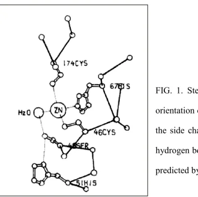
Alcohol dehydrogenase (ADH) and aldehyde dehydrogenase (ALDH) are ubiquitous enzymes that are widely distributed among several genera. Horse liver ADH has been much researched, and its catalytic mechanism is well known. X-ray crystallography study of horse liver ADH has revealed that the residues within its catalytic domain provide ligands to the catalytic zinc atom; Cys46, His67, and Cys174. The catalytic zinc atom is at the bottom of this active pocket with zinc-bound water molecule. This water molecule is hydrogen-bonded to the side chain of Ser48, which in turn is hydrogen-bonded to His51 (Fig. 1). The deprotonation of the catalytic zinc-water is facilitated by His51 residue acting through a hydrogen-bonded network to relay the proton to solvent (1, 2). The second zinc atom of the subunit is liganded by four sulfur atoms from Cys97, Cys100, Cys103, and Cys111.
Aldehydes are highly reactive and generated from multitudes of endogenous and exogenous sources. They have a variety of effects on the biological systems. Although some aldehydes such as retinaldehyde have beneficial effects, several aldehydes have deleterious effects such as cytotoxicity, mutagenicity, and carcinogenicity. Diverse enzymes have evolved to metabolize aldehydes to their less reactive forms. Among the most effective pathways for aldehyde metabolism is their oxidation to carboxylic acids by ALDHs (3). Crystallographic analyses have revealed the catalytic mechanism of ALDH. The overall mechanism of the reaction catalyzed by the hydrolytic ALDHs involves three main steps: (i) nucleophilic attack of the thiol group of the catalytic cysteine on the carbonyl carbon of the aldehyde substrate; (ii) hydride transfer from the tetrahedral thiohemiacetal intermediate to the pyridine ring of NAD(P)+; and (iii) hydrolysis of the resulting thioester intermediate (deacylation) (4).
2
FIG. 1. Stereo diagram showing the spatial orientation of the zinc-bound water molecule, the side chains of Ser48 and His51, and the hydrogen bond system between these groups, predicted by Eklund et al (1).
Acetic acid bacteria are gram-negative bacteria belonging to the group of
Proteobacteria. They are capable of oxidizing ethanol and sugars to corresponding organic
acids under aerobic condition (5). The kinetic study suggested that ADH and ALDH played a major role in alcohol metabolism of acetic acid bacteria (6, 7). They contained two systems for conversion of ethanol via aldehyde to acetic acid. One system was linked to the cytoplasmic membrane; the other system was in solution of the cytoplasm (8). They have an efflux pump for acetic acid at the cytoplasmic membrane, which is specific for acetic acid and is driven by a proton motive force independently of ATP; it is responsible for acetic acid resistance (9).
Two acetic acid bacteria, Acetobacter pasteurianus and Gluconacetobacter europaeus, are generally used for industrial vinegar production because they can efficiently oxidize ethanol to acetic acid and show strong resistance to ethanol and acetic acid. A. pasteurianus has been used in the traditional fermentation. G. europaeus has been used for the production of high-acidity vinegar in submerged bioreactors because it has extremely
3
strong ethanol-oxidizing ability and ethanol/acetic acid resistance as compared with other species. Therefore, G. europaeus is a main bacterium for industrial vinegar production in the world. However, studies on molecular genetics and biochemical analysis of G.
europaeus have been limited.
A targeted gene disruption system, using uracil auxotroph as a result of pyrimidine biosynthesis pathway genes impairment, was shown to be available for several microbes (10). This approach does not require any exogenous drug resistance genes. Orotate-phosphoribosyltransferase (OPRTase; encoded by pyrE) and orotidine-5′-mono- phosphate decarboxylase (OMPdecase; encoded by pyrF) catalyze the last two steps of pyrimidine biosynthesis. A bacterial orotic acid analogue, 5-fluoroorotic acid (5-FOA) is converted to 5-fluorouridine monophosphate (5-FUMP), which exhibits a bactericidal effect, by these enzymes. Mutants with defects in either of these enzymes are resistant to 5-FOA, but they are uracil auxotroph. Recently, a selection system utilizing these properties was successfully established in G. europaeus (11).
Molecular biology studies have shown that microorganisms can be transformed with artificial plasmids containing genes necessary for a specific synthetic production. However, the major problem with these plasmids is their instability in transmitting their properties to their daughter cells in a controlled manner during the cell division of the microorganisms. As a result, a large number of daughter cells, which does not contain the plasmid of interest, are produced during the fermentation. On a laboratory scale, this loss in desired plasmid can be countered by supplementing the culture medium with an antibiotic reagent corresponding to the antibiotic resistant gene of the plasmid. However, a large-scale addition of antibiotics in fermentations is disadvantageous. Therefore, addressing recombinant plasmid stability without adding antibiotic reagents is the most critical issue for a fermentation process.
4
In Chapter 1, the rational shift of the optimum pH was performed by substituting the catalytic amino acids of ADH from Geobacillus stearothermophilus.
In Chapter 2, the thermostable ALDH gene of G. stearothermophilus was cloned and substrate specificity was investigated.
In Chapter 3, a new method for stabilizing recombinant plasmid was established and applied for tryptophan production in Escherichia coli, and a thought-provoking approach for G. europaeus has been discussed.
5
REFERENCES
(1) Eklund, H., Nordstrom, B., Zeppezauer, E., Soderlund, G., Ohlsson, I., Boiwe, T.,
Soderberg, B. O., Tapia, O., Branden, C. I., and Akeson, A.: Three-dimensional
structure of horse liver alcohol dehydrogenase at 2.4 Å resolution. J. Mol. Biol., 102, 27-59 (1976).
(2) Plapp, B. V.: Conformational changes and catalysis by alcohol dehydrogenase. Arch. Biochem. Biophys., 493, 3-12 (2010).
(3) Lindahl, R.: Aldehyde dehydrogenases and their role in carcinogenesis. Crit. Rev. Biochem. Mol. Biol., 27, 283-335 (1992).
(4) Munoz-Clares, R. A., Gonzales-Segula, L., and Diaz-Sanchez, A. G.: Crystallographic evidence for active-site dynamics in the hydrolytic aldehyde dehydrogenases. Implications for the deacylation step of the catalyzed reaction. Chem. Biol. Interact., 191, 137-146 (2011).
(5) Cleenwerck, I., De Wachter, M., Gonzalez, A., De Vuyst, L., and De Vos, P.: Differentiation of species of the family Acetobacteraceae by AFLP DNA fingerprinting: Gluconacetobacter kombuchae is a later heterotypic synonym of Gluconacetobacter hansenii. Int. J. Syst. Evol. Microbiol., 59, 1771-1786 (2009).
(6) Nakayama, T.: Studies on acetic acid bacteria Ⅲ. Purification and Properties of Coenzyme-Independent Aldehyde Dehydrogenase. J. Biochemistry, 49, 158-163 (1961).
(7) Nakayama, T.: Studies on acetic acid bacteria Ⅳ. Purification and properties of a new type of alcohol dehydrogenase. J. Biochemistry, 49, 240-251 (1961).
(8) Nakayama, T., and De Ley, J.: Localization and distribution of alcohol-cytochrome 553 reductase in acetic acid bacteria. Antonie van Leeuwenhoek, 31, 205-219 (1965). (9) Matsushita, K., Inoue, T., Adachi, O., and Toyama, H.: Acetobacter aceti possesses a
6
proton motive force-dependent efflux system for acetic acid. J. Bacteriol., 187, 4346-4352 (2005).
(10) Sato T, Fukui T, Atomi H, and Imanaka T.: Targeted gene disruption by homologous recombination in the hyperthermophilic archaeon Thermococcus
kodakaraensis KOD1. J. Bacteriol., 185, 210-220 (2003).
(11) Akasaka, N., Sakoda, H., Hidese, R., Ishii, Y., and Fujiwara, S.: An efficient method using Gluconacetobacter europaeus to reduce an unfavorable flavor compound, acetoin, in rice vinegar production. Appl. Environ. Microbiol., 79, 7334-7342 (2013).
7
CHAPTER 1
Catalytic Metabolism of the NAD-Dependent Alcohol Dehydrogenase and
Rational Shift of the Optimum pH
INTRODUCTION
Various thermostable alcohol dehydrogenases (ADH-Ts) have been analyzed for the industrial production of alcohol (1-3), including chiral alcohol (4). Geobacillus
stearothermophilus NCA1503 was found to produce an ADH-T amounting to 1 to 2% of
soluble cell protein. This strain produced ethanol from sucrose or glucose as a carbon source under anaerobic conditions at high temperatures (1, 5). Two types of ADH have been isolated from G. stearothermophilus NCA1503 and DSM2334 (6). ADH-T from NCA1503 showed enzymatic, structural, and immunological properties different from those of the ADH from DSM2334. The ADH from DSM2334 is active with primary alcohols, including methanol (6, 7). The gene for ADH from DSM2334 has been cloned in
Escherichia coli (8). In this work, the ADH-T gene (adhT) from G. stearothermophilus
NCA1503 was cloned in Bacillus subtilis.
The ADH reaction mechanism was originally studied with horse liver ADH by X-ray crystallographic analysis and a kinetic study. The catalytic domain of horse liver ADH is composed of the catalytic zinc-water, the side chain of Ser48, and the imidazole ring of His51. The deprotonation of the catalytic zinc-water is facilitated by the imidazole ring of His51 acting through a hydrogen-bonded network to relay the proton to solvent (7, 9, 10). By comparing amino acid sequences of ADH-T with horse liver ADH and other ADHs, the catalytic system of ADH-T was inferred.
Here, we describe the molecular cloning and nucleotide sequencing of the ADH-T gene (adhT) from G. stearothermophilus NCA1503, a comparison of the deduced amino
8
acid sequence with the sequences of other ADHs, prediction of the catalytic system of ADH-T based on the crystallographically determined model of the horse liver ADH, confirmation of the putative catalytic system by replacing the catalytic amino acids of ADH-T by using site-directed mutagenesis, and construction of a modified enzyme that exhibits a pH profile different from that of the wild-type ADH-T.
MATERIALS AND METHODS
Bacterial strains and plasmids.
G. stearothermophilus NCA1503 (11) was used as a DNA donor. B. subtilis MI113
(arg-15 trpC2 hsrM hsmM) (11) and M1112 (leuA8 thr-5 arg-15 recE4 hsrM hsmM) (12) were used as host cells in gene cloning. Since B. subtilis MI112 is deficient in DNA recombination, it was used as the host cell to stably carry the recombinant plasmid. A low-copy-number plasmid, pTB524 (Tcr; coding for tetracycline resistance) (13), which has a BamHI site suitable for gene cloning, was used to construct the gene bank of G.
stearothermophilus NCA1503. pTB522 (Tcr) (14), which has a HindIII site for cloning, and pTB524 were used for subcloning of the gene. E. coli TG1 (supEA (lac-proAB) hsdΔ5 F'
traD36 proAB+ lacIq lacZΔM15) (15) and M13 mpl8 and M13 mp19 (15) were used as a
host cell and phages to subclone the gene for nucleotide sequencing.
Media.
G. stearothermophilus NCA1503 was grown at 55°C in modified L broth containing
tryptone (20 g/liter), yeast extract (10 g/liter), and NaCl (5 g/liter), and the pH was adjusted to 7.3 with 2 N NaOH. B. subtilis M1113 and M1112 were grown in L broth (11) at 37°C. Solid medium contained 20 g of agar per liter for growth at 55°C and 15 g of agar per liter for growth at 37°C.
9
Transformants of B. subtilis with pTB524, pTB522, or their derivatives carrying the tetracycline (Tc) resistance gene were grown in L broth containing Tc (25 μg/ml).
Detection of ADH-producing colonies on plates.
ADH-producing colonies were selected on modified aldehyde indicator plates as described by Conway et al. (16), with slight modification. The plates were composed of antibiotic medium 3 (17.5 g/liter) (Difco Laboratories, Detroit, MI, USA) acting as a buffer (pH 7.0), ethanol (20 ml/liter), pararosaniline (50 mg/liter) (Sigma Chemical Co., St. Louis, MO, USA), and sodium hydrogen sulfite (250 mg/liter). Ethanol diffuses into cells and can be converted by ADH to acetaldehyde, which reacts with the reagent to form Schiff base intense red.
Preparation of plasmids and chromosomal DNA.
Either the rapid alkaline extraction method or CsCl-ethidium bromide equilibrium density gradient centrifugation was used to prepare plasmid DNA, whereas chromosomal DNA was prepared as described elsewhere (17, 18).
Transformation of B. subtilis.
For transformation of B. subtilis, competent cells were prepared as described previously (18). Tcr transformants were transferred on the modified aldehyde indicator plates and incubated at 37°C for 5 hours to check colony color turning intense red.
Nucleotide sequencing.
DNA was sequenced by the dideoxy method of Sanger et al. (19) with the Sequenase sequencing kit (United States Biochemical Corp., Cleveland, OH, USA). After digestion
10
with restriction enzymes, DNA fragments were subcloned into M13 mpl8 or M13 mpl9. E.
coli TG1 was used as a host cell.
Site-directed mutagenesis.
Point mutations were introduced into a gene with an oligonucleotide-directed in vitro mutagenesis system (Amersham, Buckinghamshire, UK) according to the manufacturer's instructions.
Active staining of ADH.
Active staining of ADH was performed according to the method described by Dowds et al. (8). Crude enzymes were run on a 6% polyacrylamide gel with solutions and reagents from which sodium dodecyl sulfate (SDS) was omitted. The gel was stained for ADH activity by an alcohol-dependent nitroblue-tetrazolium procedure. The gel was soaked in 500 mM Tris-HCl (pH 8.8) at 4°C for 15 min and then incubated at 37°C for 30 min in a staining solution containing 150 mM Tris-HCl (pH 8.8), NAD (0.132 mg/ml), nitroblue-tetrazolium (0.163 mg/ml), phenazine methosulfate (0.03 mg/ml), and ethanol (10 ml/liter). These reagents were purchased from Sigma Chemical Co.
Enzyme purification.
ADH-T and its derivatives were purified from the transformants of B. subtilis MI112. They were grown to the stationary phase at 37°C in L broth containing Tc (25 μg/ml), harvested by centrifugation (10,000×g, 10 min) at 4°C, and washed in 20 mM potassium phosphate buffer (pH 7.8). The cell pellet was suspended in the phosphate buffer containing lysozyme (l mg/ml) and DNase I (10 U/ml) and incubated at 37°C for 30 min. After centrifugation (55,000×g, 30 min), the supernatant was heated at 60°C for 10 min and again
11
centrifuged (20,000×g, 10 min) at 4°C. The crude enzyme was purified by DEAE-cellulose (DE52, Whatman BioSystems Ltd., Maidstone, Kent, UK) ion-exchange column chromatography, equilibrated with 20 mM potassium phosphate buffer (pH 7.8). The enzyme was eluted with a linear gradient (0 - 1 M) of potassium chloride dissolved in the phosphate buffer. Active fractions were dialyzed overnight at 4°C in 20 mM potassium phosphate buffer (pH 7.8). The final enzyme preparation was confirmed to be homogeneous by SDS polyacrylamide gel electrophoresis (PAGE).
According to the method described above, wild-type ADH-T and mutant enzymes Thr40Ser and His43Arg were purified to homogeneity.
Assay of ADH activity.
ADH activity was assayed by monitoring alcohol-dependent NAD reduction or acetaldehyde- or ketone-dependent NADH oxidation at 340 nm (6, 20, 21). ADH-activity (U) was expressed as μmol of NADH produced or decreased per min, using a molar absorption coefficient of 6.22 mM-1・cm-1. The assay was performed at 55°C in a reaction mixture containing 100 mM potassium phosphate buffer (pH 7.8), 1 mM NAD, and 100 mM each alcohol. The aldehyde or ketone reductase (reverse reaction of alcohol dehydrogenase) assay was performed at 55°C in a reaction mixture containing 100 mM potassium phosphate buffer (pH 7.8), 0.2 mM NADH, and 100 mM acetaldehyde or each ketone (20). Concentrations of substrates and coenzymes were confirmed to be excessive by measuring enzyme activity under various concentrations of substrates and coenzymes. The standard ADH assay was performed at 55°C in a reaction mixture which contained 100 mM potassium phosphate buffer (pH 7.8), 1 mM NAD, and 100 mM ethanol. To examine the pH profile of the enzyme, 100 mM potassium phosphate buffer or 100 mM glycine-KOH buffer having various pH was used.
12 Protein assay.
The protein concentration was measured by the method of Lowry et al. (22) with bovine serum albumin as the standard.
Other procedures.
Procedures for digestion of DNA with restriction endonucleases, ligation of DNA with T4 DNA ligase, agarose gel electrophoresis, and SDS-PAGE were described elsewhere (17, 23-25). Unless otherwise specified, all chemicals used in this work came from sources described in a previous paper (26).
Nucleotide sequence accession number.
The nucleotide sequence data reported in this paper will appear in the DDBJ, EMBL, and GenBank nucleotide sequence data bases under the accession number D90421.
13
RESULTS
Cloning of the ADH gene from G. stearothermophilus NCA1503.
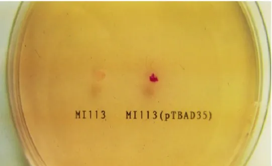
Chromosomal DNA of G. stearothermophilus was partially digested with Sau3AI, and fragments of approximately 6 kb were isolated and purified by Gene-Clean (Bio101 Inc., La Jolla, CA, USA). These fragments were ligated into the BamHI site of pTB524. The ligation mixture was used to transform B. subtilis MI113. Of 3,000 Tcr transformants of B. subtilis, one ADH-positive clone was found on the modified aldehyde indicator plates (Fig.1). The transformant carried a recombinant plasmid containing an insert of about 7 kb. A lysate of the candidate cell, subjected to electrophoresis, showed a band of ADH-active staining at the same position as that of a DNA donor strain, G. stearothermophilus NCA1503 (photograph not shown). The level of ADH-activity of the recombinant plasmid carrier (1.48 U/mg of dry cell) was about nine-fold higher than that of the DNA donor (0.17 U/mg of dry cell), whereas
FIG. 1. Detection of ADH-producing colonies on the aldehyde indicator plate.
Ethanol diffusing into cell can be converted by ADH to acetaldehyde, which reacts with the reagent (pararosaniline) to form Schiff base intense red. Host cell (B. subtilis MI113) showed a slightly pink. Transformants harboring a recombinant plasmid (pTBAD35) showed intense red.
14
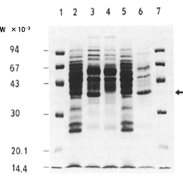
the host cell showed little activity (less than 0.002 U/mg of dry cell). The candidate produced a MW 35,000 protein, which could also be found in the DNA donor but not in the host cell, and moreover, the protein was thermostable (Fig. 2). The recombinant plasmid was designated pTBAD70. It was concluded that pTBAD70 carried the ADH-T gene (adhT) from G. stearothermophilus NCA1503.
FIG. 2. SDS-PAGE analysis of cell extracts.
Each lane contains 5 μl of cell extract. Lanes: 1 and 7, molecular weight markers; 2, G.
stearothermophilus NCA1503; 3, ADH-positive transformant of B. subtilis M1113; 4, B.
subtilis M1113; 5, cell extract of G. stearothermophilus NCA1503, heated (60°C, 10 min) and centrifuged (20,000 x g); 6, cell extract of B. subtilis transformant, heated (60°C, 10 min) and centrifuged (20,000 x g). The arrow indicates the position of ADH-T.
15 Subcloning of the adhT gene.
To analyze the location of the adhT gene, I constructed three deletion plasmids from pTBAD70. A BamHI fragment (about 3.5 kb) and a HindⅢ fragment (about 4.0 kb) from pTBAD70 were subcloned in pTB524 and pTB522, and their recombinant plasmids were designated pTBAD35 and pTBAD40, respectively. pTBAD70-, pTBAD40-, and pTBAD35- harboring cells showed ADH-activity on the aldehyde indicator plate (Fig. 1). In contrast, the strain carrying pTBAD35ΔSphI, which lacked an SphI fragment (about 0.6 kb) from pTBAD35, showed no ADH-activity. Therefore, the adhT gene was considered to be located in the 2.2-kb HindIII-BamHI fragment including the SphI fragment.
Nucleotide sequence of the adhT gene.
The nucleotide sequence of the 2.2 kb HindIII-BamHI fragment was determined. A large open reading frame was found in the 1.7-kb EcoRI-BamHI fragment (Fig. 3). It was composed of 1,011 bp corresponding to 337 amino acids. The molecular weight was estimated to be 36,098, which agreed with the result of SDS-PAGE (Fig. 2). The N-terminal amino acid sequence has been reported elsewhere (27, 28), and the first 40 amino acids were identical to the N-terminal sequence deduced from the nucleotide sequence. The amino acid composition which had been reported previously (6) was also in agreement with the sequencing result in this work. It was therefore concluded that the open reading frame encoded the ADH-T gene (adhT). A Shine-Dalgarno sequence was found 10 bases upstream from the translation start site (ATG). Since a large amount of ADH-T was produced, a strong promoter was expected. However, a typical promoter sequence was not found. The sequence resembling typical prokaryotic terminators was found downstream from the open reading frame. The highly AT-rich sequence (about 200 bp) was found at the 5'-flanking region of the open reading frame (Fig. 3).
17
FIG. 3. Nucleotide sequence of the adhT gene and the deduced amino acid sequence of the encoded protein.
The amino acid sequence is shown above the nucleotide sequence. A probable Shine-Dalgarno (SD) sequence is indicated by a solid line. The terminator and inverted repeat at the 5'-flanking region are shown by arrows. An asterisk indicates a stop codon. ←
18
Comparison of the deduced amino acid sequence with the sequences of different ADHs.
Comparison of the primary structures of enzymes with the same function but different origins is useful to determine the amino acids essential for activity, because the active site and substrate binding site are highly conserved (29). ADHs are widely distributed in different organisms and tissues. I compared the amino acid sequences of different ADHs. Most of them showed homology. The deduced amino acid sequence of ADH-T from G.
stearothermophilus was homologous (55% identity) with that of Gluconacetobacter europaeus (32), 56% with Acetobacter pasteurianus (33), 32% with Saccharomyces cerevisiae (34), 31% with human (35), 34% with horse (36), 27% with Thermococcus kodakaraensis (37).
The amino acids indispensable for the catalytic activity of horse liver ADH (7, 36) are highly conserved in the seven ADHs (Fig. 4). The catalytic zinc atom of horse liver ADH is bound by three protein ligands, one sulfur atom each from Cys38 (cysteine at position 38 of ADH-T) and Cys148 and one nitrogen atom from His61. These amino acids are completely conserved. The ligands of the second zinc atom, Cys92, Cys95, Cys98, and Cys106, are also conserved. Furthermore, one of the amino acids participating in the proton release system, a serine residue of horse liver ADH corresponding to position 40 of G. stearothermophilus ADH-T, is substituted with threonine. Serine and threonine could play the same function through their hydroxyl group. Another amino acid in the proton release system, His43 of ADH-T, is highly conserved (Fig. 4).
→
FIG. 4. Comparison of amino acid sequences of seven different ADHs
Lanes: 1, G. stearothermophilus; 2, G. europaeus; 3, A. pasteurianus; 4, S. cerevisiae; 5, human; 6, horse; 7, T. kodakaraensis. The catalytic amino acids are indicated by arrows.
20 A putative reaction mechanism for ADH-T.
According to the argument mentioned above, it is believed that these ADHs, including ADH-T, have the same reaction mechanism as that shown for horse liver ADH (7). The basic reaction mechanism of horse liver ADH is as follows. One equivalent of proton is released per equivalent of ethanol that is oxidized. This proton release is associated with NAD binding and is dissociated from the water molecule bound to catalytic zinc. This proton release from the water molecule occurs via the hydrogen bond system through the side chain (hydroxyl group) of Thr40 to the imidazole ring of His43 (Fig. 5B). Then alcohol binds to zinc as the alcoholate ion, displacing the hydroxyl ion. The zinc atom polarizes the alcoholate so that direct hydrogen transfer and subsequent rearrangement to aldehyde can occur (7, 9). A putative proton release system for ADH-T was predicted by following the mechanism of horse liver ADH.
FIG. 5. Higher structure of ADH-T predicted with SWISS-MODEL. A, Tetramer structure of ADH-T: B, Detail picture of the active pocket.
Putative mechanism for the proton release system for ADH-T was composed of a zinc-bound water molecule, Thr40, and His43. The proton release is through a hydrogen -bonded network to relay the proton to solvent.
21
Analysis of the proton release system by amino acid substitution.
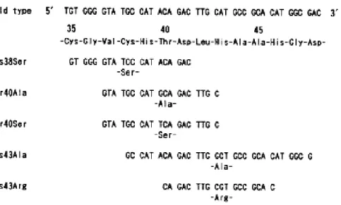
To verify the reaction mechanism of ADH-T, the putative catalytic amino acid residues, Cys38, Thr40, and His43, were substituted by site-directed mutagenesis with chemically synthesized oligonucleotides (Fig. 6). The following mutant enzymes were produced: Cys38Ser (Cys38 as a putative catalytic zinc ligand was replaced by serine), Thr40Ala, Thr40Ser, and His43Ala. Their cell lysates were used for ADH-assay and SDS-PAGE. All mutant enzymes were produced at the same level (data not shown). However, Cys38Ser, Thr40Ala, and His43Ala had no ADH-activity.
In contrast, Thr40Ser showed ADH-activity. The wild-type ADH-T and the mutant enzyme Thr40Ser were purified to homogeneity. Thr40Ser had the same pH profile as the wild-type enzyme, although the level of enzyme activity was lower (Fig. 7). These results indicate that Cys38, His43, and the hydroxyl group of Thr40 or serine residue are essential for enzyme activity and that the lower level of activity of Thr40Ser might be explained by
FIG. 6. Nucleotide sequence and deduced amino acid sequence of the catalytic site and its flanking regions. Synthetic oligonucleotides to introduce mutations are shown. Mutated amino acids are indicated below the nucleotides. Amino acid numbers are shown above the amino acid sequences.
22
a subtle change of steric conformation. The proton in the ADH-T reaction mechanism would be transported from a water molecule, through the hydroxyl group of Thr40, and released from the imidazole ring of His43 (Fig. 5B).
Change of the enzyme pH profile by site-directed mutagenesis.
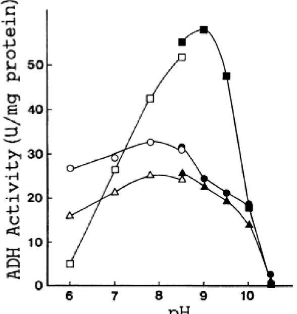
His43 was substituted by arginine to alter the pKa of the side chain (i.e., the pKa of the imidazole ring of histidine is 6.0 and that of the guanidine group of arginine is 12.5). We inferred that this mutation might disturb the pKa of the proton release group and result in pH dependence different from that of wild-type ADH-T. The mutant enzyme His43Arg was produced and purified to homogeneity. The pH dependences of ADH-T, Thr40Ser, and His43Arg were tested by using purified enzymes. Wild-type ADH-T and Thr40Ser showed maximum activity at around pH 7.8, corresponding to the pKa of 7.6 of the proton release group of horse liver ADH in the presence of NAD. In contrast, His43Arg exhibited a lower level of activity under acidic conditions but a higher level of activity under alkaline conditions than the wild-type did. The maximum activity was observed at pH 9.0. Surprisingly, the maximum activity of His43Arg was about two fold higher than that of the wild-type (Fig. 7). Thus, the optimum pH of ADH-T was shifted from neutral to alkaline by replacing the catalytic amino acid His43 with arginine.
23
FIG. 7. pH profiles for ethanol of wild-type ADH-T (○and●), Thr40Ser mutant enzyme (△and▲), and His43Arg (□and■).
Open symbols, enzyme assay in 100 mM potassium phosphate buffer; closed symbols, enzyme assay in 100 mM glycine-KOH buffer. Enzyme activity was assayed under the standard conditions described in the text, except for buffer pH. When the NAD concentration in the reaction mixtures was reduced to 0.2 mM, nearly the same results were obtained. Therefore, 1.0 mM NAD was actually excessive at different pH conditions.
24 Efficient purification of thermostable enzyme
The ADH positive mutant enzymes Thr40Ser (Thr40 was substituted by serine) and His43Arg were obtained by site-directed mutagenesis. The wild-type enzyme, ADH-T (molecular weight 36,098), and the mutants, Thr40Ser and His43Arg, were easily purified to homogeneity from the transformant of B. subtilis, respectively (Fig. 8). Since wild-type ADH-T and two mutants were thermostable at 60°C for 1 hour (data not shown), heat-treatment was a powerful step for purification.
FIG. 8. SDS-PAGE photograph of purified enzyme.
Lane: 1, the purified wild-type ADH-T; 2, molecular weight markers. The mutant enzymes, Thr40Ser and His43Arg, gave the same results (photograph not shown).
25
Amino acid substitutions for substrate specificity of the alcohol dehydrogenase
More intensive studies have been conducted with the wild-type ADH-T and the mutant enzymes, Thr40Ser and His43Arg. It was shown that the hydroxyl group of Thr40 or Ser40 is essential for ADH-activity, and that the imidazole ring of His43 can be substituted by the guanidine group of Arg43 as a catalytic side chain. The optimum pH of Thr40Ser was 7.8, like ADH-T. While that of His43Arg in contract, shifted from 7.8 to 9.0. The substitution, His43 (pKa 6.0) by arginine (pKa 12.5), would change the pKa value of the catalytic group. The pH-dependence of the enzyme should be attributed to the pKa value of the proton release group (38). Thr40Ser exhibited lower ADH-activity than wild-type ADH-T. In contrast, the maximum activity of His43Arg showed two-fold higher activity than the wild-type. These phenomena would be explained by the conformational changes of the catalytic group.
The space available in the active pocket of ADH-T for positioning the substrate was inferred to be restricted by the side chains of Thr40 and His43 as suggested by Xray analysis with horse liver ADH (7, 9). The substrate specificity of ADH-T and its mutants was systematically tested.
Wild-type ADH-T was active not only with primary alcohols, but also with secondary alcohols. As the reverse reaction, acetaldehyde and ketones were reduced by the enzyme (Table 1). The substrate specificity of Thr40Ser was nearly identical to that of wild-type ADH-T. However, the mutant enzyme exhibited higher activity for 2-methyl-l-propanol, 3-pentanol, and cyclohexanol (Table 1). Horse liver ADH (31), having a catalytic serine corresponding to Thr40 of ADH-T, is active with cyclohexanol (7). However, yeast ADH II (30), having a catalytic threonine, is not active with cyclohexanol (7). The length of the side chain of Thr40 or Ser40 must be significant for substrate binding. The substrate specificity of Thr40Ser could be explained by the expansion of the substrate binding pocket
26
by the substitution; in other words, the methyl group of Thr40 was eliminated in Ser. His43Arg exhibited higher activity for primary alcohols (except for 1-pentanol and branched alcohol such as 2-methyl-l-propanol) and acetaldehyde than wild-type ADH-T.
However, the mutant enzyme showed little activity to secondary alcohols, diols, triol, and ketones (Table 1). The drastic change in the substrate specificity of His43Arg might be
27
caused by a size reduction of the substrate binding pocket occupied by a long side chain of arginine residue. The preferential reaction for chiral (R or S) 2-butanol was not changed by these mutations (Thr40Ser, and His43Arg).
An alcohol dehydrogenase catalyzes the oxidation/reduction reaction between ethanol and acetaldehyde. Some isozymes are suitable for ethanol oxidation, the others for acetaldehyde reduction. The wild-type ADH-T and Thr40Ser had a low Km value for ethanol (Km-e), whereas His43Arg gave a much higher Km-e value. To consider the properties of the enzymes, I compared their rate constants with those of ADH I and ADH II (21) from Saccharomyces cerevisiae (Table 2). A constitutive enzyme, ADH I, has a high
Km-e value, and seems to be mainly responsible for the production of ethanol from
acetaldehyde in cells grown anaerobically. Another cytoplasmic isozyme, ADH II, has a much lower Km-e. ADH II, an inducible enzyme, is found only in aerobically grown yeast. A primary function of ADH II is the conversion of ethanol accumulated during anaerobic growth into acetaldehyde with the concomitant reduction of NAD (21, 30).
(21)
28
It should be able to use a low concentration of ethanol efficiently since its Km-e value is about 9-fold lower than that of yeast ADH I (21, 40). The wild-type ADH-T, having a smaller Km-e value than that of ADH II, should be powerful for ethanol oxidation, even at a low concentration. In contrast, His43Arg gave a large Km-e value and remarkably high
kcat value for acetaldehyde (kcat-a). It would function as a fermentative isozyme, reducing
acetaldehyde to ethanol like yeast ADH I. His43Arg showed its optimum activity at pH 9.0. Its kcat value (71.9±2.7s-1) toward ethanol at pH9.0 was much larger than that measured under pH 7.8, and the Km value (21.0±0.1 mM) at pH 9.0 was similar to that under pH 7.8. However, the values for acetaldehyde at pH 9.0 could not be measured because of the high background of the reaction mixture containing acetaldehyde under the alkaline condition, pH 9.0.
29
DISCUSSION
The thermostable alcohol dehydrogenase (ADH-T) gene (adhT) from G.
stearothermophilus was cloned in B. subtilis. Wild-type ADH-T and its derivatives were
easily purified from the transformants to homogeneity by heat treatment and DEAE-cellulose ion-exchange chromatography. Heat treatment is a powerful step to purify thermostable enzymes as shown in Fig. 2.
By site-directed mutagenesis, some adhT mutants were constructed. Studies with the mutant enzymes, which were constructed on the basis of three-dimensional structure information available for horse liver ADH, provided considerable information about ADH-T catalysis. Thr40 and His43 should be essential as the active center of ADH-T. Cys38 would be a ligand of the catalytic zinc (Fig. 5B). The pH profile of ADH-T was altered by replacing the catalytic amino acid histidine, His43, with arginine. By the substitution, the pKa of the active group, which was composed of a water molecule, Thr40, and His43, was thought to be shifted from neutral to alkaline. As a result, the alkaline enzyme His43Arg was obtained. Under acidic conditions, the mutant enzyme exhibited a lower level of activity than did wild-type ADH-T. The explanation for this might be that substitution of His43 with arginine slowed down the proton release reaction under acidic conditions.
Generally speaking, the pKa value of the active center of an enzyme can influence the pH profile. In other words, the pH profile of an enzyme could be altered by changing the
pKa value of a catalytic amino acid. For example, an active-site histidine residue of serine
protease acts as a general base in enzyme catalysis, and its pKa rules enzyme activity. Increasing the overall negative charge on the enzyme should raise the pKa of the active-site histidine by stabilizing the protonated form of the histidine, whereas increasing the positive charge should lower the pKa by destabilizing the protonated form of the histidine. Its
30
activity under acidic condition increased when the number of lysine residues of the enzyme surface was increased by site-directed mutagenesis (38).
Since enzymes are proteins containing many ionizable groups, they exist in a whole series of different states of ionization. However, only one of the ionic forms of the active center is catalytically active (41, 42). This experiment shows that the pKa value of an active site is responsible for the pH profile of an enzyme and that the optimum pH is altered by substituting a catalytic amino acid.
The alcohol dehydrogenase, involving an arginine residue in the proton release system, has never been isolated from any organism and tissue, nor has it been constructed except for this work. The substitution of catalytic histidine residue by arginine residue in other alcohol dehydrogenases would change their properties, and useful and powerful enzymes might be created. The activity of the His43Arg mutant enzyme at its optimum pH of 9.0 is about twice that of the wild-type at pH 7.8. Arg43 rather than His43 might be more sterically suitable for proton transfer from Thr40. The level of ADH-activity of Thr40Ser at its optimum pH of 7.8 is lower than that of the wild-type, perhaps because of steric hindrance.
31
SUMMARY
Using B. subtilis as a host and pTB524 as a vector plasmid, we cloned the thermostable alcohol dehydrogenase (ADH-T) gene (adhT) from G. stearothermophilus NCA1503 and determined its nucleotide sequence. The deduced amino acid sequence (337 amino acids) was compared with the sequences of ADHs from different origins. The amino acid residues responsible for the catalytic activity of horse liver ADH had been clarified on the basis of three-dimensional structure. Since those catalytic amino acid residues were fairly conserved in ADH-T and other ADHs, ADH-T was inferred to have basically the same catalytic mechanism (a proton release system) as horse liver ADH. The putative proton release system of ADH-T was elucidated by introducing point mutations at the catalytic amino acid residues, Cys38, Thr40, and His43, with site-directed mutagenesis. Cys38Ser (Cys38 was replaced by serine), Thr40Ala, and His43Ala had no ADH-activity. However, the mutant enzyme Thr40Ser showed a little lower level of activity than wild-type ADH-T did. These results indicate that the hydroxyl group of serine instead of threonine can also be used for the catalytic activity. Catalysis of the thermostable alcohol dehydrogenase from G.
stearothermophilus is performed by the proton release system involving a zinc-bound water
molecule, a hydroxyl group of Thr40, and an imidazole ring of His43.
The mutant enzyme, Thr40Ser, had a tendency toward lower activity for primary alcohols than the wild-type enzyme. However, the mutant enzyme became more active for substrates with a larger side chain, such as 2-methyl-l-propanol and cyclohexanol. This phenomenon might be explained by the fact that the methyl group of Thr40 was eliminated in serine residue.
To change the pKa value of the putative system, His43 was replaced by the more basic amino acid arginine residue. As a result, the optimum pH of the mutant enzyme His43Arg was shifted from 7.8 (wild-type enzyme) to 9.0. His43Arg exhibited a higher level of activity
32
than wild-type enzyme at the optimum pH. His43Arg exhibited higher activity to primary alcohols (except 2-methyl-1-propanol) and acetaldehyde (as a reverse reaction) than the wild-type ADH-T, but little activity for secondary alcohols and ketones. The Km value for ethanol (Km-e) of His43Arg was fifty-fold larger than that of the wild-type enzyme.
33
REFERENCES
(1) Atkinson, A., Ellwood, D. C., Evans, C. G. T., and Yeo, R. G.: Production of alcohol by Bacillus stearothermophilus. Biotech. Bioeng., 17, 1375-1377 (1975).
(2) Rothstein, D. M.: Clostridium thernosaccharolyticum strain deficient in acetate production. J. Bacteriol., 165, 319-320 (1986).
(3) Zeikus, J. G.: Chemical and fuel production by anaerobic bacteria. Annu. Rev. Microbiol., 34, 423-464 (1980).
(4) Lamed, R. J., Keinan, E., and Zeikus, J. G.: Potential applications of an alcohol-aldehyde/ketone oxidoreductase from thermophilic bacteria. Enzyme Microb. Technol., 3, 144-148 (1981).
(5) Runswick, M. J., and Harris, J. I.: Purification of alcohol dehydrogenase from Bacillus
stearothermophilus by affinity chromatography. FEBS Lett., 92, 365-367 (1978).
(6) Sheehan, M. C., Bailey, C. J., Dowds, B. C. A., and McConnell, D. J.: A new alcohol dehydrogenase, reactive towards methanol, from Bacillus stearothermophilus. Biochem. J., 252, 661-666 (1988).
(7) Branden, C.-I., Jornvall, H., Eklund, H., and Furugren, B.: Alcohol dehydrogenases, p. 103-190. In P. D. Boyer (ed.), The enzymes, vol. 11. Oxidation-reduction, part A. Academic Press, Inc., New York . (1975).
(8) Dowds, B. C. A., Sheehan, M. C., Bailey, C. J., and McConnell, D. J.: Cloning and characterization of the gene for a methanol-utilising alcohol dehydrogenase from
Bacillus stearothermophilus. Gene, 68, 11-22 (1988).
(9) Eklund, H., Nordstrom, B., Zeppezauer, E., Soderlund, G., Ohlsson, I., Boiwe, T.,
and Branden, C.-I.: The structure of horse liver alcohol dehydrogenase. FEBS Lett., 44,
200-204 (1974).
34
complex of homogeneous horse liver alcohol dehydrogenase and nicotiniumamide adenine dinucleotide. Acta Chem. Scand., 21, 1903-1920 (1967).
(11) Takagi, M., Takada, H., and Imanaka, T.: Nucleotide sequence and cloning in
Bacillus subtilis of the Bacillus stearothermophilus pleiotropic regulatory gene degT. J.
Bacteriol., 172 ,411-418 (1990).
(12) Tanaka,T.:recE4-independent recombination between homologous deoxyribonucleic acid segments of Bacillus subtilis plasmids. J. Bacteriol., 139, 775-782 (1979).
(13) Nakamura, K., and Imanaka, T.: Expression of the insecticidal protein gene from
Bacillus thuringiensis subsp. aizawai in Bacillus subtilis and in the thermophile Bacillus stearothermophilus by using the α-amylase promoter of the thermophile. Appl. Environ.
Microbiol., 55, 3208-3213 (1989).
(14) Imanaka, T., Himeno, T., and Aiba, S.: Effect of in vitro DNA rearrangement in the NH2-terminal region of the penicillinase gene from Bacillus licheniformis on the mode of expression in Bacillus subtilis. J. Gen. Microbiol., 131, 1753-1763 (1985).
(15) Sambrook, J., Fritsch, E. F., and Maniatis, T.: Molecular cloning: a laboratory manual, 2nd ed. Cold Spring Harbor Laboratory, Cold Spring Harbor, N.Y., (1989). (16) Conway, T., Sewell, G. W., Osman, Y. A., and Ingram, L. O.: Cloning and
sequencing of the alcohol dehydrogenase II gene from Zymomonas mobilis. J. Bacteriol.,
169, 2591-2597 (1987).
(17) Imanaka, T., Fujii, M., Aramori, I., and Aiba, S.: Transformation of Bacillus
stearothermophilus with plasmid DNA and characterization of shuttle vector plasmids
between Bacillus stearothermophilus and Bacillus subtilis. J. Bacteriol., 149, 824-830 (1982).
(18) Kuriki, T., Okada, S., and Imanaka, T.: New type of pullulanase from Bacillus
35
J. Bacteriol., 170, 1554-1559 (1988).
(19) Sanger, F., Nicklen, S., and Coulson, A. R.: DNA sequencing with chain-terminating inhibitors. Proc. Natl. Acad. Sci. USA, 74, 5463-5467 (1977).
(20) Lamed, R. J., and Zeikus, J. G.: Novel NADP-linked alcohol-aldehyde/ketone oxidoreductase in thermophilic ethanologenic bacteria. Biochem. J., 195, 183-190 (1981).
(21) Wills, C: Production of yeast alcohol dehydrogenase isoenzymes by selection. Nature (London), 261, 26-29 (1976).
(22) Lowry, O. H., Rosebrough, N. J., Farr, A. L., and Randall, R. J.: Protein measurement with the Folin phenol reagent. J. Biol. Chem., 193, 265-275 (1951).
(23) Aiba, S., Kitai, K., and Imanaka, T.: Cloning and expression of thermostable α-amylase gene from Bacillus stearothermophilus in Bacillus stearothennophilus and
Bacillus subtilis. Appl. Environ. Microbiol., 46, 1059-1065 (1983).
(24) Fujii, M., Takagi, M. Imanaka, T., and Aiba, S.: Molecular cloning of a thermostable neutral protease gene from Bacillus stearothermophilus in a vector plasmid and its expression in Bacillus stearothermophilus and Bacillus subtilis. J. Bacteriol., 154, 831-837 (1983).
(25) Imanaka, T., Fujii, M., and Aiba, S.: Isolation and characterization of antibiotic resistance plasmids from thermophilic bacilli and construction of deletion plasmids. J. Bacteriol., 146, 1091-1097(1981).
(26) Sakoda, H., and Imanaka, T.: A new way of stabilizing recombinant plasmids. J. Ferment. Bioeng., 69, 75-78 (1990).
(27) Bridgen, J., Kolb, E., and Harris, J. I.: Amino acid sequence homology in alcohol dehydrogenase. FEBS Lett., 33, 1-3 (1973).
36
amino acid residue modified in Bacillus stearothermophilus alcohol dehydrogenase by the NAD analogue 4-(3-bromoacetylpyridinio) butyldiphosphoadenosine. Eur. J. Biochem., 93, 57-64 (1979).
(29) Imanaka, T., Shibazaki, M., and Takagi, M.: A new way of enhancing the thermostability of proteases. Nature (London), 324, 695-697 (1986).
(30) Russell, D. W., Smith, M., Williamson, V. M., and Young, E. T.: Nucleotide sequence of the yeast alcohol dehydrogenase II gene. J. Biol. Chem., 258, 2674-2682 (1983). (31) Jornvall, H.: Horse liver alcohol dehydrogenase. The primary structure of the protein
chain of the ethanol-active isoenzyme. Eur. J. Biochem., 16, 25-40 (1970).
(32) Akasaka, N., Sakoda, H., Hidese, R., Ishii, Y., and Fujiwara, S.: An efficient method using Gluconacetobacter europaeus to reduce an unfavorable flavor compound, acetoin, in rice vinegar production. Appl. Environ. Microbiol., 79, 7334-7342 (2013).
(33) Azuma, Y., Hosoyama, A., Matsutani, M., Furuya, N., Horikawa, H., Harada, T.,
Hirakawa, H., Kuhara, S., Matsushita, K., Fujita, N., and Shirai, M.:
Whole-genome analyses reveal genetic instability of Acetobacter pasteurianus. Nucl. Acid. Res. 37, 5758-5783 (2009).
(34) Valencia, E., Larroy, C., Ochoa, W. F., Pares, X., Fita, I., and Biosca, J. A.: Apo and holo structure of an NADP(H)-dependent cinnamyl alcohol dehydrogenase from
Saccharomyces cerevisiae. J. Mol. Biol. 341, 1049-1062 (2004).
(35) Xie, P., Parsons, S. H., Speckhard, D. C., Bosron, W. F., and Hurley, T. D.: X-ray structure of human classⅣσσ alcohol dehydrogenase: Structural basis for substrate specificity. J. Biol. Chem., 272, 18558-18563 (1997).
(36) Eklund, H., Nordstrom, B., Zeppezauer, E., Soderlund, G., Ohlsson, I., Boiwe, T.,
Soderberg, B. O., Tapia, O., Branden, C. I., and Akeson, A.: Three-dimensional
37
27-59 (1976).
(37) Fukui, T., Atomi, H., Kanai, T., Matsumi, R., Fujiwara, S., and Imanaka, T.: Complete genome sequence of the hyperthermophilic archaeon Thermococcus
kodakaraensis KOD1 and comparison with Pyrococcus genomes. Gemome Res., 15,
352-363 (2005).
(38) Russell, A. J., and Fersht, A. R.: Rational modification of enzyme catalysis by engineering surface charge. Nature (London), 328, 496-500 (1987).
(39) Lineweaver, H., and Burk, D.: The determination of enzyme dissociation constants. J. Am. Chem. Soc., 56, 658-666 (1934).
(40) Ganzhorn, A.J., Green, D. W., Hershey, A. D., Gould, R. M., and Plapp, B.V.: Kinetic characterization of yeast alcohol dehydrogenases. J. Biol. Chem., 262, 3754-3761 (1987).
(41) Dixon, M., Webb, E. C., Thorne, C. J. R., and Tipton, K. F.: In Enzymes. Longman Group Ltd., London., (1979).
(42) Michaelis, L., and Davidsohn, H.: Die Wirkung der Wasserstoffionen auf das Invertin. Biochem. Z., 35, 386-412 (1911).
39
CHAPTER 2
Efficient Expression of the Gene Coding for Thermostable Aldehyde
Dehydrogenase from Geobacillus stearothermophilus, and
Characterization of the Enzyme
INTRODUCTION
Aldehyde dehydrogenase (ALDH) is a ubiquitous enzyme which participates in alcohol metabolism and the biological oxidation of aldehyde compounds. In higher eukaryotes, at least two isozymes of ALDH, one cytoplasmic and the other mitochondrial, have been identified and shown to differ in their enzymatic properties, including Km values for coenzyme (NAD or NADP) and acetaldehyde, response to inhibitors, and substrate specificity (1-3). The reports for ALDH primary structures deduced from DNA sequence analyses from yeast (4), mammals (5), Escherichia coli (6), and other organisms (7-9) led to the prediction of important amino acid residues. Out of these residues, cysteine and glutamate at the middle of the amino acid sequence of ALDH, were extraordinarily conserved (4, 9), suggesting that they are catalytic amino acid residues. However, the catalytic mechanism of ALDH, which has been mainly investigated using animal liver ALDH, was not completely elucidated (10, 11).
Generally speaking, ALDH is very unstable because of the natural oxidation. Therefore, it is considerably difficult to analyze the characteristics of the enzyme, and most of the properties of the enzyme are still unknown. By the way, thermostable enzymes were shown to be superior in stability as described in chapter 1. Thermostable ALDH (ALDH-T) from Geobacillus stearothermophilus would be stable and useful for understanding the catalytic mechanism of ALDH. In this chapter, the molecular cloning and nucleotide sequence of the aldhT gene coding ALDH-T from G. stearothermophilus are described. I
40
also report here an efficient production of ALDH-T in E. coli, and purification and characterization of the enzyme. Since the adhT gene coding thermostable alcohol dehydrogenase (ADH-T) has already been cloned from G. stearothermophilus (12) and described in chapter 1 of this thesis, further knowledge about both ADH and ALDH would help clarify the mechanism of alcohol metabolism in thermophilic bacteria.
MATERIALS AND METHODS
Bacterial strains, plasmids, and media
The bacterial strains and plasmids used in this study are listed as follows. G.
stearothermophilus SIC1 was used as a DNA donor. E. coli JM109 and plasmid pUC19
(13) were used to construct the gene bank of G. stearothermophilus SIC1. E. coli TG1 and M13 mpl8 or mpl9 were used to subclone DNA fragments for nucleotide sequencing. A low-copy-number plasmid, pTB523 (14), was used as a vector plasmid for Bacillus subtilis MIll3 and G. stearothermophilus SIC1 (16).
E. coli and B. subtilis MIll3 were grown at 37°C in L-broth (10 g/l Bacto-tryptone, 5
g/l yeast extract, and 5 g/l NaCl, adjusted to pH 7.3). G. stearothermophilus SIC1 was grown at 55°C in 2L-broth (20 g/l Bacto-tryptone, 10 g/l yeast extract, and 5 g/l NaCl, adjusted to pH 7.3) (15). Tetracycline was added to the culture broth for transformants of B.
subtilis (25 μg/ml) and G. stearothermophilus (5 μg/ml), respectively. Transformants of E. coli with pUC19 or its derivatives were grown in L-Ap broth containing 50 μg/ml of
ampicillin. M9 broth (17) was used as a minimal medium for E. coli. Agar plates were solidified with 20 g/l and 15 g/l agar for cell growth at 55°C and 37°C, respectively.
DNA manipulation
41
equilibrium density gradient ultracentrifugation (18). Chromosomal DNA was prepared from a sarcosyl lysate of cells as described previously (19). Restriction endonucleases, and T4 DNA ligase (Toyobo Co., Osaka) were handled according to the manufacturer's instructions.
Transformation
Transformation of E. coli with plasmid DNA was conducted according to the method of Hanahan (20). Transformation of competent B. subtilis cells was performed as previously described (21), while the transformation of G. stearothermophilus was carried out using the protoplast procedure described previously (22).
Construction of a gene library from G. stearothermophilus and library screening
The purified chromosomal DNA was digested with restriction endonuclease and ligated to digested pUC19 using T4 DNA ligase. To construct a gene library, E. coli JM109 was transformed with the resulting ligation mixture, and selected on L-Ap agar containing 47.6 μg/ml of isopropyl-β-D-thio-galactopyranoside (IPTG) (Sigma Chemical Co., St. Louis, MO, USA) and 40 μg/ml of 5-bromo-4-chloro-3-indolyl-,-D-galactoside (X-gal)
(Sigma). White colonies among the transformants were selected, screened by colony hybridization, grown on L-Ap agar at 37°C for 5 hours, and replicated onto a nylon membrane (Amersham, Buckinghamshire. UK) (23). Colony hybridization was performed according to the manufacturer's instructions.
Nucleotide sequencing
Nucleotide sequencing was carried out according to the dideoxynucleotide chain terminating method of Sanger et al. (24), using the Sequenase sequencing kit (United States
42
Biochemical Co., Cleveland, OH, USA). Single-stranded DNA from subclones was prepared using M13 phage. Chemically synthesized oligonucleotide was used as an inner probe when necessary.
Enzyme purification
E. coli JM109 cells carrying the ALDH-positive recombinant plasmid, pUALD27R,
were grown in L-broth containing 50 μg/ml of ampicillin and then harvested by centrifugation (10,000×g, 5 min). All subsequent procedures were carried out at 4°C unless otherwise stated. Cells were washed with 50 mM Tris-HC1 buffer (pH 7.8) containing 10 mM 2-mercaptoethanol and 50 mM KCI, suspended in the same buffer (1/10 volume of original culture broth) and disrupted by sonication. The sonicated suspension was centrifuged (15,000×g, 30 min) to obtain a supernatant which was used as a cell-free extract. The cell-free extract was heated at 60°C for 10 min in order to separate the thermostable ALDH-T from other E. coli proteins. The heat-denatured proteins were removed by centrifugation (20,000×g, 30 min). The supernatant was saturated to 30% with ammonium sulfate by adding solid ammonium sulfate and allowing the mixture to stand for 4 hours. After centrifugation (15,000×g, 30 min), ammonium sulfate was added to the supernatant to achieve 60% saturation, and the solution was kept at 4°C overnight. The precipitate was collected by centrifugation (15,000×g, 30 min), dissolved in a minimum volume of 50 mM Tris-HCI buffer (pH 7.8) containing 10 mM 2-mercaptoethanol and 100 mM KC1, and dialyzed against the same buffer. The enzyme sample was applied to a Toyopearl HW55 gel column (Tosoh Corp., Tokyo) that had been equilibrated with the above mentioned buffer. The enzyme was eluted with the same buffer and then fractionated. ALDH fractions were pooled and applied to a DEAE-BioGel A (Bio-Rad, Richmond, CA, USA) column that had been equilibrated with 50 mM Tris-HCl buffer (pH 7.8) containing
43
10 mM 2-mercaptoethanol. Elution of the enzyme was performed with a linear gradient of potassium chloride (0 - 0.5 M) in the same buffer. The active fractions containing ALDH-T were pooled. ALDH-T was stable within the pH range of 7 to 9. Buffer with a pH of 7.8 was used throughout the enzyme purification procedures.
Assay of ALDH-activity and protein concentration
ALDH-activity was measured spectrophotometrically by tracing NADH formation at 340 nm. ALDH-activity was expressed as μ mole of NADH produced per min, with a molar coefficient of 6.22 mM-1・cm-1. The standard assay was performed at 55℃ in a reaction mixture containing 0.1 M potassium phosphate buffer (pH 7.8), I mM NAD and 0.1 M acetaldehyde. Substrate inhibition was observed at a high acetaldehyde concentration (1.0 M), but not at 0.1 M. Reaction was started by adding the enzyme solution to the prewarmed reaction mixture, and absorbance at 340 nm was monitored. Protein concentration was determined with BCA protein assay reagent (Pierce Chemical Co., Rockford, IL, USA), using bovine serum albumin as a standard protein.
Steady-state enzyme kinetics
The Michaelis constant and maximum activity of the enzyme were determined by extrapolation to zero-order condition with respect to each substrate (aldehydes) as described by Dalziel (25). This was accomplished by drawing a Lineweaver-Burk plot (26) with a fixed concentration of one substrate, which was then varied to obtain several lines. Two kinds of secondary plot were produced from this data. The first is a plot of the ordinate intercept versus the reciprocals of fixed substrate concentration, while the second is a plot of the slope versus the reciprocals of a fixed substrate concentration. All of the kinetic constants were evaluated from the secondary plot. Since the lag phase occurred in the early
44
stage of the enzyme reaction, the initial velocities were measured at a linear portion of the increase in absorbance at 340 nm which was maintained for several minutes after the lag was over.
Estimation of molecular weight
The molecular weight of native ALDH-T was estimated by gel filtration on Fast Protein Liquid Chromatography (FPLC) using a 12HR 10/30 column (Pharmacia Fine Chemicals, Uppsala, Sweden). The column was equilibrated and the proteins were eluted with 50 mM Tris-HC1 (pH 7.8), 10 mM 2-mercaptoethanol, and 100 mM KCI at a flow rate of 12 ml/h. The enzyme was detected by absorption at 280 nm and by assaying ALDH-activity. Standard proteins used for calibration were cyanocobalamin (MW 1,350), myoglobin (MW 17,000), ovalbumin (MW 44,000), bovine serum albumin (MW 66,000), aldolase (MW 158,000), catalase (MW 232,000), ferritin (MW 440,000), and thyroglobulin (MW 669,000). Blue dextran (MW 2,000,000) was used to determine the void volume. The subunit molecular weight of ALDH-T was estimated by polyacrylamide (7.5%) gel electrophoresis under denaturing conditions in the presence of sodium dodecylsulfate (SDS-PAGE) as described by Laemmli (27).
Nucleotide sequence accession number
The nucleotide sequence data reported in this paper will appear in the DDBJ, EMBL, and GenBank nucleotide sequence data bases under the accession number D13846, and D14575.
45
RESULTS
Cloning of the gene coding for aldehyde dehydrogenase
A highly conserved amino acid sequence would be the most useful candidate as a probe for colony hybridization. Highly conserved regions containing putative catalytic amino acids (cysteine and glutamate at the middle of the amino acid sequence of ALDH) were proposed by comparing with published ALDH sequences. Two types of mix-oligonucleotides corresponding to the above amino acid sequences were synthesized for gene cloning. These oligonucleotides were used as a probe for Southern hybridization against each restriction enzyme-digest of G. stearothermophilus SIC1 chromosomal DNA. An EcoRI-digest of about 2.5 kb was intensely hybridized with one of the probes, which corresponds to the amino acid sequence containing the glutamate. The 2.5 kb EcoRI fragment was cloned in E. coli JM109 using colony hybridization. The recombinant plasmid was named pUALD25. Various fragments were subcloned in Ml3 mpl8 and mp19 phages in order to sequence both strands. Nucleotide sequence analysis revealed that the DNA fragment lacked a sequence coding for the C-terminal portion of ALDH. Colony hybridization was performed using ALDH coding region as a probe.
Nucleotide sequence of the aldhT gene
The nucleotide sequence of the aldhT gene and the flanking regions is shown in Fig. 1. A large open reading frame (ORF) was observed, composed of 1,464 bp corresponding to 488 amino acid residues. The molecular weight was estimated to be 52,912. A Shine-Dalgarno (SD) sequence, a probable ribosome binding site, was located 9 bp upstream from the initiation codon (ATG). A typical prokaryotic promoter sequence (28, 29) was located 33 bp upstream from the initiation codon. A nucleotide sequence resembling typical prokaryotic terminators was found downstream from the ORF. And
46
another promoter sequence resembling the consensus sequence of sigma 32 promoter (30) was found at the 5’-flanking region of the ORF.
The deduced amino acid sequence of the protein coded in this ORF exhibited significant homology (identity 36%) with Gluconacetobacter europaeus (31), 38% with
Acetobacter pasteurianus (32), 47% with Saccharomyces cerevisiae (33), 43% with human
(34), 42% with horse (35), 30% with Thermococcus kodakaraensis (36) (Fig. 2). The catalytic amino acids, Glu255 (glutamic acid position at 255), Cys289, were fairly conserved.
The overall reaction mechanism of the ALDH-T would be three main steps as mentioned by crystallography study of different ALDHs: (i) nucleophilic attack of the thiol group of the catalytic cysteine, Cys289, on the carbonyl carbon of the aldehyde substrate; (ii) hydride transfer from the tetrahedral thiohemiacetal intermediate to the pyridine ring of NAD(P)+; and (iii) hydrolysis of the resulting thioester intermediate (deacylation). The catalytic glutamate, Glu55, would be the general base that activates the hydrolytic water molecule in the deacylation step (37).
→ FIG. 1. Nucleotide sequence of the aldhT gene and the deduced amino acid sequence of the coded protein.
A probable Shine-Dalgarno (SD) sequence and promoter sequence are indicated by solid lines. The speculated promoter recognized with sigma 32 is indicated by red lines. The sequences resembling the cyclic AMP receptor protein (CRP) binding site are also shown by dotted lines. The inverted repeat of the terminator is shown by an arrow. The amino acid sequence (one-letter code) is shown above the nucleotide sequence. An asterisk indicates a stop codon.
47
49
FIG. 2. Comparison of amino acid sequences of seven different ALDHs
Lanes: 1, G. stearothermophilus; 2, G. europaeus; 3, A. pasteurianus; 4, S. cerevisiae; 5, human; 6, horse; 7, T. kodakaraensis. The catalytic amino acids are indicated by arrows. ←
Expression of the aldhT gene in E. coli and G. stearothermophilus
A transformant of E. coli JMl09 carrying pUALD27R produced a large amount of ALDH-T. Even though the aldhT gene was inserted downstream to the pUC19 lac promoter, gene expression was not induced by isopropyl-,β-D-thio-galactopyranoside (IPTG). The
aldhT gene seems to be expressed using an original promoter. E. coli JMl09 carrying
pUALD27R was cultured at 37°C for 16 hours in minimal medium containing 0.2% glucose or ethanol as the sole carbon source. The ALDH-activities of the cell-free extracts are shown in Table 1. The transformant grown in the ethanol minimal medium exhibited 10-fold higher activity (10.5 U/mg protein) than that in glucose minimal medium (1.1 U/mg protein). , and 2-fold higher than that in L-broth (5.0 U/mg protein). The E. coli transcriptional factor sigma 32 might promote transcription of aldhT gene from G.
stearothermophilus to survive cell at high ethanol condition.
G. stearothermophilus SIC1 showed very low ALDH-activity. To examine the aldhT
expression in G. stearothermophilus SIC1, a low copy number plasmid-vector, pTB523, was used to transform the thermophile. The recombinant plasmid, pTBALD27, carrying the