Hepatitis E Virus in Cambodia: Prevalence
among the General Population and Complete
Genome Sequence of Genotype 4
Hiroko Yamada1, Kazuaki Takahashi2, Olline Lim3, Somana Svay3, Channarena Chuon1,
Sirany Hok3, Son Huy Do1,4, Mayumi Fujimoto1, Tomoyuki Akita1, Noboru Goto5, Keiko Katayama1, Masahiro Arai2, Junko Tanaka1*
1 Department of Epidemiology, Infectious Disease Control and Prevention, Institute of Biomedical and Health Sciences, Hiroshima University, Hiroshima, Japan, 2 Department of Medical Sciences, Toshiba General Hospital, Tokyo, Japan, 3 Ministry of Health, Phnom Penh, Cambodia, 4 Binh Thuan Medical College, Phan Thiet City, Binh Thuan Province, Vietnam, 5 Department of Management Studies, Graduate School of Social Sciences, Hiroshima University, Hiroshima, Japan
Abstract
Hepatitis E virus (HEV) is a growing public health problem in many countries. In this study, we investigated HEV seroprevalence among the general population in the Siem Reap prov-ince, Cambodia, and performed HEV genetic analysis with the aim to develop an HEV pre-vention strategy. This seroepidemiological cross-sectional study conducted from 2010 to 2014 included 868 participants from four different locations in Siem Reap province, Cambo-dia. They answered questionnaires and provided blood samples for the analysis of hepatitis virus infections. Among the participants (360 men and 508 women; age range, 7–90 years), the prevalence of anti-HEV IgG was 18.4% (95% confidence interval: 15.9–21.0); HEV RNA was detected in two participants (0.23%) and was classified as genotype 3 and 4. Full-length genome of the genotype 4 isolate, CVS-Sie10, was sequenced; it contained 7,222 nucleotides and three ORFs and demonstrated high sequence identity with the swine China isolates swGX40 (95.57%), SS19 (94.37%), and swDQ (91.94%). Multivariate logistic regression analysis revealed that men, elderly people, and house workers were risk groups significantly associated with the positivity for anti-HEV IgG. This is the first report on the detection of HEV genotype 4 in humans in Cambodia and on the complete genome sequence of HEV genotype 4 from this country. Our study demonstrates that new HEV infection cases occur frequently among the general population in Cambodia, and effective preventive measures are required.
Introduction
World Health Organization (WHO) statistics indicates that approximately 20 million people are hepatitis E virus (HEV)-infected, over 3 million have acute hepatitis E, and 70,000 die of
OPEN ACCESS
Citation: Yamada H, Takahashi K, Lim O, Svay S, Chuon C, Hok S, et al. (2015) Hepatitis E Virus in Cambodia: Prevalence among the General Population and Complete Genome Sequence of Genotype 4. PLoS ONE 10(8): e0136903. doi:10.1371/journal.pone.0136903
Editor: Ashok Chauhan, University of South Carolina School of Medicine, UNITED STATES
Received: May 5, 2015 Accepted: August 9, 2015 Published: August 28, 2015
Copyright: © 2015 Yamada et al. This is an open access article distributed under the terms of the Creative Commons Attribution License, which permits unrestricted use, distribution, and reproduction in any medium, provided the original author and source are credited.
Data Availability Statement: Full-length sequence of the HEV genome obtained in this study files will be available from the DDBJ/EMBL/GenBank database (accession number LC042232). The dataset used in this manuscript is available upon request for an ethical restriction. To request the data, contact Professor Junko Tanaka, Department of Epidemiology, Infectious Disease Control and Prevention, Institute of Biomedical and Health Sciences, Hiroshima University, Hiroshima, Japan. Funding: This work was supported by JSPS KAKENHI Grant Number 24590793 and a Health
hepatitis E every year worldwide [1,2]. HEV is transmitted mainly through the fecal-oral route because of fecal contamination of drinking water; therefore, low sanitation standards increase the risk of HEV infection [1] which is a common cause of hepatitis outbreaks in the developing world [2]. In Cambodia, one of the developing countries in Asia, HEV infection can be an important health problem.
According to genome sequence, HEV has been classified into four genotypes; recently, new HEV genotype 5 has been identified in a wild boar in Japan [3]. HEV genotypes differ in their epidemiology and severity of infection. Genotype 1 is usually detected during hepatitis E out-breaks in developing countries in Asia, Africa, and South America; genotype 2 has been identi-fied in Mexico, Chad, and Nigeria, while genotype 3 is more common in the developed countries, and genotype 4 has been found mainly in Asia, including Japan, China, and Taiwan [1,4]. HEV genotype is one of the important risk factors associated with the disease severity [5–7]. In Cambodia, HEV RNA of genotype 3 has been detected in river water [8] and swine [9]; HEV genotype 1 has also been identified in swine [9]; in patients, anti-HEV IgG and IgM have been found [10,11]. However, the full-length genome sequence of HEV isolated in Cam-bodia has not yet been submitted in the DDBJ/EMBL/GenBank database. We have been con-ducting a seroepidemiological survey on hepatitis virus infections among the general
population in Cambodia since 2010 and have reported the seroprevalence and genotype distri-bution of hepatitis B and C virus among adults in this country [12]. In the current study con-ducted in collaboration with the Ministry of Health in Cambodia, we investigated the prevalence of HEV infection among the general population in Siem Reap province and sequenced full-length genome of the HEV isolate recovered from an HEV RNA-positive individual.
Materials and Methods
Study design
We conducted a cross-sectional study among the general population in Siem Reap province, Cambodia. Based on anticipated anti-HEV IgG rate of 15%, relative precision of 15%, confi-dence coefficient of 95% and the population size of approximately 3,000 (information from the village/commune chiefs), sample size was calculated to be 755. Therefore, intended sample size was determined to be 800.
Participants
Seroepidemiological surveys were performed eight times: in February and August, 2010; Febru-ary and July, 2011; FebruFebru-ary and August, 2012; June, 2013; and June, 2014 among the general population of Chrey village, Sasar Sdam commune, Krabei Riel commune, and Rohal village in Siem Reap, a province in northwestern Cambodia. The proportion of main activity of general population in Cambodia was 51.8% of employed, 24.7% of student from the data of general population census of Cambodia 2008[13]. Then, we selected Sasar Sdam commune including elementary school according to the characteristic of the population, and three locations which have different background; Chrey was a new urban village, Krabei Riel was an old commune, and Rohal was a sightseeing craft village. On the day of the survey, a duty officer of the Ministry of Health, Cambodia, explained the study protocol to the participants or parents of elementary school students before they were enrolled in the study. The participants who were the residents of Chrey village (333 of total 2034; 16.4%), Krabei Riel commune (189 of total 447; 42.3%), and Rohal village (49 of total 100; 49.0%) were enrolled in this study by the village/commune chiefs. In Sasar Sdam commune, the participants included 240 of total 282 (85.1%) third-year elemen-tary school students (as of 2011) and 57 people living around the school.
Sciences Research Grant for Policy Research for Hepatitis Measures from the Japanese Ministry of Health, Labour and Welfare. The funders had no role in study design, data collection and analysis, decision to publish, or preparation of the manuscript. Competing Interests: The authors have declared that no competing interests exist.
Ethical permission
This study, which was based on questionnaires and blood sample analyses for hepatitis virus infections, was approved by the Ethics Committees for epidemiological research of Hiroshima University, Japan, and the Ministry of Health, Cambodia. Written informed consent was obtained from all the participants or parents of elementary school students. We informed the participants about the results of serological tests for hepatitis virus infections and provided the pamphlet with healthcare information approximately six months later.
Questionnaires
Questionnaires were used to determine participants’ characteristics such as age, sex, occupa-tion, current health status, current periodic treatment, history of disease or a major injury, his-tory of injection or infusion, operation, blood transfusion, tattoo and holes for pierced earrings.
Serological tests
About 10 ml of blood drawn from each participant was carefully centrifuged, and the serum samples were transported to Hiroshima University in Japan, where they were tested for hepati-tis virus markers to determine the prevalence of hepatihepati-tis virus infection. HEV infection was identified by the presence of anti-HEV IgG and IgM antibodies detected using the enzyme immunoassay(EIA) with IgG/IgM anti-HEV EIA (Institute of Immunology Co., Ltd, Tokyo, Japan), and anti-HEV IgA was detected using IMMUNIS IgA anti-HEV EIA (Institute of Immunology). Hepatitis B virus (HBV) was identified based on seropositivity for hepatitis B surface antigen (HBsAg) detected by the reversed passive hemagglutination assay with Mycell II HBsAg (Institute of Immunology), hepatitis B surface antibody (anti-HBs) detected by pas-sive hemagglutination (PHA) using Mycell II anti-HBs (Institute of Immunology) or Chemilu-minescence Immunoassay (CLIA) using Architect Osabu (Abbott, Tokyo, Japan), or hepatitis B core antibody (anti-HBc) detected by PHA using Mycell anti-rHBc (Institute of Immunol-ogy) or CLIA with Architect HBc II (Abbott, Tokyo, Japan); a sample was considered HBV infection if either HBsAg or anti-HBc were detected with or without anti-HBs. Hepatitis C virus (HCV) infection was confirmed by seropositivity for anti-HCV antibodies by the particle agglutination test using Ortho HCV Ab PA test II (Ortho-Clinical Diagnostics, Inc., Tokyo, Japan). Hepatitis A virus (HAV) infection was detected by CLIA with Architect HAVAB- G (Abbott). HIV infection was determined by the gelatin-particle agglutination test with Serodia HIV-1/2 (Fujirebio Inc., Tokyo, Japan) and the presence of HIV RNA detected by reverse tran-scription (RT)-PCR [14].
Detection of HEV RNA
Nucleic acids were extracted from serum samples using Smitest EX-R & D (Medical & Biologi-cal Laboratories Co., Ltd. Nagano, Japan). HEV RNA was determined in each anti-HEV IgG-positive sample and in pooled sera of every 10 anti-HEV IgG-negative samples by nested RT-PCR with HE5 primers targeting ORF1 of the HEV genome [15].
HEV full-length genome sequencing
HEV genomic RNA was reverse transcribed and cDNA was amplified by PCR using primers specific for 12 overlapping regions in the HEV genome (Table 1). Reverse transcription and first-round PCR were conducted using the PrimeScript II High Fidelity One Step RT-PCR Kit (Takara Bio, Inc., Shiga, Japan); second-round PCR was conducted using PrimeSTAR GXL
Table 1. Hepatitis E Virus-specific oligonucleotide primers used in this study.
Primers Stage-polarity Nucleotide sequence (5'-3')
Primer set A 1st sense GCAGACCACGTATGTGGTCG
2nd sense CCACGTATGTGGTCGACGCC
1st antisense ATRGACACATCATGRTTRTA
2nd antisense CCGGCACTRGARTCNCCCTC
Primer set B 1st sense GCGGARGCNATGGCYCGYCA
2nd sense GGCATGACYCGGYTSTAYGC
1st antisense TARTCACGSCCRGAYTTYTC
2nd antisense CARCTRTARAGGCGYGTTAT
Primer set C 1st sense AAGTCNACATTTCAYGCCGT
2nd sense GTGCAYATATGGGAYAGGCT
1st antisense CCTCCRATRAGRGARTGCCG
2nd antisense TGRACRCAGCGRTTRAACCT
Primer set D 1st sense AYTWTGGGYAATAARACYTT
2nd sense CCCAGYTRGAGGYCAAYGG
1st antisense TTRGAYGCATTRACCAGCCA
2nd antisense CCRTCAGGRTARGTRTGRAG
Primer set E 1st sense CTAAYCCYTTYTGYGGKGA
2nd sense CTYTAYACYCGSACYTGGTC
1st antisense AGYGANGGGGCCTCRTCRAT
2nd antisense GGTGTRTARGCTGCRAACCC
Primer set F 1st sense AGYTTYGATGCYTGGGARCG
2nd sense CCAGCYATAGCYTGGTTYGA
1st antisense ATRCCNACCTCRCGRAGRAG
2nd antisense GCATCRACMACCACRCAYTT
Primer set G 1st sense GTYCATGARGCYCARGGYGC
2nd sense TTYACTGAGACYACRATTAT
1st antisense TTYTCAATAGCRCGRAACCA
2nd antisense GTYTTRCTCCAYGCRGATAT
Primer set H 1st sense AAYGTYACYACCTGYGAGCT
2nd sense GAGCTYGTRGAGGCYATGGT
1st antisense TGCGAAACAACATCMACACA
2nd antisense GCCACATTMGTTARCTTTCGCA
Primer set I 1st sense AAYACYGTYTGGAAYATGGC
2nd sense GGGGATGAYTCTGTTGTRCT
1st antisense CGGCGAAGCCCCAGCTGGGG
2nd antisense AGCGGCGGGGCGCTGGGAYTG
Primer set J 1st sense GTGGTTTCTGGGGTGACCGG
2nd sense TTCTCAGCCCTTCGCCCTCC
1st antisense TTAGTRTARGARTTYACAGG
2nd antisense CCATGRATRCARAGCATRAC
Primer set K 1st sense TCYATYTCYTTYTGGCCYCA
2nd sense CCRACGTCYGTNGAYATGAA
1st antisense ACAGTRTCAGARACATACAT
2nd antisense AGCCARAGYACRTCATTRGC
Primer set L 1st sense GTCTCAGCCAATGGCGAGC
2nd sense GTYGAGAAYGCYCAGCAGGA
1st antisense CARAATAAATCAATACTCCCG
2nd antisense TACCCACCTTCATYTTRAGACG
DNA Polymerase (Takara Bio, Inc.). The 30-Full RACE Core Set (Takara Bio, Inc.) was used to amplify core 30sequences. Final products were sequenced using a 3730xl DNA sequencer and the BigDye Terminator v3.1 Cycle Sequencing Kit (Applied Biosystems, Foster City, CA, USA).
Molecular evolutionary analyses
The number of nucleotide substitutions per site was estimated by a six-parameter modeling method [16], and phylogenetic trees were constructed by the neighbor-joining method [17] based on the number of substitutions. To confirm the credibility of phylogenetic analyses, bootstrap resampling tests were carried out 1,000 times [18]. The analyses were performed using the GENETYX-MAC version 17 software (Genetyx Corporation, Tokyo, Japan).
Statistical analysis
The data were analyzed using the JMP 10 software (SAS Institute Inc., Cary, NC, USA). Pro-portions were estimated with the 95% confidence interval (CI);χ2test or Fisher’s exact test and Mantel-extension test for trend were performed to evaluate the difference in the prevalence of viral markers among sex, age, residential, and occupational groups. Univariate analysis using χ2test or Fisher’s exact test and multivariate logistic regression analysis were performed to
identify potential risk factors for HEV infection by calculating odds ratios (ORs) and 95% CI. The explanatory variables were sex (reference: woman), age group (reference: 7–19 years old), location (reference: Chrey village), occupation (reference: farmer), and HBV or HCV infection (reference: positive). For all analyses, ap-value < 0.05 was considered statistically significant.
Results
Characteristics of the participants
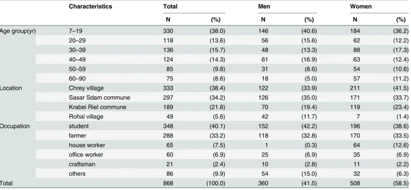
Participants’ characteristics are shown inTable 2. In total, there were 868 people, 360 men (41.5%) and 508 women (58.5%); age distribution was from 7 to 90 years (mean, 30.5 ± 18.8; median, 29 years) as of 2014. Among the participants, 38.4% lived in Chrey village, 34.2% in Sasar Sdam commune, 21.8% in Krabei Riel commune, and 5.6% in Rohal village, Siem Reap Province. In terms of occupation, most of the participants were students (40.1%), followed by farmers (33.2%), house workers (7.5%), office workers (6.9%), and craftsmen (2.4%). Other results of the questionnaire are shown inS1 Table.
HEV infection prevalence
The results of serologic testing are shown inTable 3. Overall, the prevalence of anti-HEV IgG was 18.4% (160/868; [95% CI: 15.9–21.0%]). Anti-HEV IgG positivity was significantly higher in men than in women (21.9% vs. 15.9%;p = 0.0247) and showed statistically significant corre-lation with older age (p < 0.0001). Anti-HEV IgG prevalence differed significantly among the four analyzed locations (p < 0.0001). There was also significant difference among occupa-tional/professional groups (p < 0.0001) (Table 3).
HEV RNA was detected in two participants (0.23% [0–0.55%]) (Table 3). There were no sig-nificant differences in the positivity rate of HEV RNA among sex, age, residential, and occupa-tional groups. Full-length HEV genome could be sequenced for one of the two samples and was classified as genotype 4. Another HEV RNA positive isolate was classified as genotype 3 based on partial ORF1 sequence with HE5 primers [15].
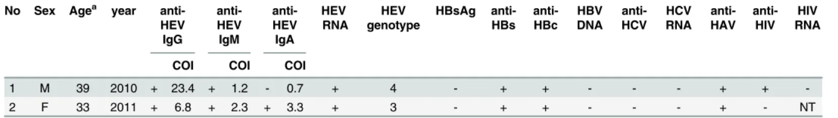
The two HEV RNA-positive participants were analyzed for other hepatitis viruses and HIV (Table 4). They were found positive for anti-HEV IgG and IgM, and one of them was positive
for anti-HEV IgA, as well as anti-HBs and anti-HBc, indicating previous HBV infection. More-over, one of them was positive for anti-HIV antibody, but HIV RNA was not detected.
Full-length sequence of the HEV genome
The full-length genome sequence of HEV isolate was recovered from a 39-year-old man at the time of the survey. The isolate designated as CVS-Sie10 (DDBJ/EMBL/GenBank accession number LC042232) had the genome of 7,222 nucleotides (nt) containing three ORFs: ORF1 [1–5,115 nt; 1,705 amino acids (aa)], ORF2 (5,115–7,136 nt; 674 aa), and ORF3 (5,143–5,484 nt; 114 aa), a 30UTR, and a poly-A tail.
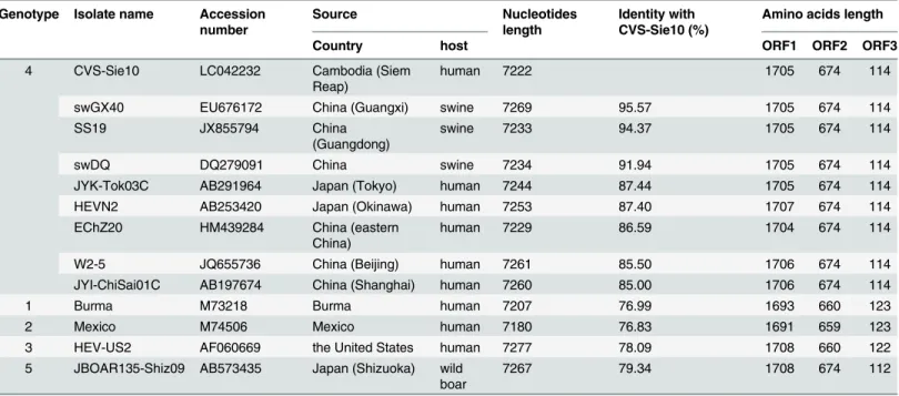
The alignment of the CVS-Sie10 genome with published HEV genotype 4 sequences showed that this isolate was close to the swine China isolates swGX40, SS19, and swDQ with sequence identities of 95.57%, 94.37%, and 91.94%, respectively (Table 5), and the same length of ORF1, ORF2, and ORF3. CVS-Sie10 displayed weak homology with other HEV genotypes. Thus, it demonstrated 76.99%, 76.83%, 78.09%, and 79.34% identity with the Burma isolate of genotype 1, the Mexico isolate of genotype 2, the HEV-US2 isolate of genotype 3, and the JBOAR135--Shiz09 isolate of genotype 5, respectively.
Based on full-length genome sequences of the CVS-Sie10 and other HEV isolates, we con-structed a phylogenetic tree (Fig 1), which showed that CVS-Sie10 clustered on a branch separate from the other genotype 4 sequences, and close to the China isolates swGX40, SS19, and swDQ.
Potential risk factors for HEV infection
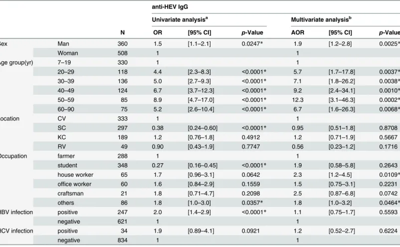
The odds ratios andp-values of potential risk factors for anti-HEV IgG positivity in univariate analysis and multivariate logistic regression model are shown inTable 6. In multivariate analysis, men (AOR: 1.9 [1.2–2.8] p = 0.0025) and older age (if the 7–19-year-old population group is taken as baseline) were significantly associated with anti-HEV IgG seropositivity. There were no significant differences among the four locations. The odds ratio of anti-HEV IgG seropositivity
Table 2. Characteristics of participants.
Characteristics Total Men Women
N (%) N (%) N (%) Age group(yr) 7–19 330 (38.0) 146 (40.6) 184 (36.2) 20–29 118 (13.6) 56 (15.6) 62 (12.2) 30–39 136 (15.7) 48 (13.3) 88 (17.3) 40–49 124 (14.3) 61 (16.9) 63 (12.4) 50–59 85 (9.8) 31 (8.6) 54 (10.6) 60–90 75 (8.6) 18 (5.0) 57 (11.2)
Location Chrey village 333 (38.4) 122 (33.9) 211 (41.5)
Sasar Sdam commune 297 (34.2) 126 (35.0) 171 (33.7)
Krabei Riel commune 189 (21.8) 70 (19.4) 119 (23.4)
Rohal village 49 (5.6) 42 (11.7) 7 (1.4) Occupation student 348 (40.1) 152 (42.2) 196 (38.6) farmer 288 (33.2) 118 (32.8) 170 (33.5) house worker 65 (7.5) 1 (0.3) 64 (12.6) office worker 60 (6.9) 25 (6.9) 35 (6.9) craftsman 21 (2.4) 10 (2.8) 11 (2.2) others 86 (9.9) 54 (15.0) 32 (6.3) Total 868 (100.0) 360 (41.5) 508 (58.5) doi:10.1371/journal.pone.0136903.t002
for house workers was more than twice higher than that for farmers (AOR: 2.3 [1.2–4.5]; p = 0.0109), and that for others was significantly higher than farmers (AOR: 1.8 [1.0–3.2]; p = 0.0464). On the other hand, HBV and HCV infections were not associated with anti-HEV IgG seropositivity.
Discussion
In this study, we performed a seroepidemiological survey for HEV infection in Cambodia. As a result, we identified the first human case of HEV genotype 4 in Cambodia and performed
Table 3. Sex-, age-, location- and occupation-specific prevalence of anti-HEV IgG and HEV RNA among the general population in Cambodia.
anti-HEV IgG positive HEV RNA positive
N (%) [95% CI] p-Value N (%) [95% CI] p-Value
Total 868 160 (18.4) [15.9–21.0] 2 (0.23) [0–0.55] Sex Man 360 79 (21.9) [17.7–26.2] 0.0247*a 1 (0.28) [0–0.82] 0.8065a Woman 508 81 (15.9) [12.8–19.1] 1 (0.20) [0–0.58] Age group 7–19 330 19 (5.8) [3.2–8.3] <0.0001*c 0 (0.0) [0–1.1] 0.3839b 20–29 118 25 (21.2) [13.8–28.6] 0 (0.0) [0–3.1] 30–39 136 32 (23.5) [16.4–30.7] 1 (0.74) [0–2.2] 40–49 124 36 (29.0) [21.0–37.0] 1 (0.81) [0–2.4] 50–59 85 30 (35.3) [25.1–45.5] 0 (0.0) [0–4.3] 60–90 75 18 (24.0) [14.3–33.7] 0 (0.0) [0–4.9] Location CV 333 74 (22.2) [17.8–26.7] <0.0001*a 1 (0.30) [0–0.89] 1.0000b SC 297 29 (9.8) [6.4–13.1] 1 (0.34) [0–1.0] KC 189 47 (24.9) [18.7–31.0] 0 (0.0) [0–2.0] RV 49 10 (20.4) [9.1–31.7] 0 (0.0) [0–7.5] Occupation farmer 288 62 (21.5) [16.8–26.3] <0.0001*a 0 (0.0) [0–1.3] 0.0712b student 348 24 (6.9) [4.2–9.6] 0 (0.0) [0–1.1] house worker 65 21 (32.3) [20.9–43.7] 1 (1.5) [0–4.5] office worker 60 18 (30.0) [18.4–41.6] 0 (0.0) [0–6.1] craftsman 21 7 (33.3) [13.2–53.5] 0 (0.0) [0–17.6] others 86 28 (32.6) [22.7–42.5] 1 (1.2) [0–3.4]
CV: Chrey village, SC: Sasar Sdam commune, KC: Krabei Riel commune, RV: Rohal village, CI: Confidence Interval * statistically significant variables.
aχ2 test
bFisher’s exact test
cMantel-extension test for trend
doi:10.1371/journal.pone.0136903.t003
Table 4. HEV RNA positives among the general population in Cambodia. No Sex Agea year
anti-HEV IgG anti-HEV IgM anti-HEV IgA HEV RNA HEV genotype HBsAg anti-HBs anti-HBc HBV DNA anti-HCV HCV RNA anti-HAV anti-HIV HIV RNA
COI COI COI
1 M 39 2010 + 23.4 + 1.2 - 0.7 + 4 - + + - - - + +
-2 F 33 2011 + 6.8 + 2.3 + 3.3 + 3 - + + - - - + - NT
M: Male, F: Female, NT: Not tested, COI: Cut off index
full-length genome sequencing of the isolate. One case of HEV genotype 3 was also detected. In developing countries, HEV infection is usually caused by genotype 1 viruses [1]; however, the data obtained in our study shows different genotype. In previous studies in Cambodia, HEV genotype 3 was detected in river water [8], and genotypes 1 and 3 were recovered from swine [9]. Partial sequences of HEV genotype 1 (accession number: DQ145797) and genotype 3 (accession number: DQ145792) from humans have been determined, but genotype 4 has not been reported. In this study, we sequenced, for the first time, the full-length genome of human HEV genotype 4 isolated in Cambodia.
Two HEV RNA-carrying participants were also positive for anti-HEV IgM and had a prior history of HBV infection as evidenced by the presence of anti-HBs and anti-HBc antibodies; one of the participants was also anti-HIV-positive. The detection of anti-HEV IgM indicates that both individuals had early stages of HEV infection. HEV genotype 4 has been reported to cause higher rate of aggravation from viral infection than genotype 3 [5,6,19], however, according to their answers to our questionnaire, both participants (genotype 3 and genotype 4) did not receive periodic treatment in hospitals and did not have serious problems with health. Moreover, they were confirmed asymptomatic during the interview 6 months after a blood test. Both of them had a prior history of HBV infection, which is not a rare case for Cambodia, where adult population has high anti-HBc positive rate of 38.5% [12]. Our multivariate analysis revealed no association between HEV and HBV infections.
Aggravation due to HEV infection is infrequent, and was not observed in this study. HEV genotype 4 is more likely to cause aggravation, and sometimes cause death [20,21]; in recent years, the risk of HEV triggering chronic hepatitis in immunocompromised patients, including HIV carriers [22] and organ transplant recipients [23,24] has been reported. These data under-score the importance of investigating the prevalence and performing genetic analysis of HEV infection in Cambodia, which is the part of Asia believed to be heavily affected by HEV infec-tion, and the necessity of developing preventive measures against HEV spread.
Table 5. Identity of full-genome sequences of HEV isolates known. Genotype Isolate name Accession
number
Source Nucleotides
length
Identity with CVS-Sie10 (%)
Amino acids length
Country host ORF1 ORF2 ORF3
4 CVS-Sie10 LC042232 Cambodia (Siem
Reap)
human 7222 1705 674 114
swGX40 EU676172 China (Guangxi) swine 7269 95.57 1705 674 114
SS19 JX855794 China
(Guangdong)
swine 7233 94.37 1705 674 114
swDQ DQ279091 China swine 7234 91.94 1705 674 114
JYK-Tok03C AB291964 Japan (Tokyo) human 7244 87.44 1705 674 114
HEVN2 AB253420 Japan (Okinawa) human 7253 87.40 1707 674 114
EChZ20 HM439284 China (eastern
China)
human 7229 86.59 1704 674 114
W2-5 JQ655736 China (Beijing) human 7261 85.50 1706 674 114
JYI-ChiSai01C AB197674 China (Shanghai) human 7260 85.00 1706 674 114
1 Burma M73218 Burma human 7207 76.99 1693 660 123
2 Mexico M74506 Mexico human 7180 76.83 1691 659 123
3 HEV-US2 AF060669 the United States human 7277 78.09 1708 660 122
5 JBOAR135-Shiz09 AB573435 Japan (Shizuoka) wild boar
7267 79.34 1708 674 112
In our survey, the overall rate of anti-HEV IgG positivity among 868 participants was 18.4%. There has been no prior study on the prevalence of HEV infection in the general popu-lation in Cambodia; however, the rate of anti-HEV IgG positivity in patients with high aspar-tate transaminase and alanine transaminase levels was determined as 5.5% [10], and the rate of anti-HEV IgM positivity in feverish patients was 11.1% [11]. Other studies have indicated that the rate of anti-HEV IgG positivity among blood donors in Japan was 3.4% [25] and in the general population of the Mekong River basin in Vietnam it was 9% [26]; among the gen-eral population of the East China Sea coast in the same country it was 28.1% (143/509 [95% CI: 24.2–32.0%]) (our unpublished data). In China, Taiwan, India, and Thailand, HEV IgG
Fig 1. Phylogenetic tree constructed based on HEV full-length genomes using the neighbor-joining method. Each of HEV genotypes 1, 2, 3, and 5 is represented by a single isolate, while for genotype 4, all the isolates with reported complete or near-complete genome sequence are presented. GenBank accession numbers are shown in parentheses; scale bar indicates nucleotide substitutions per site.
doi:10.1371/journal.pone.0136903.g001
Table 6. Univariate and multivariate analysis of positivity for anti-HEV IgG among the general population in Cambodia. anti-HEV IgG
Univariate analysisa Multivariate analysisb
N OR [95% CI] p-Value AOR [95% CI] p-Value
Sex Man 360 1.5 [1.1–2.1] 0.0247* 1.9 [1.2–2.8] 0.0025* Woman 508 1 1 Age group(yr) 7–19 330 1 1 20–29 118 4.4 [2.3–8.3] <0.0001* 5.7 [1.7–17.8] 0.0037* 30–39 136 5.0 [2.7–9.3] <0.0001* 7.1 [1.8–26.2] 0.0038* 40–49 124 6.7 [3.7–12.3] <0.0001* 9.2 [2.4–34.1] 0.0010* 50–59 85 8.9 [4.7–17.0] <0.0001* 12.3 [3.1–46.3] 0.0002* 60–90 75 5.2 [2.6–10.4] <0.0001* 6.7 [1.6–26.3] 0.0068* Location CV 333 1 1 SC 297 0.38 [0.24–0.60] <0.0001* 0.95 [0.51–1.8] 0.8708 KC 189 1.2 [0.76–1.8] 0.4912 1.2 [0.71–1.9] 0.5667 RV 49 0.90 [0.43–1.9] 0.7747 0.56 [0.23–1.2] 0.1716 Occupation farmer 288 1 1 student 348 0.27 [0.16–0.45] <0.0001* 1.9 [0.58–5.8] 0.2643 house worker 65 1.7 [0.96–3.1] 0.0642 2.3 [1.2–4.5] 0.0109* office worker 60 1.6 [0.84–2.9] 0.1559 1.5 [0.75–3.1] 0.2231 craftsman 21 1.8 [0.71–4.7] 0.2098 2.5 [0.87–6.8] 0.0742 others 86 1.8 [1.0–3.0] 0.0357* 1.8 [1.0–3.2] 0.0464* HBV infection positive 247 2.0 [1.4–2.9] <0.0001* 1.1 [0.75–1.7] 0.5593 negative 621 1 1 HCV infection positive 34 1.9 [0.89–4.1] 0.0921 1.2 [0.52–2.7] 0.6224 negative 834 1 1
OR: Odds Ratio, AOR: Adjusted Odds Ratio, CI: Confidence Interval, CV:Chrey village, SC: Sasar Sdam commune, KC: Krabei Riel commune, RV: Rohal village
aχ2test or Fisher’s exact test
bLogistic regression analysis: R2= 0.1113, Model p<0.0001*, N = 868
* statistically significant variables doi:10.1371/journal.pone.0136903.t006
positivity rate was 20.2%, 10.7%,4.0%, and 2.8–7%, respectively, while in urban and rural Malaysia, it was 2% and 44–50%, respectively [27]. Compared to these data, the rate of anti-HEV IgG positivity detected in this study is slightly higher than average. There might be possi-ble selection bias because we could not perform the random sampling. However, the screening for hepatitis virus infections does not conducted among the general population in Cambodia. On the other hand, the sensitivity of detection of anti-HEV IgG differs according to the assay. We detected by the kit used the purified recombinant HEV ORF2 protein in EIA same as the method described previously [28]. The results of the detection of anti-HEV IgG using this recombinant HEV ORF2 protein showed that the positivity of anti-HEV IgG was as high as 98% among the totally 57 acute hepatitis samples with positive for HEV RNA (56/57 samples: anti-HEV IgM/IgG +/+, HEV RNA +) [28–30](unpublished data). Furthermore, the sensitivity of the assay was not depend on genotype, and it also showed the high positivity among the swine [31]. Therefore, we considered that the sensitivity of detection of anti-HEV IgG by our used assay was high.
Our present results demonstrate that men have a significantly higher rate of HEV infection than women; similar association has been observed in Japan among blood donors [25], and reported by the National Epidemiological Surveillance of Infectious Diseases [32]. Moreover, similar correlation has been shown in the United Kingdom [33,34], the United States [35], and China [36]. The reason for such an association is not clear; given that HEV infection is primar-ily transmitted via the oral route, it can be hypothesized that game meat consumption and other food preferences may be involved.
Higher prevalence of anti-HEV IgG in older age groups indicates age-related risk. Even after the adjustment for other factors, older people demonstrate significantly higher rates of anti-HEV IgG positivity, if the 7–19-year-old population group is taken as baseline. In Vietnam [26] and Indonesia [37], it has been reported that the rate of anti-HEV positivity rises with age. Conceivable reason for the correlation of anti-HEV IgG positivity with age is because currently occurring new infections cause the number of people with a prior history of HEV infection to increase with age. Although the period when HEV RNA is detectable in the blood after HEV infection is brief constituting 28.3 days [38], two HEV RNA-positive participants (0.23%: 2/ 868 [95%CI: 0–0.55%]) were identified in our cross-sectional study of 868 people. Assuming that HEV RNA detection period is about four weeks, HEV incidence is estimated at 3.00/100 person-years (0–7.2/100 person-years). Among rural Chinese population, the rate of anti-HEV IgG positivity was 38%, with the incidence of 2.8/10,000 person-years [36], and in rural Bangla-desh regarded as HEV endemic region, the prevalence of anti-HEV total Ig was 22.5% and the incidence was 63.9/1,000 person-years [39]. Compared to these data, HEV incidence deter-mined in our study can be regarded as high, indicating that infection control measures are required.
HEV infection is often foodborne in developed countries, including Japan and Europe [4, 6]; in developing countries, contaminated water is considered to be a major cause [40–42]. Thus, there has been an outbreak of HEV infection in southwestern Vietnam along the Hau river, adjacent to Cambodia [43], and HEV has been detected in the water of Siem Reap River, Cambodia [8], indicating a possibility that HEV outbreak could also occur in Cambodia. In turn, HAV positive rate among the participants in this study was 88.8% (767/864; [86.7– 90.9%]): adults (18 years or older) had 99.6% (552/554; [99.1–100.0%]) and minors (17 years or younger) had 69.4% (215/310; [64.2–74.5%]), indicating age-related effect.
In Cambodia, nearly everyone is assumed to have HAV exposure by the time they reach adulthood. As with HEV infection, HAV is transmitted via the fecal-oral route, and by adult-hood, nearly everyone is HAV-infected via contaminated food or water, indicating serious issues with health management. To prevent HEV infection in Cambodia, proper water hygiene
is regarded as the first necessary measure. Multivariate analysis also showed that house workers had a significantly higher rate of anti-HEV IgG positivity than farmers, office workers, stu-dents, and craftsmen, which suggested possible problems with water hygiene for cooking or washing the cloth, or handling with raw stuff [44].
The HEV isolate fully sequenced in this study is most closely related to the strains isolated from swine in Guangxi and Guangdong, both in southern China. The cause of HEV infection for the participants with detected HEV RNA (including the CVS-Sie10 isolate) is unknown; however, HEV is a zoonotic pathogen that infects pigs, wild boars, and other animals, and causal relationship between consumption of contaminated meat and hepatitis E onset has been confirmed [29]. In Japan, the majority of foodborne infections are presumed to be related to meat consumption [32], which can be also true in Cambodia; therefore, it is possible that the consumption of undercooked meat or drinking water contaminated by animal waste may be the cause of HEV infection for the participants in this study.
In Japan, HEV infection by blood transfusion has been reported in Hokkaido [45,46], which is regarded as a region with increasing danger of HEV infection; therefore, it is the only place in the world where donor blood is screened for HEV RNA. In recent years, HEV infection through blood transfusion has been regarded as a growing problem also in the West, and the pros and cons of HEV blood screening have been considered. In this study, we identified HEV genotype 4 characterized with high post-infection aggravation rate, and also found that new HEV infections occur very frequently in Cambodia, raising concerns about HEV infection through blood transfusion and suggesting that it may be necessary to enhance the safety of blood supply.
The present study is the first to detect HEV genotype 4 in human blood in Cambodia and to report sequencing of genotype 4 full-length genome. Our survey revealed high HEV prevalence among Cambodian general population, including frequent cases of early HEV infection, sug-gesting that measures to prevent HEV infection, such as improving water and food safety and spreading health and hygiene education in school, are urgently required.
Supporting Information
S1 Table. Results of the questionnaire. This table shows the answers of eight questions. Ques-tion 3 and 4 were not asked to elementary school students.
(DOCX)
Author Contributions
Conceived and designed the experiments: HY OL SS SH SHD JT. Performed the experiments: HY KT KK MA. Analyzed the data: HY KT TA KK JT. Contributed reagents/materials/analysis tools: NG JT. Wrote the paper: HY KT JT. Survey for blood sampling: HY OL SS CC SH SHD MF TA NG JT. Coordinate the survey between Japan and Cambodia: SS JT.
References
1. WHO. Hepatitis E (Fact sheet No. 280): World Health Organization; 2014 [updated June cited 2014 December 24]. Available from:http://www.who.int/mediacentre/factsheets/fs280/en/.
2. WHO. Prevention and Control of Viral Hepatitis Infection: Framework for Global Action: World Health Organization; 2012 [cited 2014 December 24]. Available from:http://www.who.int/csr/disease/hepatitis/ GHP_Framework_En.pdf.
3. Takahashi K, Terada S, Kokuryu H, Arai M, Mishiro S. A wild boar-derived hepatitis E virus isolate pre-sumably representing so far unidentified "genotype 5". Kanzo. 2010; 51(9):536–538.
4. Dalton HR, Bendall R, Ijaz S, Banks M. Hepatitis E: an emerging infection in developed countries. Lan-cet Infect Dis. 2008 Nov; 8(11):698–709. doi:10.1016/S1473-3099(08)70255-XPMID:18992406
5. Mizuo H, Yazaki Y, Sugawara K, Tsuda F, Takahashi M, Nishizawa T, et al. Possible risk factors for the transmission of hepatitis E virus and for the severe form of hepatitis E acquired locally in Hokkaido, Japan. J Med Virol. 2005 Jul; 76(3):341–349. PMID:15902701
6. Takahashi M, Okamoto H. Features of hepatitis E virus infection in humans and animals in Japan. Hepatol Res. 2014 Jan; 44(1):43–58. doi:10.1111/hepr.12175PMID:23721425
7. Okamoto H. Genetic variability and evolution of hepatitis E virus. Virus Res. 2007 Aug; 127(2):216– 228. PMID:17363102
8. Kitajima M, Matsubara K, Sour S, Haramoto E, Katayama H, Ohgaki S. First detection of genotype 3 hepatitis E virus RNA in river water in Cambodia. Trans R Soc Trop Med Hyg. 2009 Sep; 103(9):955– 957. doi:10.1016/j.trstmh.2009.04.004PMID:19446859
9. Caron M, Enouf V, Than SC, Dellamonica L, Buisson Y, Nicand E. Identification of genotype 1 hepatitis E virus in samples from swine in Cambodia. J Clin Microbiol. 2006 Sep; 44(9):3440–3442. PMID: 16954296
10. Buchy P, Monchy D, An TT, Srey CT, Tri DV, Son S, et al. [Prevalence of hepatitis A, B, C and E virus markers among patients with elevated levels of Alanine aminotransferase and Aspartate aminotransfer-ase in Phnom Penh (Cambodia) and Nha Trang (Central Vietnam)]. PMID:Bull Soc Pathol Exot. 2004 Aug; 97(3):165–171.
11. Kasper MR, Blair PJ, Touch S, Sokhal B, Yasuda CY, Williams M, et al. Infectious etiologies of acute febrile illness among patients seeking health care in south-central Cambodia. Am J Trop Med Hyg. 2012 Feb; 86(2):246–253. doi:10.4269/ajtmh.2012.11-0409PMID:22302857
12. Yamada H, Fujimoto M, Svay S, Lim O, Hok S, Goto N, et al. Seroprevalence, genotypic distribution and potential risk factors of hepatitis B and C virus infections among adults in Siem Reap, Cambodia. Hepatol Res. 2015 Apr; 45(4):480–487. doi:10.1111/hepr.12367PMID:24905888
13. Genaral Population Census of Cambodia 2008: National Institute of Statistics, Ministry of Planning Phnom Penh, Cambodia; [cited 2014 Jan 11]. Available from:http://camnut.weebly.com/uploads/2/0/3/ 8/20389289/2009_census_2008.pdf.
14. Imai M, Kondo M, Sudo K, Saito T, Sato H, Takebe Y, et al. [Simple method for typing human immuno-deficiency virus type I (B, E) by PCR and chronological change of distribution of subtype (B to E) in Japan]. Kansenshogaku Zasshi. 1997 Sep; 71(9):918–923. PMID:9339629
15. Takahashi K, Kang JH, Ohnishi S, Hino K, Mishiro S. Genetic heterogeneity of hepatitis E virus recov-ered from Japanese patients with acute sporadic hepatitis. J Infect Dis. 2002 May 1; 185(9):1342– 1345. PMID:12001054
16. Felsenstein J. Confidence-Limits on Phylogenies—an Approach Using the Bootstrap. Evolution. 1985; 39(4):783–791.
17. Gojobori T, Ishii K, Nei M. Estimation of average number of nucleotide substitutions when the rate of substitution varies with nucleotide. J Mol Evol. 1982; 18(6):414–423. PMID:7175958
18. Saitou N, Nei M. The neighbor-joining method: a new method for reconstructing phylogenetic trees. Mol Biol Evol. 1987 Jul; 4(4):406–425. PMID:3447015
19. Abe T, Aikawa T, Akahane Y, Arai M, Asahina Y, Atarashi Y, et al. Demographic, epidemiological, and virological characteristics of hepatitis E virus infections in Japan based on 254 human cases collected nationwide. Kanzo. 2006; 47(8):384–391.
20. Okamoto H, Takahashi M, Nishizawa T. Features of hepatitis E virus infection in Japan. Intern Med. 2003 Nov; 42(11):1065–1071. PMID:14686743
21. Peron JM, Bureau C, Poirson H, Mansuy JM, Alric L, Selves J, et al. Fulminant liver failure from acute autochthonous hepatitis E in France: description of seven patients with acute hepatitis E and encepha-lopathy. J Viral Hepat. 2007 May; 14(5):298–303. PMID:17439518
22. Dalton HR, Bendall RP, Keane FE, Tedder RS, Ijaz S. Persistent carriage of hepatitis E virus in patients with HIV infection. N Engl J Med. 2009 Sep 3; 361(10):1025–1027. doi:10.1056/NEJMc0903778 PMID:19726781
23. Kamar N, Selves J, Mansuy JM, Ouezzani L, Peron JM, Guitard J, et al. Hepatitis E virus and chronic hepatitis in organ-transplant recipients. N Engl J Med. 2008 Feb 21; 358(8):811–817. doi:10.1056/ NEJMoa0706992PMID:18287603
24. Haagsma EB, van den Berg AP, Porte RJ, Benne CA, Vennema H, Reimerink JH, et al. Chronic hepati-tis E virus infection in liver transplant recipients. Liver Transpl. 2008 Apr; 14(4):547–553. doi:10.1002/ lt.21480PMID:18383084
25. Takeda H, Matsubayashi K, Sakata H, Sato S, Kato T, Hino S, et al. A nationwide survey for prevalence of hepatitis E virus antibody in qualified blood donors in Japan. Vox Sang. 2010 Nov; 99(4):307–313. doi:10.1111/j.1423-0410.2010.01362.xPMID:20576022
26. Hau CH, Hien TT, Tien NT, Khiem HB, Sac PK, Nhung VT, et al. Prevalence of enteric hepatitis A and E viruses in the Mekong River delta region of Vietnam. Am J Trop Med Hyg. 1999 Feb; 60(2):277–280. PMID:10072151
27. Worm HC, van der Poel WH, Brandstatter G. Hepatitis E: an overview. Microbes Infect. 2002 May; 4 (6):657–666. PMID:12048035
28. Mizuo H, Suzuki K, Takikawa Y, Sugai Y, Tokita H, Akahane Y, et al. Polyphyletic strains of hepatitis E virus are responsible for sporadic cases of acute hepatitis in Japan. J Clin Microbiol. 2002 Sep; 40 (9):3209–3218. PMID:12202555
29. Tei S, Kitajima N, Takahashi K, Mishiro S. Zoonotic transmission of hepatitis E virus from deer to human beings. Lancet. 2003 Aug 2; 362(9381):371–373. PMID:12907011
30. Shrestha A, Lama TK, Karki S, Sigdel DR, Rai U, Rauniyar SK, et al. Hepatitis E epidemic, Biratnagar, Nepal, 2014. Emerg Infect Dis. 2015 Apr; 21(4):711–713. doi:10.3201/eid2104.141512PMID: 25811975
31. Takahashi M, Nishizawa T, Miyajima H, Gotanda Y, Iita T, Tsuda F, et al. Swine hepatitis E virus strains in Japan form four phylogenetic clusters comparable with those of Japanese isolates of human hepatitis E virus. J Gen Virol. 2003 Apr; 84(Pt 4):851–862. PMID:12655086
32. National Institute of Infectious Diseases and Tuberculosis and Infectious Diseases Control Division MoH, Labour and Welfare. Infectious Agents Surveillance Report. 2014; 35(1):1–14.
33. Dalton HR, Stableforth W, Hazeldine S, Thurairajah P, Ramnarace R, Warshow U, et al. Autochtho-nous hepatitis E in Southwest England: a comparison with hepatitis A. Eur J Clin Microbiol Infect Dis. 2008 Jul; 27(7):579–585. doi:10.1007/s10096-008-0480-zPMID:18299907
34. Lewis HC, Boisson S, Ijaz S, Hewitt K, Ngui SL, Boxall E, et al. Hepatitis E in England and Wales. Emerg Infect Dis. 2008 Jan; 14(1):165–167. doi:10.3201/eid1401.070307PMID:18258100
35. Kuniholm MH, Purcell RH, McQuillan GM, Engle RE, Wasley A, Nelson KE. Epidemiology of hepatitis E virus in the United States: results from the Third National Health and Nutrition Examination Survey, 1988–1994. J Infect Dis. 2009 Jul 1; 200(1):48–56. doi:10.1086/599319PMID:19473098
36. Zhu FC, Huang SJ, Wu T, Zhang XF, Wang ZZ, Ai X, et al. Epidemiology of zoonotic hepatitis E: a com-munity-based surveillance study in a rural population in China. PLoS One. 2014; 9(1):e87154. doi:10. 1371/journal.pone.0087154PMID:24498033
37. Corwin A, Putri MP, Winarno J, Lubis I, Suparmanto S, Sumardiati A, et al. Epidemic and sporadic hep-atitis E virus transmission in West Kalimantan (Borneo), Indonesia. Am J Trop Med Hyg. 1997 Jul; 57 (1):62–65. PMID:9242320
38. Takahashi M, Tanaka T, Azuma M, Kusano E, Aikawa T, Shibayama T, et al. Prolonged fecal shedding of hepatitis E virus (HEV) during sporadic acute hepatitis E: evaluation of infectivity of HEV in fecal specimens in a cell culture system. J Clin Microbiol. 2007 Nov; 45(11):3671–3679. PMID:17728471 39. Labrique AB, Zaman K, Hossain Z, Saha P, Yunus M, Hossain A, et al. Epidemiology and risk factors of
incident hepatitis E virus infections in rural Bangladesh. Am J Epidemiol. 2010 Oct 15; 172(8):952–961. doi:10.1093/aje/kwq225PMID:20801864
40. Naik SR, Aggarwal R, Salunke PN, Mehrotra NN. A large waterborne viral hepatitis E epidemic in Kan-pur, India. Bull World Health Organ. 1992; 70(5):597–604. PMID:1464145
41. Bile K, Isse A, Mohamud O, Allebeck P, Nilsson L, Norder H, et al. Contrasting roles of rivers and wells as sources of drinking water on attack and fatality rates in a hepatitis E epidemic in Somalia. Am J Trop Med Hyg. 1994 Oct; 51(4):466–474. PMID:7943574
42. Rab MA, Bile MK, Mubarik MM, Asghar H, Sami Z, Siddiqi S, et al. Water-borne hepatitis E virus epi-demic in Islamabad, Pakistan: a common source outbreak traced to the malfunction of a modern water treatment plant. Am J Trop Med Hyg. 1997 Aug; 57(2):151–157. PMID:9288807
43. Corwin AL, Khiem HB, Clayson ET, Pham KS, Vo TT, Vu TY, et al. A waterborne outbreak of hepatitis E virus transmission in southwestern Vietnam. Am J Trop Med Hyg. 1996 Jun; 54(6):559–562. PMID: 8686771
44. Inoue G, Michitaka K, Takahashi K, Abe N, Oka K, Nunoi H, et al. A case of acute hepatitis E developed in a housewife who had cooked and eaten wild boar meat a month before. Kanzo. 2006; 47(10):459– 464.
45. Matsubayashi K, Nagaoka Y, Sakata H, Sato S, Fukai K, Kato T, et al. Transfusion-transmitted hepatitis E caused by apparently indigenous hepatitis E virus strain in Hokkaido, Japan. Transfusion. 2004 Jun; 44(6):934–940. PMID:15157263
46. Matsubayashi K, Kang JH, Sakata H, Takahashi K, Shindo M, Kato M, et al. A case of transfusion-transmitted hepatitis E caused by blood from a donor infected with hepatitis E virus via zoonotic food-borne route. Transfusion. 2008 Jul; 48(7):1368–1375. doi:10.1111/j.1537-2995.2008.01722.xPMID: 18651907