Title
Heart Rate-Associated Left Ventricular Morphologic Changes
Observed in Two Heart Failure Patients
Author(s)
Kinjo, Kaori; Inoue, Taku; Tamashiro, Masahiro; Miiji, Asuka
Citation
琉球医学会誌 = Ryukyu Medical Journal, 37(1-4): 91-95
Issue Date
2018
URL
http://hdl.handle.net/20.500.12001/24355
Corresponding Author: Taku Inoue, Cardiovascular Medicine, Nambu Hospital, 870 Maesato, Itoman Okinawa 903-0362, Japan. Tel: + 81 98 994 0501; Fax: + 81 98 994 0506, E-mail: [email protected]
Introduction:
Several lines of evidence, including basic experiments and cohort studies, indicate that elevated resting heart rate (HR) is an independent risk factor for a poor prognosis. A clear prognostic significance of HR-lowering therapy has been demonstrated in patients with heart failure 1), and HR is considered to be a therapeutic target in this spectrum of patients. Elevated resting HR is associated with every stage of the cardiovascular continuum 2, 3). Left ventricular hypertrophy (LVH) is also an independent risk factor for a cardiovascular event 4, 5). Previous cross sectional analysis evaluating patients without heart failure, however, revealed that resting HR is negatively associated with LVH 6-8). We report two cases of congestive heart failure exhibiting characteristic LV morphologic changes associated with HR-lowering therapy.
Case Presentation
Cases 1: A 93-year-old female patient was referred to our hospital after diagnosis and treatment of end-stage heart failure with severe aortic stenosis, atrial fibrillation, and implantation of a DDD pacemaker. The patient had a baseline physical status of New York Heart Association (NYHA) class IV, blood pressure of 100/60mmHg, and heart rate (HR) of 117beats/min. An electrocardiogram (ECG) revealed atrial fibrillation with a complete left branch block. Cardiac pacemaker program analysis indicated atrial fibrillation with all ventricular sensing. Cardiac ultrasound revealed diffuse hypokinesis with an ejection fraction of 41% and an aortic valve area of 0.52cm2 (Table). After medical therapy, including diuretics and a beta blocker, her HR was reduced to 60beats/min and her general condition improved remarkably to a physical status of NYHA class II. ECG showed atrial fibrillation with all
ABSTRACT
We report two heart failure cases in which the left ventricular (LV) mass increased after heart rate (HR)-lowering therapy. The initial LV mass index was 98 and 121g/m 2, respectively. After HR-lowering therapy, the HR was reduced from 120 and 100beats/ min to 70 and 60beats/min, respectively. After HR reduction and clinical stabilization, the LV mass index was 110 and142 g/m2, respectively, with LV chamber shrinking. Elevated resting HR was considered to indicate deteriorating target organ damage and worse prognosis. The present findings contrast with the previous concept that HR-lowering is good for patientsʼ health. Ryukyu Med. J., 37 (1~4) 91~96, 2018
Key words: heart rate, left ventricular hypertrophy, left ventricular mass, remodeling 1)Clinical Laboratory Department, Nambu Hospital
2)Cardiovascular Medicine, Nambu Hospital 3)Cardiovascular Medicine, Tomishiro Central Hospital
(Received on June 29, 2017, accepted on September 8, 2017)
Kaori Kinjo 1), Taku Inoue 2), Masahiro Tamashiro 3), Asuka Miiji 2),
Osamu Arasaki 3), Tetsuji Shinjo 2)
Heart Rate-Associated Left Ventricular Morphologic
Changes Observed in Two Heart Failure Patients
ventricular pacing beats.
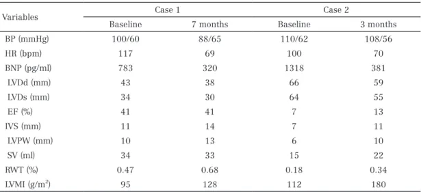
Cases 2: An 80-year-old male patient was referred to our hospital for cardiac rehabilitation with a diagnosis of chronic heart failure due to ischemic cardiomyopathy, implantation of a DDD pacemaker, and paroxysmal atrial fibrillation. The patient had a baseline physical status of NYHA class II, blood pressure of 110/62mmHg, and HR of 100beats/min. ECG showed all ventricular pacing rhythm. Despite cardiac rehabilitation, his general condition gradually worsened to a physical status of NYHA class III. Cardiac ultrasound revealed diffuse hypokinesis and a dilated LV with an ejection fraction of 7%. ECG monitoring showed that his HR was unchanged before and after exercise. Cardiac pacemaker program analysis indicated atrial fibrillation followed by ventricular pacing beats at the upper tracking rate of 100paces/min. After adjusting the pacemaker setting in DDDR mode with rate response, his HR was reduced to a resting HR of 60 to 70beats/min, and his general condition was remarkably improved to a physical status of NYHA class II. Serial changes of the cardiac ultrasound findings in both cases are summarized in Figure 1 and the table. Figure 1 shows the serial echocardiographic changes in both cases, demonstrating increased wall thickness during the follow-up period. The LV morphologic change was revealed based on the relative wall thickness (RWT) and left ventricular mass index (LVMI). RWT is a phenotype for quantifying LVH, and
is represented as the ratio of LV chamber size and LV wall thickness. This method allows for further classification of the LV mass increase as either concentric hypertrophy (RWT>0.42) or eccentric hypertrophy (RWT≦0.42). RWT and LV mass are calculated by the following formula: RWT=2 x IVSd / LVDd, LV mass (g)=0.8{1.04[([LVDd + IVSd + PWd)3 - LVEDd3)]} + 0.6. LVMI (g/m2) was defined as LV mass divided by body surface area (m 2) 9). After the HR was lowered, the LV chamber decreased in size with an increase in the wall thickness, resulting in a higher LVMI and a higher RWT, and the plots shifted to the right (Figure 2).
Discussion:
Evidence obtained from both basic and clinical research demonstrates that resting HR is associated with all steps of the cardiovascular continuum, including cardiometabolic risks, inflammation, endothelial dysfunction, target organ damage, and cardiovascular events 2). Resting HR is associated with target organ damage such as chronic kidney disease 10, 11), arterial stiffness12), and impaired cognitive function 13). Fácila et al. evaluated 560 patients with essential hypertension and demonstrated that the odds ratio for target organ damage in patients with a resting HR of 65 beats/min and above was 2.4 14). In other words, a lower resting HR is good for patient health. The association between
Variables Case 1 Case 2
Baseline 7 months Baseline 3 months
BP (mmHg) 100/60 88/65 110/62 108/56 HR (bpm) 117 69 100 70 BNP (pg/ml) 783 320 1318 381 LVDd (mm) 43 38 66 59 LVDs (mm) 34 30 64 55 EF (%) 41 41 7 13 IVS (mm) 11 14 7 11 LVPW (mm) 10 13 6 10 SV (ml) 34 33 15 22 RWT (%) 0.47 0.68 0.18 0.34 LVMI (g/m2) 95 128 112 180
BP: blood pressure; HR: heart rate; BNP: brain natriuretic peptide; LVDd: left ventricular diastolic dimension; LVDs: left ventricular systolic dimension; IVS: interventricular septum; LVPW: left ventricular posterior wall; SV: stroke volume; RWT: relative wall thickness; LVMI: left ventricular mass index
Table Serial changes in echocardiographic and other
clinical parameters in the two heart failure cases
Relative wall thickness
Left ventricular mass index (g/m2)
0.84
0.42
0.00
0 106 212
Figure 2 Serial left ventricular morphologic changes in the two heart failure cases. In both cases, the relative
wall thickness and left ventricular mass index increased over time, shifting the plot to the right.
Figure 1 Echocardiograms of both cases examined at baseline (left) and during the follow-up period (right).
Case 1
resting HR and LVH, however, does not support this notion. Cross sectional results evaluating normotensive and hypertensive patients without heart failure indicate that resting HR is negatively associated with left ventricular mass 6-8). The LIFE study 15) demonstrated that angiotensin receptor blocker (losartan)-based antihypertensive therapy afforded a better prognostic outcome compared with beta blocker (atenolol)-based antihypertensive therapy. The results of the LIFE study suggest that beta blocker-derived HR-lowering might partly contribute to a worse prognosis through the development of or sustained LVH. Consistent with these findings, our previous result indicated that lower resting HR accelerates the new onset of ECG-LVH in healthy subjects without heart failure 16). As we demonstrate in the present two cases, HR-lowering decreased the LV chamber size, but increased LVMI in patients with heart failure. These findings support the notion that lower HR or HR-lowering might be a risk factor for LVH. A possible mechanism connecting the resting HR and LVH in the present two cases includes stroke volume and stroke work. Stroke work was estimated as systolic blood pressure times stroke volume and was converted in gram-meters (g-m) by multiplying by 0.0144 17). When cardiac output remains the same, there is a negative association between resting HR and stroke volume. Based on these assumptions, HR-lowering therapy increases stroke volume to maintain cardiac output, and subsequent stroke work increases, resulting in an increased LV mass. There might be another causal relationship between HR and LVH. Hypertensive patients with LVH have an enhanced parasympathetic tone compared with normal subjects 18). In patients with heart failure with a reduced ejection fraction, HR-lowering therapy improves the LV chamber size 19) and prognosis 20). In an animal model of LV dysfunction, lowering HR is essential for recovering cardiac function 21) and HR is the therapeutic target for patients with LV dysfunction. On the other hand, resting HR might increase LV mass regardless of cardiac function. The clinical benefit of HR-lowering should be reconsidered.
Acknowledgement: The authors deeply ac-knowledge to Ms. Tamashiro and Dr. Yogi for re-trieving the data.
Funding Acknowledgement: This research received no specific grant from any funding agency in the public, commercial, or not-for-profit sectors.
Conflict of interest statement: The authors declare no conflict of interest in preparing this article.
References
1) Hori M, Okamoto H. Heart rate as a target of treatment of chronic heart failure. J Cardiol 2012; 60:86-90.
2) Custodis F, Reil J-C, Laufs U, Böhm M. Heart rate: A global target for cardiovascular disease and therapy along the cardiovascular disease continuum. J Cardiol 2013; 62:183-187.
3) Inoue T, Iseki K, Ohya Y. Heart rate as a possible therapeutic guide for the prevention of cardiovascular disease. Hypertens Res 2013; 36:838-44.
4) Levy D, Garrison RJ, Savage DD, Kannel WB, Castelli WP. Prognostic implications of echocar-diographically determined left ventricular mass in the Framingham Heart Study. N Engl J Med 1990; 322:1561-6.
5) Mathew J, Sleight P, Lonn E, Johnstone D, Pogue J, Yi Q, et al. Reduction of cardiovascular risk by regression of electrocardiographic markers of left ventricular hypertrophy by the angiotensin-converting enzyme inhibitor ramipril. Circulation 2001; 104:1615-21.
6) Palatini P, Visentin P, Mormino P, Mos L, Canali C, Dorigatti F, et al. Structural abnormalities and not diastolic dysfunction are the earliest left ventricular changes in hypertension. HARVEST Study Group. Am J Hypertens 1998; 11:147-54. 7) Saba MM, Ibrahim MM, Rizk HH. Gender and
the relationship between resting heart rate and left ventricular geometry. J Hypertens 2001; 19:367-73.
8) Zakopoulos NA, Ikonomidis I, Vemmos KN, Manios E, Spiliopoulou I, Tsivgoulis G, et al. Twenty-four-hour heart rate and blood pressure are additive markers of left ventricular mass in hypertensive subjects. Am J Hypertens 2006; 19:170-7.
9) Lang RM, Bierig M, Devereux RB, Flachskampf FA, Foster E, Pellikka PA, et al. Recommendations for Chamber Quantification: A Report from the American Society of Echocardiographyʼs Guidelines and Standards Committee and the Chamber Quantification Writing Group, Developed in Conjunction with the European Association of Echocardiography, a Branch of the European Society of Cardiology. J Am Soc Echocardiogr 2005; 18:1440-1463.
10) Inoue T, Iseki K, Iseki C, Ohya Y, Kinjo K, Takishita S. Heart rate as a risk factor for developing chronic kidney disease: longitudinal analysis of a screened cohort. Clin Exp Nephrol 2009; 13:487-93.
11 Bohm M, Reil J, Danchin N, Thoenes M, Bramlage P, Volpe M. Association of heart rate with micro-albuminuria in cardiovascular risk patients: data from I-SEARCH. J Hypertens 2008; 26:18-25.
12) Benetos A, Adamopoulos C, Bureau J-MM, Temmar M, Labat C, Bean K, et al. Determinants of accelerated progression of arterial stiffness in normotensive subjects and in treated hypertensive subjects over a 6-year period. Circulation 2002; 105:1202-7.
13) Böhm M, Cotton D, Foster L, Custodis F, Laufs U, Sacco R, et al. Impact of resting heart rate on mortality, disability and cognitive decline in patients after ischaemic stroke. Eur Hear J 2012; 33:2804-12.
14) Fácila L, Pallarés V, Peset A, Pérez M, Gil V, Montagud V, et al. Twenty-four-hour ambulatory heart rate and organ damage in primary hypertension. Blood Press 2010; 19:104-109. 15) Dahlöf B, Devereux RB, Kjeldsen SE, Julius S,
Beevers G, de Faire U, et al. Cardiovascular morbidity and mortality in the Losartan Intervention For Endpoint reduction in hypertension study (LIFE): a randomised trial against atenolol. Lancet 2002; 359:995-1003. 16) Inoue T, Iseki K, Iseki C, Katsumata Y, Kinjo K.
Impact of resting heart rate on the development of left ventricular hypertrophy in a healthy screened cohort: findings from the OGHMA study. Eur Heart J 2013; 34:3683-3683.
17) de Simone G, Devereux RB, Kimball TR, Mureddu GF, Roman MJ, Contaldo F, et al. Inter-action between body size and cardiac workload: influence on left ventricular mass during body growth and adulthood. Hypertension 1998; 31:1077-82.
18) Chakko S, Mulingtapang RF, Huikuri H V, Kessler KM, Materson BJ, Myerburg RJ. Alterations in heart rate variability and its circadian rhythm in hypertensive patients with left ventricular hypertrophy free of coronary artery disease. Am Hear J 1993; 126:1364-72. 19) Peng L, Yan B, Song A, Li L, Zeng L, Wang G.
Ivabradine significantly improves cardiac function in patients with ischemic heart disease: A meta-analysis of randomized controlled trials. Int J Cardiol 2013; 168:3007-3010.
20) Böhm M, Swedberg K, Komajda M, Borer JS, Ford I, Dubost-Brama A, et al. Heart rate as a risk factor in chronic heart failure (SHIFT): the association between heart rate and outcomes in a randomised placebo-controlled trial. Lancet 2010; 376:886-94.
21) Nagatsu M, Spinale FG, Koide M, Tagawa H, DeFreitas G, Cooper G, et al. Bradycardia and the Role of beta-Blockade in the Amelioration of Left Ventricular Dysfunction. Circulation 2000; 101:653-659.