Sporadic Medullary Thyroid Carcinoma in Grave
’s disease : Report of a case
著者
ARIMA Hideo, NAKAJO Akihiro, KIJIMA Yuko,
YOSHINAKA Heiji, HIRATA Munetsugu, NAKANO
Shizuo, ARIMA Miyako, ISHIGAMI Sumiya, OKUMURA
Hiroshi, NATSUGOE Shoji
journal or
publication title
Medical journal of Kagoshima University
volume
67
number
1-3
page range
19-25
year
2015-12
別言語のタイトル
甲状腺気機能亢進に合併した孤発髄用甲状腺がん
URL
http://hdl.handle.net/10232/00030360
Article type: Case Report
Sporadic Medullary Thyroid Carcinoma in Grave’s disease -Report of a
case-Hideo Arima
1 *), Akihiro Nakajo
1), Yuko Kijima
1), Heiji Yoshinaka
1), Munetsugu Hirata
1),
Shizuo Nakano
2), Miyako Arima
3), Sumiya Ishigami
1), Hiroshi Okumura
1), Shoji Natsugoe
1).
1) Department of Digestive Surgery, Breast and Thyroid Surgery, Kagoshima University Graduate School of Medical and Dental
Science, 8-35-1 Sakuragaoka, Kagoshima 890-8520, Japan
2) Nakano Clinic, 26-29 Uearata-chou, Kagoshima 890-0055, Japan 3) Kaneko Clinic, 8-6 Uearata-chou, Kagoshima 890-0055, Japan
(Received Feb. 12; Revised Mar. 20; Accepted May. 18) * Address to Correspondence
Hideo Arima
Department of Digestive Surgery, Breast and Thyroid Surgery, Kagoshima University Graduate School of Medical and Dental Science, 8-35-1 Sakuragaoka, Kagoshima 890-8520, Japan
Tel.: +81-99-275-5361; Fax: +81-99-265-7426 E-mail: [email protected]
Abstract
A 60-year-old man who noted numbness and dyskinesia of his left-hand fingers was admitted to a hospital. After anticoagulant therapy and medication with L-3, 4-dihydroxyphenylalanine (L-DOPA) were started, his symptoms didn’t improve. He was introduced to our institution with gait disturbance and difficulty of articulating properly. L-DOPA was administered under a diagnosis of multiple system atrophy by magnetic resonance imaging. He was also diagnosed as Graves’ disease with hyperthyroidism and expression of TSH receptor antibodies. Cervical ultrasonography revealed diffuse thyroid enlargement with abundant blood flow, which was consistent with Graves’ disease. A nodule of 6.3 x 8.3 x 9.2mm was also detected in the middle of the right lobe. The nodule was diagnosed as medullary thyroid carcinoma (MTC) by fine needle aspiration cytology (FNAC). The serum calcitonin and CEA level were elevated. We diagnosed sporadic MTC because lacking of pituitary or adrenal tumor, and family history of MTC. After medication for Graves’ disease, total thyroidectomy with central lymph nodes dissection was performed. Histopathological examination diagnosed as a MTC the diameter of tumor was 9 mm in the middle of the right lobe without extra thyroidal extension. Most of the thyroid cancers with Graves’ disease have been reported to be papillary thyroid cancer. The case of MTC with Graves’ disease is very rare with only 12 cases having been reported. We report the case of sporadic MTC with Graves’ disease which accidentally diagnosed by episode of multiple system atrophy.
Med. J. Kagoshima Univ., July, 2015 〔20〕
Introduction
Graves’ disease is the most common cause of hyperthyroidism, usually presenting with nodules of thyroid. In patients with both Graves’ disease and nodular thyroid disease, thyroid cancer was detected in 2-6 % after surgical treatment 1, 2).
Medullary thyroid carcinoma (MTC) comprises only 1.4-8.7% of all thyroid cancers 3, 4). In 73 patients who
underwent surgery for Graves’ disease in our institution from January 2000 through October 2012, the incidence of differentiated thyroid carcinoma (DTC) was 17.8% (13 cases). Histologically, nine patients had papillary thyroid cancer (PTC), two PTC of follicular variant, one follicular thyroid cancer (FTC), respectively. Only one patient (7.6%) had MTC.
We herein report the case of MTC with Grave’s disease and also review the literature of such diseases.
Case Report
A 60-year-old man who noted numbness and dyskinesia of his left-hand fingers was admitted to a hospital. Anticoagulant therapy and medication with L-3, 4-dihydroxyphenylalanine (L-dopa) were started, because magnetic resonance imaging (MRI) revealed cerebral infarction. Electromyogram and orthopedics examination were normal. Two months later, gait disturbance occurred and one more month later, he was unable to articulate properly. He was introduced to our
institution for further examination and treatment. Although L-dopa was administered, his symptoms didn’t improve. His temperature was 36.3°C, heart rate 68 beats per minute with blood pressure of 117/77 mmHg. Heart rate was regular and atrial fibrillation was not observed. His body weight and height were 71.1kg and 170cm, respectively. Cervical palpation revealed a soft and non-tender thyroid gland without nodules. No exophthalmos or skin changes were found. There were no clinical features other than left-side-dominant extrapyramidal symptoms and no special family history. T2 image on MRI revealed high intensity in the lateral area of the putamen. L-DOPA was administered under a diagnosis of multiple system atrophy.
Serum levels of free triiodothyronine, free thyroxine, and thyroid-stimulating hormone (TSH) were 6.4pg/ml (normal range 2.3-4.0), 2.51ng/ml (normal range 0.9-1.7), and <0.016.4 µIU/ml (normal range 0.5-5.0), respectively. The value of TSH receptor antibodies (TRAb) was high (35, normal range <1.0). Preoperative calcitonin was 220 pg/ml (reference range, 15-86), CEA was 4.0 ng/ml (reference range, 0-5), and thyroglobulin was 50.9 ng/ml (reference range, 0-32), respectively.
Ultrasonography revealed diffuse thyroid enlargement with abundant blood flow, which was consistent with Graves’ disease. A nodule of 6.3 x 8.3 x 9.2mm was also detected in the middle of the right lobe (Figure 1). Enlarged lymph nodes weren’t detected. Fine needle aspiration cytology (FNAC) based on Papanicolau stain revealed hyper-cellular tumor with round or elongated nuclei, quite different from
Figure 2: The tumor cells revealed round or elongated nuclei with non-papillary and non-follicular structures, and amyloid substance was recognized without necrosis.
papillary and follicular structures, and an amyloid substance was partly recognized without necrosis (Figure 2). In these findings, the nodule was diagnosed as MTC cytologically. Computed tomography (CT) of the cervix, chest and abdomen didn’t show any metastases or tumors. We diagnosed sporadic MTC because lacking of pituitary or adrenal tumor and family history of MTC. RET gene mutation analysis was not performed because he didn’t consent.
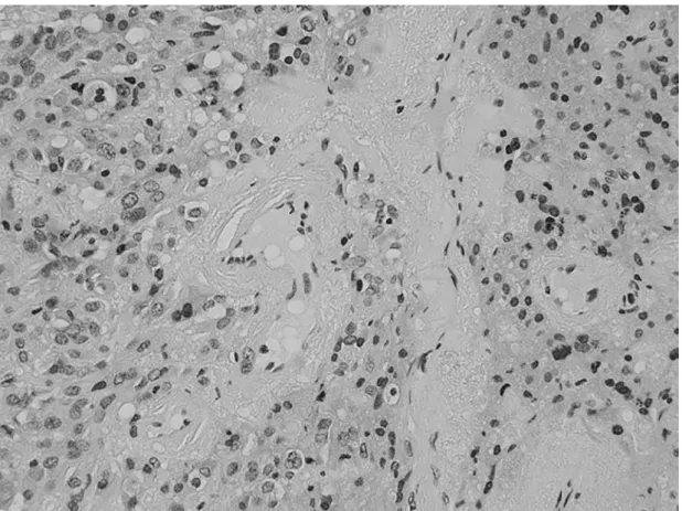
Since Graves’ disease was probably diagnosed with hyper thyroid state and TRAb positive, thyroid scintigraphy wasn’t performed. After medication, total thyroidectomy with central lymph nodes dissection was performed. The diameter of tumor was 9 mm in the middle of the right lobe without extra thyroidal extension (Figure 3). Histopathological examination showed MTC composed of small nests, sheets, and single cells characterized by moderate pleomorphism and hyperchromasia (Figure 4A). Amyloid was present in the background, confirmed by direct fast scarlet staining (Figure 4B). The tumor cells were positive for calcitonin
by immunohistochemistry (Figure 4C). Non-neoplastic thyroid parenchyma was almost normal without features of lymphocytic thyroiditis or hyperplasia. The serum calcitonin was <0.1ng/l at 55 months after surgery.
Discussion
Overt Graves’ disease and DTC are relatively rarely associated. It has been reported that the incidence of DTC with Graves’ disease ranged from 0.76% to 9.8% 5-8). Thyroid
nodules with Graves’ disease are reportedly only palpable in the range of 15% to 25% 6, 9). The risk of cancer in palpable
thyroid nodules is controversial. The majority studies reported that there was no significant difference in DTC incidence between Graves’ disease and other toxic nodular goiter10-18).
FNAC is a useful tool to evaluate thyroid nodules. The majority of DTC with Graves’ disease are PTC, followed by FTC, as in the general population. According to our survey, only 12 cases of MTC combined with Graves’ disease have
Med. J. Kagoshima Univ., July, 2015 〔22〕
Figure 3: The tumor was 9mm in diameter in the middle of the right lobe without extrathyroidal extension.
Figure 4A: Histopathological examination showed a medullary carcinoma composed of small nests, sheets, and single cells characterized by moderate pleomorphism and hyperchromasia.
been reported in the literature19-28). Although MTC with
Graves’ disease is very rare, MTC may be also suspected by FNAC. Immunohistochemical staining against calcitonin and Congo red (direct fast scarlet) staining for amyloid substance should be considered when the cytological diagnosis suggests MTC. FNAC, measurement of serum calcitonin and CEA are useful for the diagnosis of MTC. In the case of a normal upper limit of serum calcitonin or CEA, measurement of calcitonin in wash-out fluid from fine needle aspiration is useful29, 30).
The patient was incidentally diagnosed as having MTC with Graves’ disease. He had no certain clinical features suggesting multiple endocrine neoplasia such as diarrheal syndrome, flushing or a specific family history. Neither
thyroid nodule nor neck lymph nodes were palpable. Although MTC with Graves’ disease is uncommon, FNAC was able to detect such a rare disease. Left-side-dominant extrapyramidal symptoms he first noted numbness and dyskinesia were not improved with the surgical treatment. In conclusion, meticulous examination by ultrasound is also needed in Graves’ disease as well as hormonal examination; and if abnormal nodules are found, FNAC is strongly recommended without overlooking thyroid malignancy combined with Graves’ disease.
Figure 4B: Amyloid was present in the background, confirmed by Congo red (direct fast scarlet) staining.
Med. J. Kagoshima Univ., July, 2015 〔24〕
References
1) Pacini F, Elisei R, Di Coscio GC, Anelli S, Macchia E, Concetti R, et al. Thyroid carcinoma in thyrotoxic patients treated by surgery. J Endocrinol Invest 1988; 11: 107-112.
2) Hales IB, McElduff A, Crummer P, Clifton-Bligh P, Delbridge L, Hoschl R, et al. Does Graves’ disease or thyrotoxicosis affect the prognosis of thyroid cancer. J Clin Endocrinol Metab 1992; 75: 886-889.
3) Ezaki H, Ebihara S, Fujimoto Y, Iida F, Ito K, Kuma K, et al. Analysis of thyroid carcinoma based on material registered in Japan during 1977-1986 with special reference to predominance of papillary type. Cancer 1992; 70: 808-814.
4) Saad MF, Ordonez NG, Rashid RK, Guido JJ, Hill CS, Jr., Hickey RC, et al. Medullary carcinoma of the thyroid. A study of the clinical features and prognostic factors in 161 patients. Medicine (Baltimore) 1984; 63: 319-342. 5) Belfiore A, Garofalo MR, Giuffrida D, Runello F, Filetti
S, Fiumara A, et al. Increased aggressiveness of thyroid cancer in patients with Graves’ disease. J Clin Endocrinol Metab 1990; 70: 830-835.
6) Dobyns BM, Sheline GE, Workman JB, Tompkins EA, McConahey WM, Becker DV. Malignant and benign neoplasms of the thyroid in patients treated for hyperthyroidism: a report of the cooperative thyrotoxicosis therapy follow-up study. J Clin Endocrinol Metab 1974; 38: 976-998.
7) Rieger R, Pimpl W, Money S, Rettenbacher L, Galvan G. Hyperthyroidism and concurrent thyroid malignancies. Surgery 1989; 106: 6-10.
8) Terzioglu T, Tezelman S, Onaran Y, Tanakol R. Concurrent hyperthyroidism and thyroid carcinoma. Br J Surg 1993; 80: 1301-1302.
9) Kraimps JL, Bouin-Pineau MH, Mathonnet M, De Calan L, Ronceray J, Visset J, et al. Multicentre study of thyroid nodules in patients with Graves’ disease. Br J Surg 2000; 87: 1111-1113.
10) Pazaitou-Panayiotou K, Perros P, Boudina M, Siardos G, Drimonitis A, Patakiouta F, et al. Mortality from thyroid cancer in patients with hyperthyroidism: the Theagenion Cancer Hospital experience. Eur J Endocrinol 2008; 159: 799-803.
11) Linos DA, Karakitsos D, Papademetriou J. Should the primary treatment of hyperthyroidism be surgical? Eur J Surg 1997; 163: 651-657.
12. Vaiana R, Cappelli C, Perini P, Pinelli D, Camoni G, Farfaglia R, et al. Hyperthyroidism and concurrent thyroid cancer. Tumori 1999; 85: 247-252.
13) Zanella E, Rulli F, Sianesi M, Sciacchitano S, Danese D, Pontecorvi A, et al. Hyperthyroidism with concurrent thyroid cancer. Ann Ital Chir 2001; 72: 293-297.
14) Gabriele R, Letizia C, Borghese M, De Toma G, Celi M, Izzo L, et al. Thyroid cancer in patients with hyperthyroidism. Horm Res 2003; 60: 79-83.
15) Miccoli P, Minuto MN, Galleri D, D’Agostino J, Basolo F, Antonangeli L, et al. Incidental thyroid carcinoma in a large series of consecutive patients operated on for benign thyroid disease. ANZ J Surg 2006; 76: 123-126. 16) Pradeep PV, Agarwal A, Baxi M, Agarwal G, Gupta SK,
Mishra SK. Safety and efficacy of surgical management of hyperthyroidism: 15-year experience from a tertiary care center in a developing country. World J Surg 2007; 31: 306-312; discussion 313.
17) Senyurek Giles Y, Tunca F, Boztepe H, Kapran Y, Terzioglu T, Tezelman S. The risk factors for malignancy in surgically treated patients for Graves’ disease, toxic multinodular goiter, and toxic adenoma. Surgery 2008; 144: 1028-1036; discussion 1036-1027.
18) Gul K, Di Ri Koc A, Ki Yak G, Ersoy PE, Ugras NS, Ozdemi D, et al. Thyroid carcinoma risk in patients with hyperthyroidism and role of preoperative cytology in diagnosis. Minerva Endocrinol 2009; 34: 281-288. 19) Habra MA, Hijazi R, Verstovsek G, Marcelli M.
Medullar y thy roid carcinoma associated with hyperthyroidism: a case report and review of the literature. Thyroid 2004; 14: 391-396.
20) Igarashi K, Ishikawa K, Akasu H, Shimizu K. A case of medullary thyroid cancer with Graves’ disease. The Medical Association of Nippon Medical School 2007; 3: 39-43.
21) Brandle M, Galeazzi RL, Diener PA, Schmid C. Medullary thyroid carcinoma in Graves’ disease. Clin Endocrinol (Oxf) 1999; 50: 545-546.
22) Diklic A, Paunovic I, Batev N, Malenkovic V, Havelka M, Jankovic R. Occurrence of thyroid gland carcinoma in Graves-Basedow disease. Acta Chir Iugosl 1991; 38: 61-68.
23) Ardito G, Mantovani M, Vincenzoni C, Guidi ML, Corsello S, Rabitti C, et al. Hyperthyroidism and carcinoma of the thyroid gland. Ann Ital Chir 1997; 68: 23-27; discussion 27-28.
month. Association between medullary thyroid cancer and type 3 autoimmune thyroiditis: discussion of an unusual case. Rev Med Liege 2000; 55: 817-820.
25) Nakamura S, Saio Y, Ishimori M, Shima H. Incidental medullary thyroid carcinoma in a case of Graves’ disease. Intern Med 2002; 41: 323-324.
26) McFa rl a n d K F, H awk sle y VC , Re y n old s JC. Hyperthyroidism and medullary carcinoma of the thyroid. South Med J 1980; 73: 1661-1663.
27) Mazziotti G, Rotondi M, Manganella G, Franco R, Capone P, Colantuoni V, et al. Medullary thyroid cancer, papillary thyroid microcarcinoma and Graves’ disease: an unusual clinical coexistence. J Endocrinol Invest 2001; 24: 892-896.
28) Schwartz RW, Kenady DE, Bensema M, McGrath PC, Flueck J. Medullary thyroid cancer and Graves’ disease. Surgery 1989; 105: 804-807.
29) Boi F, Maurelli I, Pinna G, Atzeni F, Piga M, Lai ML, et al. Calcitonin measurement in wash-out fluid from fine needle aspiration of neck masses in patients with primary and metastatic medullary thyroid carcinoma. J Clin Endocrinol Metab 2007; 92: 2115-2118.
30) Kudo T, Miyauchi A, Ito Y, Takamura Y, Amino N, Hirokawa M. Diagnosis of medullary thyroid carcinoma by calcitonin measurement in fine-needle aspiration biopsy specimens. Thyroid 2007; 17: 635-638.
和文要約
症例は 60 歳男性。左手指のしびれ・ジズキネシアを主訴に近医入院。脳梗塞やパーキンソン病を疑われ治療をされ るも改善が認められなかった。その後、歩行障害の出現と徐々にろれつが回りにくいことに気付き、当院紹介受診。頭 部 MRI 施行され多系統萎縮症を疑われ内服加療となった。採血にて甲状腺機能亢進・TRAb 陽性にてバセドウ病(Graves’ disease)の診断となった。頚部超音波検査では、血流豊富で腫大した甲状腺が描出され、バセドウ病が疑われた。また、 甲状腺右葉中部に 6.3 x 8.3 x 9.2mm の低エコー結節を認めた。超音波下穿刺細胞診まで施行し甲状腺髄様癌の診断と なった。カルシトニン及び CEA の上昇も認められた。家族歴はなく、全身検索も行ったがその他の腫瘍性病変は認めず、 散発性甲状腺髄様癌の診断となり甲状腺全摘術、リンパ節郭清を施行した。病理診断にて甲状腺右葉中部に 9x7mm の 限局型髄様癌を認めるのみでリンパ節転移も認めなかった。現在まで再発・転移は認めていない。甲状腺髄様癌は全甲 状腺癌の中でも 2 ~ 3% と稀な疾患で、バセドウ病における甲状腺癌の合併は乳頭癌が一般的に多く、髄様癌の報告は これまで 12 例のみである。今回我々は、多系統萎縮症の精査の際に偶然バセドウ病を合併した散発性甲状腺髄様癌が 診断された、貴重な 1 例を経験したので報告する。