T itle
Phosphorylation of the synaptonemal complex protein S Y P-1
promotes meiotic chromosome segregation
A uthor(s )
S ato-C arlton, A ya; Nakamura-T abuchi, C hihiro; C hartrand,
S tephane K azuki; Uchino, T omoki; C arlton, Peter Mark
C itation
T he J ournal of C ell B iology (2017)
Is s ue D ate
2017-12-08
UR L
http://hdl.handle.net/2433/228869
R ig ht
©
2018 S ato-C arlton et al. http://www.rupress.org/terms/
https://creativecommons.org/licenses/by-nc-sa/4.0/ T his article
is distributed under the terms of an
A ttribution‒Noncommercial‒S hare A like‒No Mirror S ites
license for the first six months after the publication date (see
http://www.rupress.org/terms/). A fter six months it is available
under a C reative C ommons L icense
(A ttribution‒Noncommercial‒S hare A like 4.0 International
license, as described at
https://creativecommons.org/licenses/by-nc-sa/4.0/).
T ype
J ournal A rticle
JCB: Article
T
H
E J
O
U
R
N
A
L O
F C
E
L
L B
I
O
L
O
G
Y
The Rockefeller University Press
Introduction
The partitioning of single haploid genomes from a replicated diploid genome in meiosis requires that the four linked chro-matids of each homologous chromosome pair come apart from each other in two successive divisions. Throughout these two divisions, chromosomes are held together by sister chromatid cohesion. Cohesion must therefore be released in two discrete steps, so that chromosomes remain linked between the irst and second division. Organisms with monocentric chromosomes irst release cohesion from chromosome arms in meiosis I but protect cohesion at the centromere using the protein Shu-goshin (Kitajima et al., 2004); cohesion at the centromere is only released in the second division. Many organisms, includ-ing Caenorhabditis elegans, have holocentric chromosomes (Dernburg, 2001; Melters et al., 2012), in which kinetochores are not restricted to a single locus but instead spread over the entire length of the chromosome. This arrangement presents a challenge to the two-step loss of chromosome cohesion, be-cause predeined centromere and chromosome arm domains do not exist. In C. elegans meiosis, two-step cohesion loss is achieved by the facultative creation on each chromosome of two functionally distinct domains separated by the single crossover (CO; Martinez-Perez et al., 2008). Because the CO has a re-liably off-center position (Barnes et al., 1995), these domains have different lengths and are termed the short arm, which loses cohesion in meiosis I, and the long arm, which retains cohe-sion until meiosis II (Lui and Colaiácovo, 2013). The mecha-nisms that determine the functional state of these domains in a length-dependent manner are not understood.
The synaptonemal complex (SC) is a macroassembly that plays critical roles in holding homologous chromosomes
together (Zickler and Kleckner, 1999) and ensuring the cor-rect distribution of COs (Hayashi et al., 2010) during meiotic prophase. The SC consists of axial elements, located on the long axis of each replicated chromosome, and the central element, which bridges the two axial elements (called lateral elements after synapsis). Previous studies have shown that several SC proteins disassemble asymmetrically from either short or long arms in diplotene and diakinesis, the prophase substages imme-diately before the irst meiotic division. In wild-type animals, all the central element proteins (SYP-1, SYP-2, SYP-3, and SYP-4) disassemble from the long arms of bivalents in diplo-tene, remain on short arms through early diakinesis, and disap-pear completely by the −2 position (proceeding distally from the spermatheca, oocyte precursors in diakinesis are designated as stage −1, −2, −3, etc., oocytes; Nabeshima et al., 2005). Conversely, two of the four axial element proteins (HTP-1 and HTP-2) disassemble from short arms in diplotene and remain on long arms in diakinesis, whereas the remaining two (HTP-3 and HIM-3) persist on both short and long arms (Martinez-Perez et al., 2008). This asymmetric disassembly depends on the activity of Polo-like kinase 2 (PLK-2; Harper et al., 2011). PLK-2 irst localizes to the meiotic pairing centers (PCs), DNA sequences that promote pairing in cis (Herman and Kari, 1989; McKim et al., 1993; Villeneuve, 1994; MacQueen et al., 2002; Phillips et al., 2005) in the leptotene/zygotene transition zone (TZ), and then relocalizes to the SC from early pachytene. PLK-2 itself becomes enriched on the short arm at late pachytene
Chromosomes that have undergone crossing over in meiotic prophase must maintain sister chromatid cohesion some-where along their length between the irst and second meiotic divisions. Although many eukaryotes use the centromere as a site to maintain cohesion, the holocentric organism Caenorhabditis elegans instead creates two chromosome do-mains of unequal length termed the short arm and long arm, which become the irst and second site of cohesion loss at meiosis I and II. The mechanisms that confer distinct functions to the short and long arm domains remain poorly under-stood. Here, we show that phosphorylation of the synaptonemal complex protein SYP-1 is required to create these do-mains. Once crossover sites are designated, phosphorylated SYP-1 and PLK-2 become cooperatively conined to short arms and guide phosphorylated histone H3 and the chromosomal passenger complex to the site of meiosis I cohesion loss. Our results show that PLK-2 and phosphorylated SYP-1 ensure creation of the short arm subdomain, promoting disjunction of chromosomes in meiosis I.
Phosphorylation of the synaptonemal complex protein
SYP-1 promotes meiotic chromosome segregation
Aya Sato-Carlton, Chihiro Nakamura-Tabuchi, Stephane Kazuki Chartrand, Tomoki Uchino, and Peter Mark Carlton
Graduate School of Biostudies, Kyoto University, Kyoto, Japan
© 2018 Sato-Carlton et al. This article is distributed under the terms of an Attribution– Noncommercial–Share Alike–No Mirror Sites license for the irst six months after the publication date (see http ://www .rupress .org /terms /). After six months it is available under a Creative Commons License (Attribution–Noncommercial–Share Alike 4.0 International license, as described at https ://creativecommons .org /licenses /by -nc -sa /4 .0 /).
Correspondence to Peter Mark Carlton: [email protected]
on January 9, 2018
jcb.rupress.org
Downloaded from
http://doi.org/10.1083/jcb.201707161
Supplemental material can be found at:
on January 9, 2018
jcb.rupress.org
Downloaded from
on January 9, 2018
jcb.rupress.org
Downloaded from
on January 9, 2018
jcb.rupress.org
Downloaded from
on January 9, 2018
jcb.rupress.org
Downloaded from
on January 9, 2018
jcb.rupress.org
Downloaded from
on January 9, 2018
jcb.rupress.org
(Pattabiraman et al., 2017). Although PLK-2 localization to the SC was shown to be dependent on SYP-1 (Harper et al., 2011), PLK-2 becomes conined to short arms earlier than SYP-1 does (Pattabiraman et al., 2017), leaving the mechanism of PLK-2 recruitment to the SC unexplained.
Previous studies have found SC-interacting proteins that also localize to chromosomes asymmetrically as SC proteins disassemble from either short or long arms. The protein LAB-1 forms a complex with lateral element proteins and protects co-hesion on long arms at meiosis I (de Carvalho et al., 2008). LAB-1 localizes to the entire length of the SC from the TZ through pachytene and becomes conined to long arms in diplo-tene as SC components disassemble (de Carvalho et al., 2008). LAB-1 binds to protein phosphatase 1 (PP1), represented by orthologues gsp-1 and gsp-2 in C. elegans, and promotes the activity of GSP-1/2 (PP1) on the SC, antagonizing Aurora B kinase (de Carvalho et al., 2008; Tzur et al., 2012). Aurora B (C. elegans AIR-2) functions in a protein complex called the chromosomal passenger complex (CPC) together with INC ENP (ICP-1), Borealin (CSC-1), and Survivin (BIR-1), which to-gether play a crucial role in triggering cohesin cleavage during mitosis and meiosis (Carmena et al., 2012, 2014). The CPC also plays multiple roles in activating the spindle assembly check-point and destabilizing erroneous microtubule attachment to the kinetochore to ensure correct orientation of chromatids at cell division. Previous studies in C. elegans have shown that AIR-2 (Aurora B) localizes to short arms right before the mei-osis I division and to the interface between sister chromatids before the meiosis II division (Kaitna et al., 2002; Rogers et al., 2002). AIR-2 phosphorylates the meiotic cohesin REC-8 to trigger cohesin removal and recruits spindle assembly check-point proteins (Kaitna et al., 2002; Rogers et al., 2002; Dumont et al., 2010). Because the CPC dictates the site of cohesion loss and chromosome separation, its localization is strictly regulated by multiple feedback loops (Carmena et al., 2012). In diverse eukaryotic cells, two histone marks, phosphorylated histone H3 threonine 3 (H3T3ph) and phosphorylated H2A threonine 120 (H2AT120ph) are bound by the CPC and CPC-interacting Shu-goshin in mitosis (Kelly et al., 2010; Yamagishi et al., 2010). H3T3 is phosphorylated by Haspin kinase (Wang et al., 2010, 2011), and this is counterbalanced by the phosphatase activity of PP1 (Qian et al., 2011), whereas H2AT120 is phosphorylated by Bub1 kinase (Kawashima et al., 2010). Thus, chromatin carrying both phosphorylated histones functions as a docking site for the CPC. Although the mechanisms localizing the CPC to centromeres during mitosis in monocentric organisms have been well studied, how the CPC localizes to meiotic chromo-somes in holocentric organisms is not well understood.
Posttranslational modiication of the SC has been reported as a major aspect of its regulation (Fukuda et al., 2012; Jordan et al., 2012; Leung et al., 2015; Gao et al., 2016; Nadarajan et al., 2017). Here, we report a role for C-terminal phosphor-ylation of SYP-1 in establishing the functions of the short and long arms in stepwise cohesion loss in meiosis. Phosphorylated SYP-1 promotes PLK-2 localization to the SC, facilitating its departure from the PCs and progression of meiotic prophase. Upon CO designation, PLK-2 is required to enrich phosphory-lated SYP-1 at the short arm, which in turn leads to restriction of PLK-2 itself to the short arm as well. Phosphorylation of SYP-1 precedes and is required for the asymmetric disassembly of SC components in late prophase and for the enrichment of CPC-recruiting histone marks on the short arms. Loss of SYP-1
phosphorylation therefore prevents the formation of asymmet-ric chromosome domains, leading ultimately to the mislocaliza-tion of the CPC and failures of the irst meiotic division. This work establishes SYP-1 phosphorylation as a key upstream fac-tor in the speciication of chromosomal domains important for meiotic chromosome segregation.
Results
C-terminal phosphorylation of SYP-1 is required for meiotic competence
To gain insight into the possible roles of SC phosphorylation, we performed a phosphoproteomics analysis using mass spec-trometry of phosphoprotein-enriched protein lysates from adult
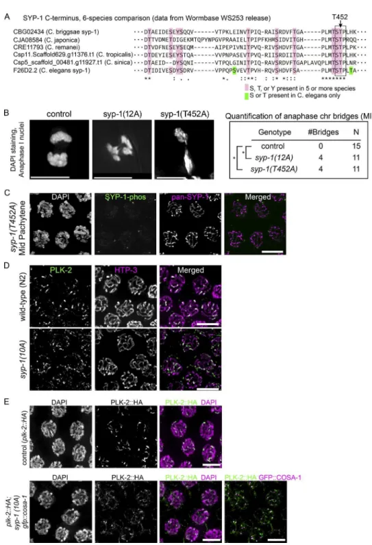
C. elegans, in which roughly half of the cells are oocyte precur-sor cells in meiotic prophase. We identiied 12 phosphorylation sites at the C terminus of SYP-1 (Fig. 1 A and Table S1), 10 of which show conservation in other Caenorhabditis species (Fig. S1 A). To test whether phosphorylation of these residues is im-portant for SYP-1 function in meiosis, we constructed a strain expressing a transgene with a nonphosphorylatable SYP-1 al-lele termed 12A, with all 12 potential phosphoresidues changed to alanine. In a background lacking the endogenous syp-1 gene, the 12A allele displayed reduced viability (59.5% viable), and a high incidence of male progeny (Him) phenotype (6%; Fig. 1 B). The Him phenotype relects meiotic X chromosome nondisjunction in XX hermaphrodite self-progeny, because worms with a single X (XO) develop as males. The syp-1(me17)
null allele shows severely reduced viability (5% viable) and 38% males in the surviving self-progeny (MacQueen et al., 2002), indicating that syp-1(12A) is a partial loss-of-function allele. A wild-type SYP-1 transgene integrated at the same site as the 12A allele restored fertility to wild-type levels in the presence of syp-1(me17), showing that the defects in the 12A allele are speciic to the introduced mutations. In oocytes un-dergoing the irst meiotic division, we found anaphase chromo-some bridges between separating chromatin masses (Fig. S1 B) in 12A and T452A mutants, but not in wild-type animals: 36% of anaphase I nuclei had chromosome bridges (n = 11 for both 12A and T452A), whereas no bridges were detected in the wild type (n = 15). This observation suggests that phosphorylation of the SYP-1 C terminus is required for proper chromosome segregation during meiosis.
Phosphorylation of SYP-1 Thr 452 is crucial to the function of SYP-1
To dissect the role of the SYP-1 phosphorylation sites, we made a series of transgenic worms expressing SYP-1 proteins with nonphosphorylatable mutations. First, we made syp-1(4A)
mutants in which the irst four phosphosites were converted to alanine, as well as syp-1(8A) mutants in which the last eight phosphosites were converted to alanine. The transgenic line expressing SYP-1(4A) showed no progeny inviability or Him phenotype, whereas the line expressing SYP-1(8A) showed in-viability and male production comparable to the line expressing SYP-1(12A) (Fig. 1 B). The most strongly conserved region within these eight residues contains a putative S-[pS/pT]-P/X Polo-box domain (PBD)–binding motif, in which phosphoryla-tion of the central Ser/Thr residue by a priming kinase (Elia et al., 2003) permits subsequent binding of PLK. Our phosphopro-teomics analysis detected phosphorylated Thr452 in this putative
on January 9, 2018
jcb.rupress.org
PBD-binding motif (Ser451-Thr452-Pro453) at a higher level (55
peptide counts total) than the other phosphosites (ranging from 1 to 24 counts; Table S1). To test whether this putative PBD-binding motif is important for SYP-1 function, we gener-ated syp-1(T452A) mutants in which Thr452 in the PBD-binding
motif was converted to alanine, as well as a syp-1(10A) mutant in which the 10 phosphosites outside the PBD-binding motif were converted to alanine. The syp-1(T452A) mutants showed progeny inviability and male production similar to the 12A and 8A mutants. In contrast, syp-1(10A) mutants were fully viable and displayed only a weak Him phenotype (Fig. 1 B). This suggests that phosphorylation at the PBD-binding motif is cru-cial for SYP-1 function.
Phosphorylated SYP-1 concentrates on short arms of chromosomes after COs are designated
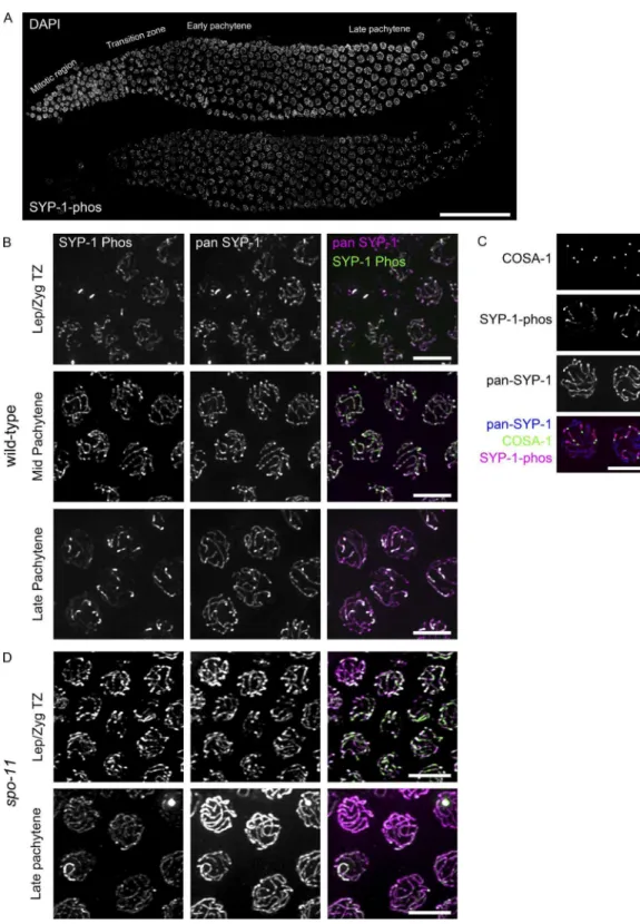
To investigate the timing and location of SYP-1 phosphoryla-tion, we generated two different phospho-speciic antibodies: one against a peptide from SYP-1’s C terminus with phos-pho-threonine at Thr452 (1-phos antibody) and another against the same peptide phosphorylated at three residues (Thr450, Thr452, and Thr455 [3-phos antibody]). Immunoluorescence staining showed similar patterns with both antibodies but higher background with the 3-phos antibody; thereafter, the 1-phos antibody was consistently used to detect phosphorylated SYP-1. The speciicity of the antibody to phospho-SYP-1 was conirmed by lack of staining in T452A mutants (Fig. S1 C). Using this antibody, we irst observed phosphorylated SYP-1 signals in the TZ, coextant with pan-SYP-1 signals along the entire length of the SC (Fig. 2 A). As meiocytes progress into late pachytene, and the CO designation marker COSA-1 starts to appear, SYP-1-phos staining concentrates on short arms and decreases on long arms, whereas pan-SYP-1 signals are still detected along the entire SC (Fig. 2, B and C). In diplotene, SYP-1-phos signal intensity increases further on short arms, whereas pan-SYP-1 starts to disassemble from long arms, as previously shown (Martinez-Perez et al., 2008). The obser-vation that SYP-1-phos signals begin to accumulate on short arms shortly after GFP::COSA-1 foci are detected suggested that COs trigger the coninement of SYP-1-phos to short arms. To test this hypothesis, we examined SYP-1-phos in gonads of
spo-11(me44) mutants, which cannot initiate programmed dou-ble-strand breaks (DSBs) and therefore lack COs (Dernburg et al., 1998). In the absence of SPO-11, SYP-1-phos signals start to appear in the TZ but remain along the entire length of the SC through late pachytene and eventually disappear from the SC at the end of pachytene or diplotene (Fig. 2 D), providing evidence that DSBs are required for the asymmetric distribution of phosphorylated SYP-1.
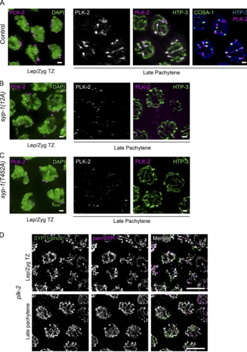
The coninement of SYP-1-phos signals to short arms was reminiscent of the previously reported localization of PLK-2. PLK-2 irst localizes to the PCs during zygotene by binding to HIM-8 and ZIM proteins, then relocates to the entire length of the SC in pachytene in a SYP-1–dependent manner, and later becomes conined to the short arms in a CO-dependent manner (Harper et al., 2011; Labella et al., 2011; Pattabiraman et al., 2017). We examined syp-1(12A) and syp-1(T452A) mu-tants and found that PLK-2 remained at PCs longer, eventu-ally disappearing from the PCs in the region corresponding to late pachytene but failing to relocate to the SC (Fig. 3). This result strongly suggests that the phosphorylated PBD-binding motif of SYP-1 is required for PLK-2 localization to the SC in pachytene and subsequent stages. In contrast, in syp-1(10A)
mutants, PLK-2 also remained at PCs longer but eventually did colocalize weakly to the SC in late pachytene (Fig. S1, D and E). Because PBD-binding motifs must be phosphorylated at the central threonine residue (Thr452 in SYP-1) to enable PLK2 recruitment, the conversion of the nearby Thr450 to alanine in
the 10A mutant might lower priming phosphorylation at Thr452
or confer weaker afinity to PLK-2 itself.
To examine whether PLK-2 acts strictly downstream of SYP-1, we visualized SYP-1-phos in plk-2 mutants. The plk-2(ok1936) null mutant shows delayed synapsis, but more than 60% of chromosomes do achieve homologous pairing and syn-apsis, and the number of COs made are suficient for 33.8% progeny viability (8.4% male; Harper et al., 2011; Labella et al., 2011). The presence of meiocytes carrying COs in plk-2 (ok1936) mutants let us examine the coninement of phosphor-ylated SYP-1 to short arms in this mutant. We found that SYP-1 phosphorylation starting from the TZ is robustly detected in
plk-2(ok1936) mutants but persists over the entire length of the SC in late pachytene, never becoming conined to short arms (Fig. 3 D). This observation suggests the existence of a
feed-Figure 1. Phosphorylation of the C terminus
of SYP-1 is required for correct chromosome segregation in meiosis. (A) SYP-1 phosphor-ylation sites identiied by mass spectrometry,
and schematic diagram of a series of syp-1
phospho mutants. Phosphorylation sites are depicted in red, and the conserved PBD-bind-ing motif [STP] is boxed. For each mutant, the indicated Ser/Thr/Tyr residues are
con-verted to Ala. (B) Percentage of viability and
males among the self-progeny of worms with the indicated genotypes.
on January 9, 2018
jcb.rupress.org
Figure 2. Phosphorylated SYP-1 localizes to the entire SC in early prophase and then becomes progressively restricted to the short arm. (A) A wild-type
gonad stained with DAPI (top) and antibodies against phosphorylated SYP-1 (bottom). Bar, 50 µm. (B) High-magniication images of SYP-1-phos (green)
and pan-SYP-1 (magenta) staining from the indicated gonad regions showing SYP-1-phos on chromosomal subdomains in late pachytene. Bars, 5 µm.
(C) Short arm restriction of SYP-1-phos (magenta) in late pachytene nuclei shown by GFP::COSA-1(green) marking of CO designation sites; pan-SYP-1 is
shown in blue. Bar, 5 µm. (D) SYP-1-phos and pan-SYP-1 staining in spo-11 (me44) mutant gonads. Bars, 5 µm.
on January 9, 2018
jcb.rupress.org
back loop wherein PLK-2 is required to conine SYP-1-phos to short arms in response to CO designation, and SYP-1-phos in turn restricts PLK-2 itself to short arms.
SYP-1 phosphorylation is required for correct timing of early meiotic prophase events
To gain insight into the chromosome segregation defects in 12A mutants, we next examined earlier steps in meiosis: chromo-some pairing, synapsis, recombination, and CO designation. Homologous chromosome pairing was assessed by staining of ZIM proteins (Phillips and Dernburg, 2006) binding the PC of chromosomes I and IV (ZIM-3) or V (ZIM-2). When chro-mosomes are fully homologously paired, the protein ZIM-3, which binds the PCs of chromosomes I and IV, is seen as two foci, whereas ZIM-2, binding to chromosome V, shows a sin-gle focus per nucleus. Chromosome pairing at these sites was found to be normal in syp-1(12A) mutants (Fig. S2 A and not
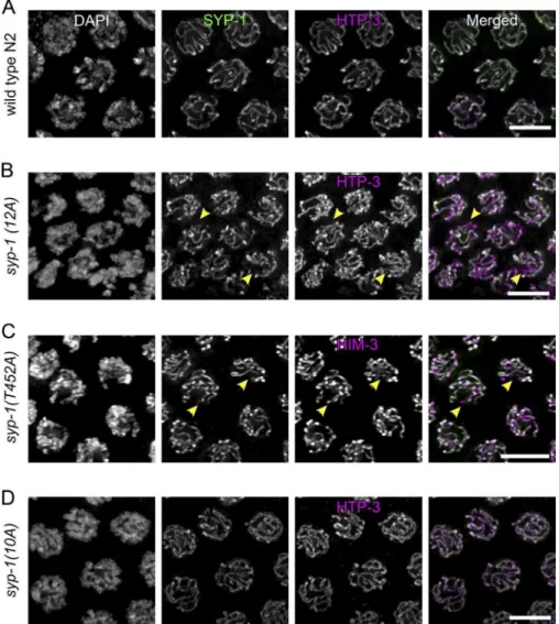
depicted). Next, we assessed chromosome synapsis by immuno-luorescence against the SC proteins. Immunostaining showed that SYP-1(12A) protein colocalized with the axial element protein HTP-3, indicating that the nonphosphorylatable protein is expressed and correctly localized similarly to wild-type SC (Fig. 4, A and B). However, in syp-1(12A) and syp-1(T452A)
mutants, we often observed unsynapsed chromosomes, which are positive for HTP-3 staining but missing SYP-1, in nu-clei from the region corresponding to wild-type mid and late pachytene (Fig. 4, B and C). In contrast to 12A and T452A mu-tants, we detected neither delayed nor partial synapsis in 10A
mutants (Fig. 4 D). This suggests that SYP-1 phosphorylation at the PBD-binding motif is necessary to promote timely and complete SC formation.
We next examined the timing of progression through mei-otic prophase in syp-1 phosphorylation mutants by measuring the proportion of the gonad occupied by each substage. Meiotic nuclei move unidirectionally through the C. elegans gonad in a
Figure 3. Phosphorylation of SYP-1 at
Thr452 (PBD-binding motif) is required for PLK-2 relocalization from the PC to the SC.
(A) Left: Wild-type (N2) oocyte precursor cells
in the leptotene/zygotene TZ immunostained
with DAPI (green) and PLK-2 (magenta); right:
oocyte precursor cells in late pachytene in
con-trol gfp::cosa-1 animals immunostained with
PLK-2 (magenta) and HTP-3 (green), or PLK-2 (magenta), HTP-3 (cyan) and COSA-1::GFP
(green) in the rightmost column. (B) As in A,
but for syp-1(12A); syp-1(me17) mutants, and
without COSA-1 staining. (C) As in B, but for
syp-1(T452A) syp-1(me17) mutants. Immu-nostaining shows PLK-2 relocalization from PCs to the SC (short arms) in control animals,
but not in the syp-1 mutants. Bars, 1 µm.
(D) SYP-1-phos and pan-SYP-1 localization in
plk-2(ok1936) mutant gonads. Bars, 5 µm. In all images, color of text label indicates color of the corresponding signal in merged images.
on January 9, 2018
jcb.rupress.org
manner that allows the physical span of a substage to serve as a proxy for its duration (Hirsh et al., 1976; Jaramillo-Lambert et al., 2007). We found that gonads of both syp-1(12A) and
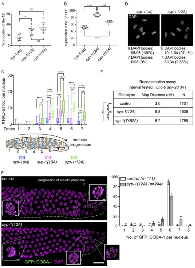
syp-1(10A) mutants showed delayed exit from the leptotene/ zygotene TZ and early pachytene stages compared with con-trol animals (Fig. 5, A and B; and Fig. S2, B and C). Previous studies have shown that meiotic checkpoints monitor the for-mation of CO intermediates and the status of synapsis (Bhalla and Dernburg, 2005; Carlton et al., 2006; Saito et al., 2012; Rosu et al., 2013; Stamper et al., 2013; Kim et al., 2015), ex-tending the time spent in the TZ and early pachytene for nuclei lacking CO intermediates. When chromosomes fail to obtain CO intermediates, the cell cycle checkpoint kinase CHK-2 is activated and continues to phosphorylate PC proteins, which keeps PLK-2 bound to PCs (Kim et al., 2015). Subsequently, PC-bound PLK-2 nucleates the SUN-1–ZYG-12 nuclear enve-lope complex to connect chromosome ends to the cytoskeleton and promotes clustering of chromosomes, a cytological marker for the TZ and early pachytene. (Harper et al., 2011; Labella et al., 2011; Woglar et al., 2013). Gonads from 12A and 10A mutants showed an extended region of the TZ, suggesting that PLK-2 persistence at PCs maintains clustering of chromosomes in these mutants (Fig. 5 A). Gonads from 12A, T452A, and 10A mutants also had an extended early pachytene stage, marked by phosphorylation of the nuclear protein SUN-1 (SUN-1 Ser8-phos; Penkner et al., 2009; Figs. 5 B and S2 D; T452A is not
depicted). Delayed exit from early pachytene could be explained by the complete or partial inability of PLK-2 to transit from PCs to the SC. In addition, delayed synapsis leading to delayed for-mation of COs in 12A and T452A mutants would activate the CHK-2–mediated meiotic checkpoint (Kim et al., 2015), which promotes retention of PLK-2 by continuous phosphorylation of PC proteins, delaying the exit from early pachytene.
Next, we examined the contribution of SYP-1 phosphor-ylation to recombination and CO formation by visualizing the recombination protein RAD-51 and formation of intact biva-lents. RAD-51 foci appeared with normal timing in syp-1(12A)
and syp-1(10A) mutants, suggesting that the initiation of pro-grammed DNA DSBs by SPO-11 is not perturbed in these mutants. Instead, the number of RAD-51 foci per nucleus was found to be prominently increased in mid and late pachytene (zones 4–7) in 12A and less prominently increased in 10A tants compared with control animals (Fig. 5 C). In 12A mu-tants, delayed synapsis would be expected to lead to delayed homologous recombination and activate the CHK-2–mediated meiotic checkpoint, extending the window during which DSBs are generated. In contrast, 10A mutants had increased levels of DSBs without obvious delays in synapsis. This suggests that persistence of PLK-2 at PCs (or absence of PLK-2 from the SC) could sufice to maintain the DSB-generating machinery in an active state with or without activating the upstream ki-nase CHK-2. In addition, recent results show that PLK-1/2–
Figure 4. Phosphorylation of SYP-1 is
re-quired for timely completion of synapsis. (A–D) Oocyte precursor cells in mid-pachy-tene immunostained with SYP-1 and HTP-3
antibodies in wild-type (A), 1(12A);
syp-1(me17) (B), syp-1(T452A); syp-1(me17) (C), and syp-1(10A); syp-1(me17) (D) animals. DNA is counterstained with DAPI. Unsynapsed chromosomes in 12A and T452A mutants are indicated by arrowheads. Bars, 5 µm.
on January 9, 2018
jcb.rupress.org
Figure 5. Phosphorylation of SYP-1 is required for timely progression through meiotic prophase and affects CO distribution. (A) The proportion of TZ nuclei in syp-1(wt); syp-1(me17), syp-1(12A); syp-1(me17), and syp-1(10A); syp-1(me17) animals (see also Materials and methods and Fig. S2). Seven gonads
were scored for each genotype. Error bars show SD. Statistical analysis was performed with a Mann–Whitney test (**, P < 0.01; ***, P < 0.001). (B) The
proportion of TZ and early pachytene nuclei marked by SUN-1 Ser8-phos staining. Error bars show SD. Seven gonads were scored for each genotype.
****, P < 0.0001, Mann–Whitney test. (C) Quantiication of recombination marker RAD-51 focus counts in each of seven equal-length zones covering the
TZ to late pachytene. RAD-51 focus numbers per nucleus are depicted as a box plot, with box indicating mean and quartiles, and whiskers indicating the
entire range of measurements. Four gonads were scored for each genotype. The syp-1(12A); syp-1(me17) and syp-1(10A); syp-1(me17) mutants showed
increased levels of RAD-51 compared with the control syp-1(wt); syp-1(me17). ****, P < 0.0001; ***, P < 0.001; **, P < 0.01, Mann-Whitney test.
(D) Representative diakinesis chromosomes from syp-1(wt); syp-1(me17) (left) and syp-1(12A); syp-1(me17) mutant (right) gonads stained with DAPI.
Sta-tistics of DAPI body observations are shown below. A total of 95 nuclei were scored for the control and 104 nuclei scored for 12A mutants. Bars, 5 µm.
(E, left) The timing of appearance of the CO designation marker GFP::COSA-1 is delayed in syp-1(12A); syp-1(me17) mutants. The direction of meiotic
prophase progression is from left to right. The gonad region in which GFP::COSA-1 foci (green) are observed is highlighted with a green dotted line for
on January 9, 2018
jcb.rupress.org
mediated phosphorylation of SYP-4 is required to turn off DSB formation (Nadarajan et al., 2017). To test whether SYP-1 phos-phorylation is required for SYP-4 phosphos-phorylation, we immu-nostained syp-1 phosphomutants and found that SYP-4-phos signals are still detected (Fig. S2 F) in both 12A and T452A mutants. This suggests that the increase in RAD-51 focus num-bers we observe in syp-1(12A) is not solely caused by loss of SYP-4 phosphorylation.
To investigate whether the increase in recombination in-termediates in 12A mutants could explain the observed meiotic chromosome segregation defects and 40% reduction in progeny viability as a consequence of incomplete recombination, we next examined bivalent formation in diakinesis by scoring the number of DAPI-staining bodies in late meiotic prophase. We found that 12A mutants form meiotic bivalents held together by chiasmata at nearly wild-type levels (97.1%, n = 104; Fig. 5 D). The fraction of diakinesis nuclei with univalents (chromosomes lacking COs) in 12A mutants was 2.9% (7 DAPI bodies: 5 biva-lents and 2 univabiva-lents, n = 104), whereas it was 0% in the wild type (n = 95). We further examined DAPI-staining bodies for the presence of intra- and interbivalent DNA bridges, morpholog-ical features suggestive of improperly resolved recombination intermediates (Saito et al., 2013). We found one intrabivalent bridge out of 31 (3.2%) examined nuclei in syp-1(12A) and one interbivalent bridge out of 11 (9.1%) examined nuclei in syp-1(T452A) compared with none in 48 wild-type nuclei (0%). This suggests although syp-1 phosphomutants show delayed meiotic progression, a relatively minor fraction of meiocytes suffer from improperly resolved or unrepaired recombination.
SYP-1 phosphorylation affects CO distribution
Previous studies have shown that SYP-1 is required for CO dis-tribution and designation along chromosomes (Hayashi et al., 2010; Libuda et al., 2013). We tested whether SYP-1 phosphor-ylation affects the timing and extent of CO designation by visu-alizing COSA-1 in late pachytene. Meiotic nuclei which obtain DSBs in mid pachytene acquire competence to load COSA-1 at eventual CO sites (Yokoo et al., 2012). We observed a signii-cant delay in COSA-1 appearance in 12A mutants, consistent with the observation that the TZ and early pachytene are ex-tended and meiotic cell cycle progression is delayed. In control animals, sites marked by GFP::COSA-1 foci start to appear in late pachytene, which corresponds to ∼40% of the meiotic re-gion of the gonad (the last 40% of the proximal end). In con-trast, 12A mutants showed GFP::COSA-1 foci appearing only at the very end of the gonad (last 10% of the proximal end), where meiotic nuclei inally exit early pachytene and show late pachytene chromosome morphology (Figs. 5 E and S2 E). In contrast to 12A mutants, 10A mutants showed wild-type timing of GFP::COSA-1 foci appearance (Fig. S2 E), indicating that entrance into late pachytene is not delayed in 10A mutants. Im-munoluorescence of 10A mutant gonads suggests that although the TZ and early pachytene (marked by SUN-1 Ser8-phos) are
prolonged in 10A mutants, entrance into late pachytene (marked by GFP::COSA-1) is not delayed, because of a compensatory loss of mid pachytene (marked by neither; unpublished data). Quantitation of GFP::COSA-1 foci revealed an increased frac-tion of nuclei with 7 COSA-1 foci in 12A mutants (14.3% in 12A compared with 0.6% in control), suggesting that CO des-ignation is perturbed in the absence of SYP-1 phosphorylation. Although we detected a slightly increased number of nuclei with only ive GFP::COSA-1 foci in 12A (13.3% in 12A com-pared with 2.9% in control), eventual completion of a sixth CO or apoptotic culling would result in a lower frequency of diak-inesis nuclei with univalents, as we observe. To further assess the extent of CO recombination in syp-1 phospho mutants, we measured the genetic distance between the unc-5 and dpy-20
genes on chromosome IV. In control animals, the distance was calculated as 3.0 cM, in agreement with the reference map dis-tance of 3.44 cM (WormBase WS260). However, the 12A and T452A alleles showed signiicantly larger distances (Fig. 5 F), suggesting either an elevated CO frequency or changes in the recombination landscape. Interestingly, the map distance mea-sured in 12A mutants (8.8 cM) was also signiicantly larger than that in T452A mutants (5.2 cM), raising the possibility that the different levels of SYP-1 phosphorylation might inluence CO designation capacity differently. These experiments show that loss of SYP-1 phosphorylation alters levels of synapsis and re-combination as well as CO designation and distribution, which together are likely to partially contribute to the inviability ob-served in the syp-1 phosphomutants.
SYP-1 phosphorylation promotes
establishment of short/long arm asymmetry
Previous studies have shown that PLK-2 plays essential roles in the establishment of short and long arm asymmetry in addition to its role in homologue pairing and synapsis (Harper et al., 2011; Labella et al., 2011; Nadarajan et al., 2017; Pattabiraman et al., 2017). The observed coninement of SYP-1-phos signals to short arms also raises the possibility that SYP-1 phosphory-lation is involved in the functional distinction of the short and long arms. To test this hypothesis, we used immunoluorescence to examine protein localization on diakinesis chromosomes. Wild-type SYP-1 departs from long arms as bivalents undergo diplotene remodeling (Fig. 6 A) and eventually departs from short arms no later than the −3 oocyte (Fig. S3). In contrast, we found that nonphosphorylatable SYP-1, as well as SYP-2, always persists on both the short and long arms of diakinesis chromosomes until the −2 or −1 oocyte in the syp-1(12A) or
(T452A) mutants (Fig. 6, B and C; and Fig. S3 D; SYP-2 is not depicted). This result shows that phosphorylation of SYP-1 is required for its timely removal from long and short arms. We next visualized proteins reported to dissociate from the short arm after CO formation: HTP-1/2, LAB-1, and COH-3/4 (de Carvalho et al., 2008; Martinez-Perez et al., 2008; Severson and Meyer, 2014). These proteins also remained on both short and long arms on at least one chromosome in the majority of
the control gfp::cosa-1 and syp-1(12A) gfp::cosa-1; syp-1(me17) gonads. DNA is counterstained by DAPI (magenta). (E) Right: quantitation of COSA-1
focus numbers from three gonads for the control gfp::cosa-1 or ten gonads for syp-1(12A) gfp::cosa-1; syp-1(me17) mutants (n = 171 nuclei scored for wild
type, 244 nuclei scored for syp-1(12A)). Bar heights are percentages of nuclei with the given number of COSA-1 foci; error bars show 95% conidence
intervals. Bars: (main) 15 µm; (insets) 1 µm. (F) Measured genetic map distances in centimorgans for the interval unc-5—dpy-20 on chromosome IV scored
on the given number (N) of unc-5 dpy-20/+ + (control), syp-1(12A); unc-5 dpy-20/+ +; syp-1(me17), and syp-1(T452A); unc-5 dpy-20/++; syp-1(me17)
self-progeny. ****, P < 0.0001; ***, P = 0.0001484; **, P = 0.001336, Fisher’s exact test.
on January 9, 2018
jcb.rupress.org
−1 oocytes in syp-1(12A) and syp-1(T452A) mutants (Fig. 6, B and C; and Fig. S3 A). These results indicate that C-terminal phosphorylation of SYP-1 promotes the establishment of short/ long chromosome arm asymmetry, as well as the timely disas-sembly of SYP-1 from the SC.
To further understand the function of SYP-1 phosphory-lation, we created alleles with all 12 phosphosites mutated to D or E (12D or 12E), an alteration that in some cases mimics phosphorylation. However, we found that viability, male progeny production, and immunostaining phenotypes of syp-1(12E) and
syp-1(12D) were indistinguishable from syp-1(12A), with SYP-1 persisting on both short and long arms in diakinesis (Fig. S2 C and
Fig. S3, B–D). From this, we conclude that SYP-1(12D) or (12E) may functionally resemble the nonphosphorylatable 12A alleles rather than mimicking constitutively phosphorylated SYP-1.
The CPC is mislocalized in syp-1 nonphos mutants
The arm asymmetry defects and anaphase bridges we observed in syp-1(12A) mutants next prompted us to examine the local-ization of the CPC. Loss of arm asymmetry caused by labo-ratory-1 or htp-1 mutations has been linked to mislocalization of AIR-2 (Aurora B) and the CPC, of which AIR-2 (Aurora B) is a part, leading to chromosome segregation defects (de
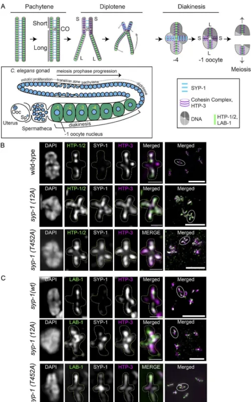
Figure 6. Phosphorylation of SYP-1 is
re-quired for the asymmetric localization of fac-tors involved in chromosome segregation. (A)
Top: Diagram of chromosome axis remodel-ing durremodel-ing meiotic prophase from pachytene through meiosis I. Cruciform axis of HTP-3/ cohesin with short arm (S) and long arm (L) is shown in diakinesis. Bottom: Diagram of
meiotic prophase substages in the C. elegans
gonad with meiotic nuclei indicated by blue circles. Ooc, oocyte nucleus; Sp, sperm pronu-cleus. Diakinesis nuclei are referred to by their position relative to the spermatheca, with the
nearest diakinesis nucleus being the −1 oocyte.
After entering the spermatheca and becoming
fertilized, the −1 oocyte nucleus goes through
the meiosis I and II divisions to generate the
oocyte pronucleus. (B and C) Partial
Z-projec-tion images of a representative chromosome
pair in −1 or −2 oocytes with immunostaining
for HTP-1/2 (green), SYP-1, and HTP-3 (ma-genta; B) or LAB-1 (green), SYP-1, and HTP-3
(magenta; C) in the syp-1(wt); syp-1(me17)
(−1 oocyte), syp-1(12A); syp-1(me17) (−1
oocyte), and syp-1(T452A); syp-1(me17) (−2
oocyte) gonads. The 12A and T452A mutant gonad shows aberrant persistence of SYP-1 on both arms (see also Fig. S3 D), and HTP-1/2
and LAB-1 on short arms, in −1 or −2 oocytes.
In B and C, representative chromosomes are presented with the color of image label indicat-ing the color of immunostainindicat-ing in the merged image. Full-projection images of each nucleus are shown in the rightmost column, with the chromosome pair shown on the left encircled. The cruciform axis of HTP-3 is visible only for chromosomes lying perpendicular to the op-tical axis. Bars: (magniied insets) 1 µm; (full projection images at right) 5 µm.
on January 9, 2018
jcb.rupress.org
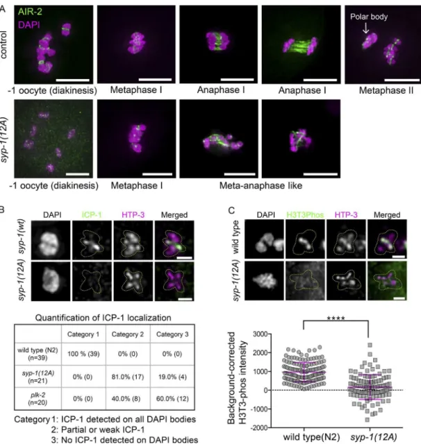
Carvalho et al., 2008; Martinez-Perez et al., 2008). To exam-ine whether syp-1(12A) mutations lead to CPC mislocalization, we looked at −1 oocytes of 12A mutants. In control animals expressing GFP::AIR-2, we observed the expected concen-tration of AIR-2 (Aurora B) on short arms in −1 oocytes, fol-lowed by its localization to the meiotic spindle in anaphase I and relocalization between sister chromatids at metaphase II. In contrast, in syp-1(12A) mutants, AIR-2 (Aurora B) was faint and disorganized in many of the corresponding oocytes, as well as on chromosomes in metaphase/anaphase I (Fig. 7 A). Similarly, the CPC component INC ENP (ICP-1 in C. elegans) shows robust localization to short arms in wild-type −1 oocyte nuclei, whereas syp-1(12A) and syp-1 (T452A) mutants had signiicantly reduced levels of ICP-1 signals in the correspond-ing nuclei: 81% of nuclei showed reduced ICP-1 signals com-pared with the wild type, whereas 19% of nuclei showed no ICP-1 signal on any chromosomes in 12A mutants (Figs. 7 B and S4). In plk-2 mutants, an even stronger loss of ICP-1 from short arms was observed in −1 oocytes: 40% of nuclei showed reduced ICP-1 signals compared with the wild type, whereas 60% of nuclei showed no ICP-1 staining on any chromosomes. We noted that the magnitude of ICP-1 reduction does not cor-respond to the level of progeny viability in syp-1(12A) (60% viability) or plk-2 (ok1936) (34% viability; Harper et al., 2011) mutants. Further examination revealed that although wild-type gonads always show ICP-1 signals on short arms from the −3 or −4 oocyte stage, some of the syp-1(12A) and plk-2 (ok1936)
mutant gonads have ICP-1 signals on prometaphase I chromo-somes, but not diakinesis chromosomes (Fig. S4 A [plk-2]; 12A is not depicted). This suggests that ICP-1 is sometimes capa-ble of rapidly accumulating on chromosomes upon entrance to the meiosis I division without signiicant prior accumulation in prophase. This is likely a result of redundant positive feed-back mechanisms that enhance CPC localization (Carmena et al., 2012) and could partially explain the signiicant progeny viability observed in syp-1(12A) and plk-2 (ok1936) mutants. Consistent with reduced localization of the CPC in syp-1(12A)
mutants, levels of H3Ser10 phosphorylation, which is mediated by AIR-2 (Aurora B) kinase (Hsu et al., 2000), were also re-duced in syp-1(12A) mutants compared with the wild type (Fig. S4 D). The mislocalization of the CPC could lead to failures in triggering cohesin cleavage and the spindle assembly check-point and could explain the anaphase chromosome bridges and loss of viability found in syp-1(12A) mutants.
Because LAB-1 has been shown to promote the phos-phatase activity of GSP-2 (PP1) at the SC in wild-type animals (Tzur et al., 2012), and because we observed mislocalization of LAB-1 and the CPC in syp-1(12A) mutants, we next exam-ined the localization of phosphorylated H3T3, which has been shown to recruit the CPC and is a substrate of PP1 in other model organisms (Qian et al., 2011). In wild-type germlines, H3T3ph signals appeared on the short arm of diakinesis chro-mosomes from the −3 to −4 oocyte stage. In contrast, H3T3ph was strikingly reduced or absent from short arms in −1 oocytes in syp-1(12A) mutants (Figs. 7 C and S4 C). This suggests that in 12A mutants, LAB-1 mislocalizes to both long and short arms, promoting dephosphorylation of H3T3 via GSP-2 (PP1) on the entire chromosome.
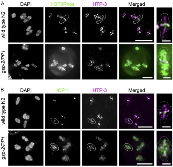
To conirm that PP1 dephosphorylates H3T3ph in C. ele-gans meiosis, we immunostained H3T3ph in worms homozy-gous for the gsp-2(PP1) deletion allele tm301. In contrast to the short arm–speciic H3T3ph staining in wild type, gsp-2(PP1)
mutants showed H3T3 phosphorylation over the entire chro-mosome from diakinesis onward (Fig. 8 A). This suggests that dephosphorylation of H3T3 by PP1 is likely to be conserved in C. elegans. Similarly to H3T3ph staining, ICP-1 was pres-ent all over chromosomes in gsp-2 (PP1) mutants by immuno-luorescence (Fig. 8 B).
Collectively, the high incidence of defects in the asym-metric disassembly of SC components and compromised CPC localization likely explain the bulk of reduced viability in syp-1(12A) and (T452A) mutants by causing chromosome segrega-tion problems at meiosis I. The loss of short/long arm asymmetry in the nonphosphorylatable SYP-1 alleles and the dynamic lo-calization of SYP-1-phos signals are two independent lines of evidence that strongly suggest that phosphorylation of SYP-1 is a key element of the molecular cascade that establishes domains of successive cohesion loss de novo in response to COs.
Discussion
Here, we have shown that phosphorylation of SYP-1 at its PBD-binding motif promotes timely synapsis and progres-sion of meiotic prophase, affects recombination rates, and is critical for early steps in establishing the short and long arm chromosome domains essential for correct meiotic dis-junction. SYP-1 phosphorylation begins at the TZ as SYP-1 proteins polymerize between axial elements. Once COs are made and meiocytes enter the late pachytene stage, phosphor-ylated SYP-1 and PLK-2 localize to short arms and vacate the long arms. This asymmetric distribution is required for the full establishment of functionally distinct short and long arms in diplotene and diakinesis. Prevention of SYP-1 phosphoryla-tion at its PBD-binding motif disrupts PLK-2 relocaphosphoryla-tion from PCs to the SC, delays synapsis and CO formation, and de-lays the exit from the TZ and early pachytene. Without SYP-1 phosphorylation, meiotic defects from early and late prophase lead to mislocalization of the CPC, resulting in chromosome missegregation and reduced progeny viability. Collectively, our results suggest that PLK-2 acts through SYP-1 (poten-tially through SYP-1’s PBD-binding motif) to ensure conine-ment of the chromosome segregation machinery to the short arm subdomain, promoting correct segregation of holocentric chromosomes in meiosis I.
PLK-2 has been shown to bind to PC proteins at nuclear envelope-associated LINC complexes to promote chromosome pairing and synapsis in the TZ (Harper et al., 2011; Labella et al., 2011). Our results raise the possibility that SYP-1 protein phosphorylated at its PBD-binding motif in the nascent SC could provide a new binding site for PLK-2. This new site could promote meiotic events in two ways: (1) by allowing PLK-2 to move off of PCs (possibly in cis), promoting the progression of the cell cycle, and (2) by recruiting PLK-2 to the SC to establish short and long arm distinction. PLK-2 bound to SYP-1-phos could then phosphorylate other proteins in the vicinity of the SC, initiating a phosphorylation cascade.
Phosphorylated SYP-1 localizes to the short arm imme-diately after COSA-1 focus formation in late pachytene, sug-gesting that this relocation is one of the earliest events to take place after recombination intermediates are committed to COs. Another SC central component, SYP-3, has been shown to dy-namically move onto and off of chromosomes during meiotic prophase. In a process dependent on PLK-2, this exchange
on January 9, 2018
jcb.rupress.org
becomes less dynamic after COs are made (Pattabiraman et al., 2017). Thus, the asymmetric relocalization of phosphorylated SYP-1 coincides with a large-scale reduction in the dynamic properties of the SC, raising the possibility that both of these PLK-2–dependent events are mechanistically connected.
The SYP-1 phosphomutants also affected COSA-1 focus number and CO recombination rate. In the 12A allele, COSA-1 foci representing CO designation sites departed from the wild-type number of six per nucleus, with ∼13.5% of nuclei having ive foci and 14.3% having seven foci. Perturbation of the SC by partial loss of SYP-1 has been shown to increase COSA-1 number (Hayashi et al., 2010; Libuda et al., 2013); perhaps the
alterations in our mutant alleles act in a similar fashion by af-fecting the level of SYP-1 incorporation or the overall integrity of the SC. In contrast, the genetic distance of the unc-5—dpy-20 interval on chromosome IV increased by more than a factor of two in 12A mutants. The disagreement between the change in COSA-1 foci and the large increase in the genetic map we observed has several nonexclusive possible explanations, in-cluding generation of additional COs not marked by COSA-1, a shift of CO formation to a central position (the unc-5—dpy-20
interval roughly bounds the central third of chromosome IV), or a bias in recovery of chromosomes with COs in this region in the surviving progeny.
Figure 7. SYP-1 phosphorylation is required for correct localization of CPC components (AIR-2 and ICP-1) and the CPC-guiding histone mark (H3T3ph).
(A) Localization of GFP::AIR-2 (green) on meiotic chromosomes ixed and stained with DAPI (magenta) at different stages preceding and during the
mei-otic divisions in control (gfp::air-2)(top) and gfp::air-2; syp-1(12A); syp-1(me17) (bottom) animals. Bars, 5 µm. (B) ICP-1 is enriched on short arms in −1
oocytes in syp-1(wt); syp-1(me17) animals (top), whereas some −1 oocytes lack ICP-1 localization in syp-1(12A); syp-1(me17) mutants (bottom; see also
the whole-nucleus image in Fig. S4). The ICP-1 localization in −1 oocytes was categorized as follows in syp-1(wt); syp-1(me17), syp-1(12A); syp-1(me17),
and plk-2(ok1936): all six DAPI bodies had robust ICP-1 staining on short arms (category 1), some DAPI bodies had partial or weak ICP-1 staining
(cate-gory 2), or no DAPI bodies had ICP-1 staining (cate(cate-gory 3). The analysis was limited to −1 oocyte nuclei carrying six bivalents. Bars, 1 µm. (C) H3T3ph
staining in −1 oocyte nuclei in N2 wild-type (top) and syp-1(12A); syp-1(me17) mutants (bottom); representative images are shown above quantitation
(see also the whole-nucleus image in Fig. S4). Bars, 1 µm. In the scatterplot below, each point is a measurement of a single DAPI body. Error bars indicate
mean and SD of all points. The number of DAPI bodies/points counted for H3T3ph is 151 for wild type and 111 for syp-1(12A) mutants. ****, P <
0.0001, Mann–Whitney test.
on January 9, 2018
jcb.rupress.org
Loss of SYP-1 phosphorylation nearly always resulted in abnormal persistence of SC or SC-interacting proteins in diplo-tene and diakinesis, and ICP-1 was never observed at wild type levels in −1 oocytes in syp-1(12A) mutants. These very pen-etrative phenotypes contrast with the relatively high viability (60%) of syp-1(12A) mutant self-progeny. If spermatogenesis and oogenesis were equally compromised, this level of viability would suggest that gamete production succeeds at least 77% of the time in the absence of SYP-1 phosphorylation. This would imply that despite the localization defects observed in prophase in syp-1(12A) mutants, redundant pathways sufice to deposit CPC components at the short arm by metaphase I in a subset of nuclei. Why some chromosomes in syp-1(12A) mutants ul-timately succeed in recruiting the CPC to short arms and suc-cessfully segregate in meiosis while others do not remains to be determined. One possible explanation is the unpredictable nature of CO position. In C. elegans, the obligatory single CO is most likely to occur in the terminal thirds of chromosomes (Barnes et al., 1995). Because establishment of short and long arm domain patterning appears to be determined by the physi-cal distance from the CO to the chromosome ends, perhaps the mechanisms behind this functional distinction could be con-founded on chromosomes that happen to receive more centrally
located COs. More generally, if COs at different positions (cen-tral, arm, or terminal) of chromosomes vary with respect to the ease or speed of subsequent long and short arm domain pattern-ing, then nuclei with an excess of “dificult” COs may be more likely to harbor chromosomes that fail in downstream steps of short and long arm establishment in a sensitized background such as syp-1(12A) mutants. Similar reasoning could account for the fact that the syp-1(10A) mutant has a slight but signii-cant increase in male production without an increase in embry-onic lethality, which suggests the X chromosome has a higher nondisjunction rate than autosomes in this mutant. Compared with autosomes, the X chromosome has a smaller region of sup-pressed crossing over near the chromosome midpoint (Rockman and Kruglyak, 2009), predicting a higher incidence of centrally located COs. Such COs, though occurring with low frequency, could result in failure to effectively establish the long and short arm domains in syp-1(10A) mutants.
The observation that synapsis is delayed in syp-1(12A) or
(T452A) mutants, but not in syp-1(10A) mutants, suggests that SYP-1 phosphorylation at its PBD-binding domain promotes the timely progression of synapsis. Although immunoluo-rescence against both native and HA-tagged PLK-2 failed to detect PLK-2 on the SC in the TZ or early pachytene in 10A
Figure 8. GSP-2/PP1 restricts the CPC-guiding histone mark H3T3ph and ICP-1 to short arms. (A and B) H3T3ph immunostaining shown in green
(A) or ICP-1 immunostaining shown in green (B) in −1 oocytes in wild type (top) and gsp-2(tm301) mutants (bottom). Short and long arms are marked
by HTP-3 staining (magenta). Representative chromosomes with cruciform HTP-3 staining indicated by circles are shown in the rightmost panels. Bars: (left) 5 µm; (right) 1 µm.
on January 9, 2018
jcb.rupress.org
mutants, a residual amount of PLK-2 at the SC below our de-tection threshold may sufice to promote timely completion of synapsis in this mutant. However, the fact that many chromo-somes achieve timely synapsis even in plk-2 mutants shows that PLK-2 is not strictly necessary for synapsis. The mecha-nism of synapsis facilitation by PLK-2 remains unknown but could involve distributive phosphorylation of SC components, which may alter SC structure or binding afinity. In addition to containing the PBD-binding motif, the C terminus of SYP-1 interacts with SYP-3 (Schild-Prüfert et al., 2011), raising the further possibility that phosphorylation of SYP-1 at T452 may modulate its binding capacity to SYP-3 and promote assem-bly of central components.
How could the localization of phosphorylated SYP-1 on the short arm lead to subsequent localization of the CPC on the same domain at the end of meiotic prophase? Our data show that SYP-1 phosphorylation controls the correct distribution of one of the CPC-guiding histone modiications, phosphorylated histone H3T3. We have shown that SYP-1 phosphorylation is critical for LAB-1 restriction to the long arms, where it nor-mally locally increases the activity of PP1 (Tzur et al., 2012), which we have shown to counteract H3T3 phosphorylation. A previous study in ission yeast demonstrated that the H3T3 ki-nase Haspin is recruited to centromeres by the cohesin regulator Pds5 (Yamagishi et al., 2010). If this mechanism is conserved in C. elegans, then we would expect Haspin to be recruited to the axis region of the SC, because cohesins and the C. elegans
orthologue of Pds5, EVL-14, associate with the lateral elements (Pasierbek et al., 2001; Kim et al., 2014). Although our detec-tion of globally high H3T3ph levels in gsp-2 mutants shows that PP1 likely suppresses Haspin activity all over chromatin during diakinesis, additional negative reinforcement may be required speciically at the axis region of the long arms where Pds5 may increase the amount of Haspin present. In addition to this potential mechanism of negative regulation on the long arms we describe, previous work has also shown that Haspin is phosphorylated and activated by PLK in Xenopus laevis ex-tracts (Ghenoiu et al., 2013). PLK-2 constrained to the SC short arm by phosphorylated SYP-1 could similarly activate Haspin, and subsequently enrich the CPC at short arms, in C. elegans.
Our indings, combined with previous results, suggest major functions of SYP-1 phosphorylation in guiding the localization of the CPC by suppressing H3T3 phosphorylation on long arms while promoting it on short arms.
Materials and methods
C. elegans strains and conditions
C. elegans strains were grown using standard procedures (Brenner, 1974) at 20°C. Wild-type worms were from the N2 Bristol strain. The following mutations, transgenes, and balancers were used in this study:
me107[plk-2::HA] (I), plk-2(ok1936)/hT2[bli-4(e937) let-?(q782) qls48](I;III), gsp-2(tm301)/eT1(unc-36)(I;III), meIs8 [Ppie-1::gfp:: cosa-1 + 119(+)] (II), icmSi44 [Psyp-1::syp-1(T452A) + unc-119(+)](II), icmSi35 [Psyp-1::syp-1(4A) + unc-unc-119(+)](II), icmSi33 [Psyp-1::syp-1(8A) + unc-119(+)](II), icmSi42 [Psyp-1::syp-1(10A) + unc-119(+)](II), icmSi25 [Psyp-1::syp-1(12A) + unc-119(+)] (II), icmSi31 [Psyp-1::syp-1(12E) + unc-119(+)] line 2 (II), icmSi32 [Psyp-1::syp-1(12E) + unc-119(+)] line 3 (II), icmSi28 [Psyp-1::s-yp-1(12D) + 119(+)] (II), icmSi24 [Psyp-1::syp-1(wild type) + unc-119(+)] (II), zim-2 (tm574) (IV), spo-11(me44)/nT1[unc-?(n754) let-?
qIs50](IV;V), syp-1(me17)/nT1[?(n754) let-?(m435)] (IV;V), unc-119(ed3) (III); ltIs14[pie-1p::GFP-TEV-STag::air-2 + unc-119(+)], unc-5 (e53), dpy-20(e1282ts) (IV).
For all mutant analyses, we used homozygous mutant progeny of heterozygous parents.
The syp-1 phospho mutants were generated using Mos-SCI (Frøkjaer-Jensen et al., 2008) with pCFJ151 plasmids and the strain EG6699. To generate syp-1 (12A), (12E), and (12D) mutants, the
syp-1 gene fragments were synthesized with respective mutations by Invitrogen GeneArt Strings and cloned into pCFJ151 using Gibson assembly cloning kit (New England BioLabs). The rest of the syp-1
phosphomutations were created by either mutating syp-1 (wt) or (12A)
genes cloned in pCFJ151 using PCR-based mutagenesis (for T452A) or by stitching syp-1 (wt) and (12A) gene fragments by Gibson assembly cloning (for 4A and 8A mutants). The following primer set was used for PCR-based mutagenesis for T452A: forward, 5′-CCA TTG ATG ACG AGC GCA CCA CTT ACC GCA GCT ACtAGA CCG TTG AAA CGA AC-3′; and reverse, 5′-GTT CGT TTC AAC GGT CTaGTA GCT GCG GTA AGT GGT GCG CTC GTC ATC AAT GG-3′. This complementary primer set introduces an additional synonymous mutation (indicated by a lowercase t in the forward primer/lowercase a in the reverse primer), because we designed this template to function with either Mos-SCI or CRI SPR/Cas9, and this synonymous mutation disrupts a potential pro-tospacer adjacent motif (PAM) sequence. At least two transgenic lines were generated for each transgene for mutant phenotype analysis. All
syp-1 transgenes contain the promoter (500 bp upstream of the gene) and the 3′ UTR (97 bp downstream of the gene). The insertion of the
syp-1 transgene was veriied by DNA sequencing.
Phosphoproteomics
Wild-type N2 and pph-4.1(tm1598)/hT2[bli-4(e937) let-?(q782) qIs48]
worms were grown on NGM plates containing 25 µg/ml carbenicillin and 1 mM IPTG spread with HT115 bacteria either carrying an empty RNAi vector (L4440; http ://www .addgene .org /1654) or a pph-4.1 -RNAi plasmid. To generate the pph-4.1 RNAi plasmid, the 695-bp region spanning the second, third, and fourth exons of pph-4.1 was ampliied from C. elegans N2 genomic DNA using primers 5′-GCT CGT GAA ATC CTA GC-3′ (forward) and 5′-CGA ATA GAT AAC CGG CTC-3′ (reverse) lanked by Not1 and Nco1 sites and cloned into L4440. First, N2 and pph-4.1(tm1598)/hT2[bli-4(e937) let-?(q782) qIs48] worms (P0) synchronized by starvation were transferred to new plates with food, and worms at the L4 larval stage were harvested in M9 + 0.01% Tween buffer, washed three times with M9 + 0.01% Tween buffer, and distributed to either control or pph-4.1 RNAi plates. Approximately 30 h later, these worms on RNAi plates were harvested in M9 + 0.01% Tween buffer and bleached to collect embryos. Collected F1 embryos were distributed to fresh RNAi plates. At time points when these F1 worms were either 1 or 3 d after L4 stage, half of the F1 plates were exposed to 10 Gy γ-rays to induce DNA damage. 4 h after irradiation, worms were harvested in M9 buffer, washed three times with M9 buffer, and frozen at −80°C. 2 ml pelleted, frozen worms prepared in this manner was thawed and dissolved in 5 ml urea lysis buffer (20 mM Hepes, pH 8.0, 9 M urea, 1 mM sodium orthovanadate, 2.5 mM sodium pyrophosphate, and 1 mM β-glycerol-phosphate), sonicated for 1 min at 30-s intervals 10 times until worm bodies were broken up. The worm lysates were spun down at 20,000 g for 15 min, and supernatants were subjected to PTMScan analysis (Cell Signaling Technology); phosphorylated peptides were enriched by phospho-(Ser/Thr) kinase substrate antibody-immobilized protein A beads and analyzed by liquid chromatography–tandem mass spectrometry using an LTQ-Orbitrap-Elite ESI-CID (Thermo Fisher). Phosphoenrichment antibodies were obtained from CST (catalog numbers 9607, 6966,
on January 9, 2018
jcb.rupress.org
8139, 8738, 9624, 6967, 5759, 9942, 10001, 9614, 9477, 8134, 2325, 5243, and 3004). Protein assignments were made using Sorcerer. Peptide counts indicated in Table S1 show pooled counts from all conditions of worms (± RNAi, irradiation, or age) used in this assay.
Microscopy, cytology, and antibodies
For all cytological preparations, we followed protocols described previously (Phillips et al., 2009). Images were acquired on a Deltavision personalDV microscope (Applied Precision/GE Healthcare) with a CoolSNAP ES2 camera (Photometrics) at 23°C, using 60× PlanApoN 1.42 NA or 100× UPlanSApo 1.4 NA oil-immersion objectives (Olympus) and immersion oil (LaserLiquid; Cargille) at a refractive index of 1.513. The Z spacing was 0.2 µm, and raw images were subjected to constrained iterative deconvolution followed by subpixel chromatic shift correction using scripted control of the Priism (Chen et al., 1996) software suite (see code chromatic-shift at https ://github .com /pmcarlton /deltavisionquant). Image acquisition was performed with the softWoRx suite (Applied Precision/GE Healthcare). Image postprocessing for publication was limited to linear intensity scaling and maximum-intensity projection using OME RO (Burel et al., 2015). The following antibodies used in the present study have been described previously: HTP-1 (Martinez-Perez et al., 2008; provided by E. Martinez-Perez, Imperial College London, London, England, UK), LAB-1 (de Carvalho et al., 2008) and phosphorylated SYP-4 (Nadarajan et al., 2017; provided by M. Colaiacovo, Harvard Medical School, Boston, MA), PLK-2 (Labella et al., 2011; provided by M. Zetka, McGill University, Montreal, Canada), ICP-1 (Oegema et al., 2001; provided by K. Oegema, Ludwig Institute for Cancer Research, San Diego, CA), HIM-8 (Phillips et al., 2005), HTP-3 (MacQueen et al., 2005), SYP-1 (Harper et al., 2011), and ZIM-3 (Phillips and Dernburg, 2006; provided by A. Dernburg, Lawrence Berkeley National Laboratory, Berkeley, CA), SUN -1 :Ser8p (Penkner et al., 2009; provided by V. Jantsch, University of Vienna, Vienna, Austria), and COH-3/4 (Severson and Meyer, 2014; provided by A. Severson, Cleveland State University, Cleveland, OH). Antibodies generated for this work were rabbit-SYP-1_1Phos antibodies generated using the phosphopeptide [SAP LMTSpTPL TAA TR], rabbit-SYP-1_3Phos antibodies generated using the phosphopeptide [SAP LM(pT) S(pT)PL(pT)AATR] by Euroins, all used at 1:100 dilution. All the phospho-speciic antibodies were afinity puriied using the SulfoLink Immobilization kit (44999; Thermo Fisher) using nonphosphorylated and phosphorylated peptides. The following commercial antibodies were used: GFP (1:500 dilution; 12600500; Roche), anti-Histone H3Thr3ph (Phospho-anti-Histone H3 [Thr3; D5G1I] mAb, 1:10,000 dilution; 13576S; Cell Signaling Technology), anti-Histone H3Ser10Phos (39254, 1:1,000 dilution), rabbit RAD-51 antibody from SDIX/Novus Biologicals (1:1,000 dilution; #29480002, lot# G3048-009A02; Active Motif), rabbit anti-HIM-3 antibody (1:500 dilution; 53470002; SDIX/Novus), anti-HA (1:500 dilution; 901501; Covance/ BioLegend), and anti–ZIM-2 (1:500 dilution; 49270002; SDIX/ Novus). Secondary antibodies used were DyLight488, DyLight594, DyLight649, or Alexa Fluor 488–conjugated AfiniPure antibodies (1:500 dilution; Jackson ImmunoResearch). All immunoluorescence was performed on adult worms at 1 d after L4.
For H3T3 phosphorylation intensity measurements, Fiji (Schin-delin et al., 2012) was used to calculate the intensity of H3T3ph on DAPI bodies. Regions of interest were drawn based on DAPI-positive pixels in −1 oocyte nuclei, and the mean H3Thr3ph pixel intensity within regions of interest was measured. To subtract background inten-sity, background regions were drawn in the same nucleus outside of the DAPI bodies, whose mean pixel intensity was taken as the background
intensity. Intensity data were statistically analyzed by the Mann–Whit-ney test. The number of DAPI bodies counted for H3T3ph is 151 for the wild type and 111 for syp-1(12A) mutants. Quantiications of the length of SUN-1 Ser8ph staining as well as the TZ (deined by clustered nu-clei without resolvable chromatids) and RAD-51 foci were performed as described previously (Sato-Carlton et al., 2014). For quantiication of RAD-51 foci per nucleus, the nuclei on the coverslip-proximal side of four gonads were scored for each genotype. The numbers of nuclei scored for zones 1–7 were 142, 163, 173, 146, 127, 111, and 92 for wt; 210, 241, 172, 129, 142, 104, and 104 for syp-1(10A); and 154, 144, 86, 75, 83, 79, and 74 for syp-1(12A).
Genetic recombination frequencies
Recombination frequencies were calculated as described previously (Zalevsky et al., 1999). p, the map distance, is calculated from the frac-tion R of recombinant self-progeny, as P = 1 − [(1 − 2R)0.5]. Males of genotype icmSi25 [Psyp-1::syp-1(12A) + unc-119(+)]; syp-1(me17)/ nT1[unc-?(n754) let-? qIs50], icmSi44 [Psyp-1::syp-1(T452A) + unc-119(+)]; syp-1(me17)/nT1[unc-?(n754) let-? qIs50] or N2 were crossed with icmSi25 [Psyp-1::syp-1(12A) + unc-119(+)]; unc-5 (e53), dpy-20(e1282ts); syp-1(me17), icmSi44 [Psyp-1::syp-1(T452A) + unc-119(+)]; unc-5 (e53), dpy-20(e1282ts); syp-1(me17) or unc-5 (e53) dpy-20(e1282ts) hermaphrodites, respectively. Hermaphrodite cross-progeny were picked to single plates, and their progeny were scored for Unc Dpy, wild type, and Unc non-Dpy or Dpy non-Unc recombinants. The assay was performed at 23°C to observe the tem-perature-sensitive phenotype of dpy-20(e1282ts). We noted that syp-1(12A); syp-1(me17) and syp-1(T452A); syp-1(me17) animals generate a small population of sick progeny (worms with abnormal morphology such as arrested development, reduced pigment, early death or Unc; 2.3% and 2.1% of total progeny, respectively) and also that syp-1(12A); syp-1(me17) animals generate a small number of Dpy progeny (0.4% of total progeny, presumably because of aneuploidy observed in these mutants). The sick progeny observed in 12A or T452A mutants could be falsely scored as Unc non-Dpy animals in the recombination assay, and indeed, we found more Unc non-Dpy animals than Dpy non-Unc in these mutants. Therefore, we limited our analysis to the class of Dpy non-Unc animals to calculate the recombination rate (i.e., multiplied the number of Dpy non-Unc recombinants by two to estimate the true number of recombinants). Because syp-1(12A); syp-1(me17) animals were seen to spontaneously generate 0.4% Dpy progeny, we subtracted this percentage from the total number of observed Dpy non-Unc prog-eny in this cross before calculating.
Statistics
Experiments in Fig. 5 (A–C), Fig. 7 C, and Fig. S2 (B, C, and E) were tested for signiicance using the unpaired Mann–Whitney U test with two-sided p-values. Conidence intervals in Fig. 5 E (95%) were cal-culated from the binomial distribution using the Clopper–Pearson method. Experiments in Fig. 5 F and Fig. S1 B were tested for signii-cance using Fisher’s exact test with two-sided p-values.
Online supplemental material
Fig. S1 shows conservation of the SYP-1 PBD-binding motif among six nematode species and characterizes syp-1 phospho mu-tants. Fig. S2 characterizes syp-1 phospho mutants. Fig. S3 shows aberrant persistence of SYP-1 and cohesin subunits COH-3/4 in diakinesis nuclei in syp-1(12A) mutants. Fig. S4 shows mislocal-ization of CPC components and CPC-related histone marks in syp-1(12A) mutants. Table S1 shows tandem mass spectrometry spectra of SYP-1 phosphopeptides.
on January 9, 2018
jcb.rupress.org