Expression analyses of insulin-like peptide 3,
RXFP2, LH receptor and 3β-HSD in testes of
normal and cryptorchid dogs
著者
Hannan Minhaj A., Kawate Noritoshi, Kubo
Yoshiaki, Pathirana Indunil N., Bullesbach
Erika E., Hatoya Shingo, Inaba Toshio,
Takahashi Masahiro, Tamada Hiromichi
journal or
publication title
Theriogenology
volume
84
number
7
page range
1176-1184
year
2015-10-15
権利
(C) 2015. This manuscript version is made
available under the CC-BY-NC-ND 4.0 license
http://creativecommons.org/licenses/by-nc-nd/4
.0/ The full-text file will be made open to
the public on 15 October 2016 in accordance
with publisher's 'Terms and Conditions for
Self-Archiving'.
URL
http://hdl.handle.net/10466/15012
Re-revised
1
Expression analyses of insulin-like peptide 3, RXFP2, LH receptor and
23β-HSD in testes of normal and cryptorchid dogs
34
Minhaj A. Hannana,1, Noritoshi Kawatea,1,*, Yoshiaki Kuboa, Indunil N. Pathiranab,
5
Erika E. Büllesbachc, Shingo Hatoyaa, Toshio Inabaa , Masahiro Takahashia and
6
Hiromichi Tamadaa
7 8
a
Department of Advanced Pathobiology, Graduate School of Life and Environmental
9
Sciences, Osaka Prefecture University, Izumisano, Osaka, Japan
10
bDepartment of Animal Science, Faculty of Agriculture, University of Ruhuna, 11
Kamburupitiya, Sri Lanka
12
cDepartment of Biochemistry and Molecular Biology, Medical University of South 13
Carolina, Charleston, South Carolina, USA
14
1These authors contributed equally.
15
*Corresponding author: N. Kawate, Department of Advanced Pathobiology, Graduate
16
School of Life and Environmental Sciences, Osaka Prefecture University, Izumisano,
17
Osaka 598-8531, Japan. Tel.: +81-72-463-5354; Fax: +81-72-463-5354.
18
E-mail address: [email protected]
1
Abstract
20
Insulin-like peptide 3 (INSL3) plays a key role in testicular descent in rodents, whereas
21
in domestic animals, many aspects of the roles of INSL3 in reproductive organs after
22
puberty are still unknown. This study was undertaken to: (1) determine the quantitative
23
changes of gene expression of testicular INSL3, its receptor (RXFP2), LH receptor and
24
3β-HSD during and after puberty in normal male dogs; (2) compare the expressions of
25
these substances in normal and cryptorchid dogs; and (3) localize the cells expressing
26
INSL3 in normal and retained canine testes. Testes were obtained from small-breed
27
normal male dogs (n=56) and cryptorchid dogs (n=22). Normal scrotal testes from the
28
normal dogs (normal testes) and retained testes from both the unilateral and bilateral
29
cryptorchid dogs (retained testes) and scrotal testes of the unilateral cryptorchid dogs
30
(cryptorchid scrotal testes) were used.We measured the concentrations of these
31
testicular mRNAs by quantitative real-time RT-PCR, and an enzyme immunoassay was
32
used for measuring INSL3 peptide. Immunohistochemistry for INSL3 peptide was done
33
in paraformaldehyde-fixed frozen testicular tissue.In the normal dogs, total amount of
34
INSL3 mRNA per testis tended to decrease (P=0.05) from pubertal (6–12 mo) to
post-35
pubertal (1–5 y) and decreased (P<0.01) to middle age (5–10 y), but total amount of
36
INSL3 peptide per testis did not change among age groups. Concentrations of INSL3
2
mRNA were higher (P<0.01) in retained testes than those in the normal testes and
38
cryptorchid scrotal testes, and similar differences were observed for INSL3 peptide.
39
Reversely, total amounts of INSL3 mRNA and peptide per retained testis were lower
40
(P<0.01) than those per normal testis, due to smaller weight of retained testes.
41
Concentrations and total amount of RXFP2 mRNA in the retained testes were almost
42
nil, and lower (P<0.01) than those in the normal testes and in the cryptorchid scrotal
43
testes. Total amount of LH receptor mRNA per retained testis was lower (P<0.01) than
44
that per normal testis.The immunohistochemical analysis revealed that INSL3 was
45
expressed only in Leydig cells of both the normal and retained canine testes. These
46
results suggest that INSL3 in retained testes of cryptorchid dogs is substantially
47
expressed per unit-weight basis, but may be produced with lower amount as a whole
48
testis. Also this study provides findings that RXFP2 gene is expressed scarcely in the
49
retained testes, but normally in cryptorchid scrotal testes.
50
Keywords: INSL3; RXFP2; Leydig cell; Cryptorchid; Testis; Dog
3
1. Introduction
52
Insulin-like peptide 3 (INSL3),also known as relaxin-like factor, is a relatively
53
newly identified peptide hormone produced by testicular Leydig cells [1–3]. Its mRNA
54
is constitutively expressed in a differentiation-dependent manner related to the postnatal
55
development of Leydig cell function [1, 4]. During the fetal period, INSL3 plays an
56
important role in the trans-abdominal phase of testicular descent in mice [5, 6] and the
57
survival of germ cells as an anti-apoptotic factor in adult humans [7] and rats [8].
58
INSL3 has also been suggested to have an important endocrine role in the males of
59
many mammalian species and can readily be measured in the peripheral plasma of
60
humans [9–11], rodents [12, 13], cattle [14] and dogs [15].
61
In male dogs, plasma INSL3 concentrations increased significantly from
pre-62
pubertal to pubertal age and then declined from pubertal to post-pubertal age [15].
63
Lower INSL3 concentrations have been detected in bilateral cryptorchid dogs compared
64
to normal and unilateral cryptorchid dogs, suggesting the diagnostic value of this
65
hormone in anticipating bilaterally retained testes [15]. However, the dynamics of the
66
expression of INSL3 at the mRNA and peptide levels associated with the development
67
of reproductive stages remain to be elucidated.
68
Relaxin family peptide receptor 2 (RXFP2; formerly known as LGR8) is the
4
specific receptor of INSL3 [16]. RXFP2 knockout mice showed intra-abdominal
70
cryptorchidism and male infertility due to the arrest of spermatogenesis [17, 18]. The
71
expression of RXFP2 in adult testes was demonstrated to be localized in germ cells in
72
seminiferous tubules and interstitial Leydig cells in humans [7], rats [7, 19] and mice [7,
73
20] by reverse transcription-polymerase chain reaction (RT-PCR) and
74
immunohistochemistry. In dogs, INSL3 and RXFP2 expression were revealed by
75
immunohistochemistry in testicular Leydig cells of both normal and cryptorchid testes,
76
with a lack of RXFP2 expression in the genital tracts of cryptorchid testes [21]. The
77
quantitative changes of this receptor during sexual development in canine testes have
78
not yet been determined.
79
Cryptorchidism, a failure of one or both testes to descend normally into the
80
scrotum, affects 2%–9% of newborn boys [22], 2%–8% of male horses [23] and 1.2%–
81
10.7% of male dogs [24, 25], with a higher risk in small breeds than in larger breeds
82
dogs [26]. INSL3/ RXFP2 signaling plays a crucial role in the process of testicular
83
descent in mice, but differences in the testicular expressions of INSL3 and RXFP2
84
between normal and cryptorchid animals have not been analyzed quantitatively in any
85
species including dog, to the best of our knowledge.
86
LH receptor and 3β-hydroxysteroid dehydrogenase (3β-HSD; a steroidogenic
5
enzyme) have also been used as a marker of testicular Leydig cells and have been
88
identified in horses [27], rats [28] and dogs [29]. These markers were used for
89
identifying normal and tumorous Leydig cells in dogs [29]. In equine testes, the
90
immuno-labeling of 3β-HSD was very weak or absent in immature Leydig cells of
pre-91
pubertal testes and increased in post-pubertal and adult testes [30]. Steroidogenesis
92
occurs primarily in Leydig cells [31], and reduced testosterone production has been
93
observed in cryptorchid mice, stallions and dogs [32–34]. To the best of our knowledge,
94
there have been no studies comparing the LH receptor and 3β-HSD gene expressions
95
among normal, scrotal and retained testes in dogs.
96
The objectives of the present study were to: (1) determine the quantitative
97
changes of the gene expressions of testicular INSL3, RXFP2, LH receptor and 3β-HSD
98
during and after puberty in normal male dogs; (2) compare the expressions of these
99
substances in retained and scrotal testes of cryptorchid dogs with those of normal testes
100
of normal dogs; and (3) localize cells expressing INSL3 in normal and cryptorchid
101
canine testes.
102 103
2. Materials and methods
104
2.1. Animals and sampling 105
6
A total of 78 male dogs were used in the present study. The dogs were presented
106
to a private animal clinic close to our university for ordinary contraception or treatment
107
of cryptorchidism. All of the dogs were privately owned, and the owners’ consent was
108
obtained before the collection of samples. The study was conducted according to the
109
regulations of the local Institutional Animal Care and Use Committee. Before surgery,
110
testicular presence was checked manually and diagnosed as normal (n=56) if both testes
111
were palpable inside the scrotum. Cryptorchidism was diagnosed (n=22) when one
112
(unilateral, n=16) or both (bilateral, n=6) testes were missing in the scrotum after 6
113
months of age [35, 36]. All dogs belonged to small breeds, and nearly 80% were Toy
114
Poodles, Chihuahuas, Miniature Dachshunds, Pomeranian and Shih Tzus. The ages of
115
the dogs ranged from 6 mo to 10 y. The range of body weights was 1.4 to 8.6 kg (4.4 ±
116
0.2 kg; mean ± SEM). Testes samples were collected after castration or
117
cryptorchidectomy and then immediately dispatched to the laboratory on ice. The testes
118
were separated from the epididymides. The weight of both testes was recorded from all
119
normal and cryptorchid dogs.
120 121
2.2. Tissue processing 122
Different testes samples were used for (1) a quantitative RT-PCR and enzyme
7
immunoassay (EIA) and (2) immunohistochemistry. Normal testes (either the right or
124
left testis) of the normal dogs were used for the RT-PCR and EIA (n=46) and for
125
immunohistochemistry (n=10). Retained testes of the unilateral and bilateral cryptorchid
126
dogs were used for the RT-PCR and EIA (n=19) and for immunohistochemistry (n=5).
127
Scrotal testes of the unilateral cryptorchid dogs (cryptorchid scrotal testes) were used
128
only for the RT-PCR and EIA (n=11). For the quantitative RT-PCR and EIA, testicular
129
tissue was cut into small pieces (approx. 1 cm3) and saved at −80°C until RNA and
130
peptide extractions.
131
For the immunohistochemistry, testicular tissues were fixed overnight in 4%
132
paraformaldehyde, followed by incubating in sucrose solutions (10%, 20% and 30%)
133
for an additional 24 h at 4°C. The tissue pieces were then embedded in OCT compound
134
(Tissue-Tek, Sakura Finetek Japan, Tokyo) and maintained at −80°C until sectioning.
135 136
2.3. RNA extraction, cDNA synthesis and real-time PCR 137
Total RNA was isolated from a small amount of frozen testicular tissue (approx.
138
20 mg) using the RNeasy Mini Kit (QIAGEN, Hilden, Germany), according to the
139
manufacturer’s instructions. RNA quantity and quality were evaluated using a
140
spectrophotometer (U-2000, Hitachi, Tokyo) at 260 nm. The isolated total RNA was
8
stored at −80°C until RT-PCR.
142
Table 1 lists the pairs of primers used to quantitate mRNAs for canine INSL3,
143
RXFP2, LH receptor, 3-HSD and 18S rRNA in testicular tissue and the expected sizes
144
of their base pairs. The primers other than18S rRNA were designed based on the canine
145
nucleotide sequence registered in GenBank. The 18S rRNA primers were used as an
146
internal standard as reported [37].
147
The mRNAs were measured by reverse transcription and quantitative real-time
148
PCR with calibration curves. For the calibration of cDNA for each targeted mRNA from
149
the total RNA, an ordinary RT-PCR was performed with a Takara RNA PCR Kit (AMV)
150
Ver. 2 (Takara, Ohtsu, Japan) according to the manufacturer’s instructions. The PCR
151
products were stored at −20 °C until these analyses. A portion of the PCR products was
152
electrophoresed through a 2.0% agarose gel containing 0.5 mg/mL ethidium bromide.
153
The band was dissected out on an UV transilluminator, and DNA was extracted from the
154
agarose gel, using a QIAEX II Extraction Kit (QIAGEN, Hilden, Germany). Purified
155
PCR products were sequenced directly using a sequencer (3730xl DNA Analyzer,
156
Applied Biosystems, Carlsbad, CA) by outsourcing (Bio Matrix Research, Chiba,
157
Japan).
158
The cDNA sequence data were compared with the registered sequences in
9
GenBank using sequence analysis software (Sequence Scanner, Applied Biosystems).
160
All five targeted cDNA sequences were identical at 100% with the registered sequences
161
in GenBank. The registered cDNA sequences in GenBank for INSL3, RXFP2, LH
162
receptor, 3β-HSD and 18S rRNA are NM_001002962, NM_001005870, XM_538486,
163
NM_001010954 and NR_046237, respectively.
164
The total RNA (0.5 μg) from canine testes was reverse-transcribed into cDNA
165
using the iScript™ Select cDNA Synthesis Kit (Bio-Rad Laboratories, Hercules, CA)
166
according to the manufacturer’s instructions. The reverse transcription was reacted in a
167
Real-Time PCR System (Bio-Rad Laboratories). The subsequent real-time PCR reaction
168
was performed using Ssofast™ EvaGreen® Supermix (Bio-Rad Laboratories) per the
169
manufacturer’s instructions. The numbers of cycle for the PCR reactions of INSL3,
170
RXFP2, LH receptor, 3-HSD and 18S rRNA were 40, 40, 34, 34 and 20, respectively.
171
The concentrations of mRNA were calculated as the threshold cycle numbers of targeted
172
mRNA for each sample divided by those of 18S rRNA. Total amount of targeted mRNA
173
per testis was calculated from the data of mRNA concentration and testicular weight.
174
The standards were checked for linearity in every assay with serial 10-fold diluted
175
calibration cDNA for each targeted mRNA. The regression coefficient (R2) value was
176
more than 0.996 in all assays.
10 178
2.4. Extraction of INSL3 from testicular tissue 179
The extraction of INSL3 from testicular tissue was carried out according to the
180
procedure described earlier for bovine plasma in our laboratory [14]. First, approx. 100
181
mg of frozen testicular tissue was placed into a tube containing 500 μL of 0.1%
182
trifluoroacetic acid (TFA). Homogenization was then performed for 1 min (20 s × 3) on
183
ice using a Polytron homogenizer (Kinematica, Littau, Switzerland). Another tube with
184
300 μL of acetonitrile was kept ready in advance, into which 500 μL of the
185
homogenized mixture was transferred immediately after homogenization. This was then
186
kept at 4°C for overnight after mixing by vortexing. Next, the mixture was centrifuged
187
at 15,000 × g for 10 min at 4°C. The resulting supernatant was then transferred into
188
another tube and concentrated by a vacuum centrifugation (Centrifugal Concentrator
189
CC-105; Tomy Seiko, Tokyo) for approx. 3 h (final volume, approx. 60 μL). Finally,
190
450 μL of 0.05 M phosphate buffer (pH 7.5) was added to the concentrated supernatant,
191
which was stored at −30°C until the assay.
192 193
2.6. INSL3 assay 194
The concentrations of INSL3 peptide were determined using an EIA. The
11
immunoassay procedure was basically similar to the previously described time-resolved
196
fluorescence immunoassay (TRFIA) [15], except that biotinylated canine INSL3 was
197
used for the EIA instead of europium-labeled human INSL3. Briefly, eight-well strips
198
were coated with 100 μL of anti-mouse IgG antibody (MP Biochemicals, Solon, OH; 5
199
μg/mL in 0.05M sodium bicarbonate; pH 9.7), and nonspecific binding sites were
200
blocked overnight with assay buffer containing 2% bovine serum albumin (BSA; Cohn
201
Fraction V, Sigma-Aldrich, St. Louis, MO), and 0.02% ProClin 950 (Sigma-Aldrich) in
202
0.01M PBS, pH 7.4.
203
Next, 50 μL of canine INSL3 standard [15] or sample medium and 50 μL of
anti-204
bovine INSL3 mouse monoclonal antibody (2-8F [14, 15]; 1:1,000,000 dilution in assay
205
buffer) were dispensed and incubated for 2 h at room temperature. After that, 50 μL of
206
biotinylated canine INSL3 (2 ng/mL in assay buffer) was added and incubated for a
207
further 1 h. The biotinylated canine INSL3 was synthesized by the same procedure used
208
for the biotinylated bovine INSL3 [14]. The wells were then washed three times with
209
saline containing 0.05% Tween 20 and incubated for 30 min with horseradish
210
peroxidase-labeled streptavidin (KPL, Gaithersburg, MD; 100 ng/mL in assay buffer).
211
The wells were then again washed three times with saline containing 0.05% Tween 20
212
and incubated for another 30 min at room temperature with 100 μL substrate solution
12
containing 3,3`,5,5`-tetramethylbenzidine (TMB). The reaction was stopped by adding
214
50 μL of 2 M sulfuric acid, and the optical density was measured at 450 nm using an
215
xMark microplate absorbance spectrophotometer (Bio-Rad Laboratories). The assay
216
detection range was from 0.05 to 10 ng/mL. The intra- and inter-assay coefficients of
217
variation were 14.7% and 16.2%, respectively. The hormonal specificity of the
anti-218
bovine INSL3 antibody (2-8F) was validated previously [14]. The INSL3 peptide
219
concentrations for each sample were normalized by protein amount in the homogenate.
220
The protein amount was measured by BCA Protein Assay Reagent Kit (Thermo
221
Scientific, Rockford, IL). Total amount of INSL3 peptide per testis was calculated from
222
the data of INSL3 concentration and testicular weight.
223 224
2.6. Immunohistochemistry 225
Testicular tissues were examined by immunohistochemistry to check the
226
expression of INSL3 peptide. Briefly, sections were cut from OCT-embedded tissue
227
using a Cryostat (Leica CM1510S, Leica Microsystems, Wetzlar, Germany) at 7 μm and
228
attached on glass slides (Platinum, Matsunami Glass, Osaka, Japan) treated with an
anti-229
stripping reagent. The slide glasses were then immersed in a bottle containing PBS for
230
washing, and the washing was repeated by transferring the slides into second and third
13
washing bottles.
232
ImmPRESS™ Reagent Kit Peroxidase Anti-Mouse Ig and the Peroxidase
233
Substrate Kit DAB (Vector Laboratories, Burlingame, CA) were used for the
234
immunohistochemistry. Each slide was then blocked with 250 μL of 2.5% normal horse
235
serum and incubated for 20 min. After the blocking solution was discarded, the sections
236
were incubated overnight with the primary antibody (Anti-bovine INSL3 antibody
[2-237
8F]; 1: 1000 dilutions). After incubation with the primary antibody, the slides were
238
incubated in 0.3% H2O2 for 30 min for quenching endogenous peroxidases. Thereafter,
239
350 μL secondary antibody (Anti-mouse Ig) was applied on slides and left to stand for
240
30 min. Finally, 380 μL of DAB solution was applied on the slides and the reaction was
241
stopped after approx. 5–10 min. All incubations were carried out at room temperature in
242
a humidified chamber except for those with primary antibody (4°C).
243
Following the incubation with primary or secondary antibody or 0.3 % H2O2, the
244
sections were washed (3 × 5 min) in 0.01 M PBS solution (pH 7.4). Staining with
245
Hematoxylin was done for the same testes specimens used for immunohistochemistry in
246
a different slide to check the cellular structures of the normal and retained testes
247
including the presence/absence of sperm. The specificity of the staining with
anti-248
bovine INSL3 antibody was confirmed in parallel sections by using assay buffer instead
14
of primary antibody, which was considered the negative control for the specificity of the
250 INSL3 immunostaining. 251 252 2.7. Data analyses 253
To evaluate the mRNA and peptide changes with the age, we categorized the
254
normal dogs (6 mo–10 y; n=46) into pubertal (6 mo–1 y; n=19), post-pubertal (1–5 y;
255
n=17) and middle age (5–10 y; n=10). Samples obtained from pubertal and
post-256
pubertal ages (n=36) were used (samples from the middle age were excluded) for the
257
comparison among the normal, retained and cryptorchid scrotal testes groups, because
258
all of the cryptorchid dogs were within the age range from pubertal to post-pubertal age.
259
Immunohistochemistry was done in pubertal (n=3), post-pubertal (n=4) and middle-age
260
(n=3) normal dogs and cryptorchid dogs (n=5).
261
We also categorized the normal dogs’ breeds into four groups: (1) Toy Poodles
262
(n=12), (2) Miniature Dachshunds (n=10), (3) Chihuahuas (n=7), and (4) others (n=17),
263
to compare breed differences by using the total testicular weight per body weight as a
264
parameter. The normal dogs that were used to monitor age-related quantitative changes
265
of mRNAs and the INSL3 peptide were analyzed, not the dogs used for the
266
immunohistochemistry. We used the Chi-square test to identify any differences in the
15
breed distribution between the normal (n=56) and cryptorchid (n=22) dogs.
268
The evaluations of INSL3, RXFP2, LH receptor, 3-HSD mRNAs and INSL3
269
peptide were performed by a two-way analysis of variance (ANOVA) using generalized
270
linear (GENLIN) models of SPSS version 22 software (IBM, Somers, NY) to assess the
271
effects of age and the testicular status of the animal (normal, retained testes or
272
cryptorchid scrotal testes). Differences in mRNAs and peptides among the various age
273
groups were compared using pairwise comparisons of the GENLIN procedure by the
274
least significant difference (LSD) post hoc test. Data are expressed as mean ± SEM.
275
Differences were considered significant at P<0.05.
276
277
3. Results
278
The mean testicular weight increased significantly (P<0.05) from the pubertal
279
(2.40 ± 0.40 g) to the post-pubertal age (3.46 ± 0.29 g), and did not change from the
280
post-pubertal to the middle-age (3.72 ± 0.47 g) dogs. The testicular weight of the
281
retained testes of the cryptorchid dogs (0.56 ± 0.06 g) was much lower (P<0.01) than
282
the scrotal testes (2.02 ± 0.41 g) of the unilateral cryptorchid dogs and the normal testes
283
(2.90 ± 0.26 g) of the normal dogs. The testicular weight of the scrotal testes tended to
284
be lower (P=0.07) than that of the normal testes. There was no significant difference in
16
the testicular weight per body weight values among the various breeds (Toy Poodles,
286
1.09 ± 0.11; Miniature Dachshunds 1.48 ± 0.19; Chihuahuas 1.06 ± 0.21; others 1.20 ±
287
0.08) of normal dogs used for the analyses of age-related changes of mRNAs and
288
INSL3 peptide concentrations. The distributions of breeds were also the same between
289
the normal and cryptorchid dogs (data not shown).
290
The INSL3 mRNA concentrations decreased (P<0.05) from pubertal to
post-291
pubertal and from post-pubertal to middle age (Fig 1A), whereas the INSL3 peptide
292
concentrations did not differ significantly among the age groups (Fig. 1B). Total amount
293
of INSL3 mRNA per testis tended to decrease (P=0.05) from pubertal to post-pubertal
294
age and decreased (P<0.05) from pubertal to middle age (Fig. 1E). However, total
295
amount of INSL3 peptide per testis did not differ significantly among the age groups
296
(Fig. 1F). The RXFP2 mRNA concentrations increased significantly (P<0.01) from
297
pubertal to post-pubertal age, but there was no difference between post-pubertal and
298
middle age (Fig. 1C). Total amount of RXFP2 mRNA per testis did not change among
299
the age groups (data not shown).
300
The LH receptor mRNA concentrations did not differ between pubertal and
post-301
pubertal age in the normal dogs, but they decreased significantly (P<0.01) from pubertal
302
to middle age (Fig. 1D). The concentrations of 3β-HSD mRNA did not differ
17
significantly among age groups in normal dogs (data not shown). Total amount of LH
304
receptor and 3-HSD mRNAs per testis did not change among the age groups (data not
305
shown).
306
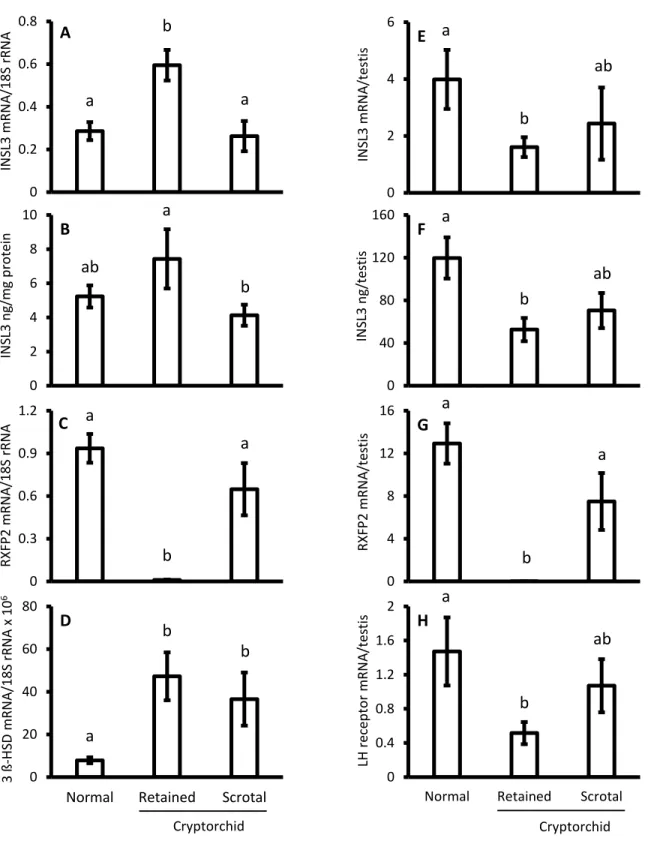
The INSL3 mRNA concentrations were significantly higher (P<0.01) in the
307
retained testes of the cryptorchid dogs compared to the normal testes of the normal dogs
308
and the scrotal testes of the unilateral cryptorchid dogs (Fig. 2A). A very similar INSL3
309
mRNA concentration was observed between the normal testes of normal dogs and the
310
scrotal testes of unilateral cryptorchid dogs (Fig. 2A). The total amount of INSL3
311
mRNA per retained testis was significantly lower (P<0.01) than that per normal testis
312
and did not differ significantly from that per cryptorchid scrotal testis (Fig. 2E). The
313
INSL3 peptide concentrations in the retained testes were significantly higher (P<0.05)
314
than those in the cryptorchid scrotal testes and tended to be higher (P=0.08) than those
315
in the normal testes (Fig. 2B). The INSL3 peptide concentrations for the scrotal testes of
316
the unilateral cryptorchid dogs did not differ from the normal testes of the normal dogs
317
(Fig. 2B). The total amount of INSL3 peptide per retained testis was significantly lower
318
(P<0.01) than that per normal testis, but did not differ significantly from that per
319
cryptorchid scrotal testis (Fig. 2F). The total amount of INSL3 peptide per cryptorchid
320
scrotal testis tended to be lower (P=0.06) than that per normal testis (Fig. 2F).
18
The RXFP2 mRNA concentrations were almost negligible in the retained testes,
322
and were much lower (P<0.001) than those in the normal testes (Fig. 2C). A similar
323
concentration of RXFP2 mRNA was observed between the normal testes of the normal
324
dogs and the scrotal testes of the unilateral cryptorchid dogs (Fig. 2C). The total amount
325
of RXFP2 mRNA per retained testis was almost nil and much lower than that per
326
normal testis (P<0.001) and cryptorchid scrotal testis (P<0.05; Fig. 2G). The total
327
amount of RXFP2 mRNA per cryptorchid scrotal testis tended to be lower (P=0.08)
328
than that per normal testis (Fig. 2G).
329
The LH receptor mRNA concentrations did not differ between the normal and
330
cryptorchid dogs (data not shown). Total amount of LH receptor mRNA per retained
331
testis was lower (P<0.01) than that per normal testis, and tended to be lower (P=0.14)
332
than that per cryptorchid scrotal testis (Fig. 2H). Significantly higher (P<0.01)
333
concentrations of 3β-HSD mRNA were observed in the retained and scrotal testes of the
334
cryptorchid dogs compared to the normal testes of the normal dogs (Fig. 2D). Total
335
amount of 3β-HSD mRNA per retained testis did not differ significantly among normal,
336
retained and cryptorchid scrotal testes (data not shown).
337
We performed immunohistochemistry to examine the specific cell type(s) that
338
shows INSL3 peptide expression in various age groups of normal testes of normal dogs
19
and retained testes of cryptorchid dogs. Only Leydig cells of both the normal (pubertal,
340
post-pubertal and middle age) and retained testes were immune-reactive to INSL3
341
antibody (shown in supplemental Fig. 1). The size of the seminiferous tubules per
342
testicular area seemed to increase from pubertal to post-pubertal and middle age.
343
The intensity of staining for INSL3 was clearly stronger in the Leydig cells of the
344
retained testes compared to those of the normal testes in all age categories. No other
345
testicular cell showed any immune reaction for INSL3 antibody (shown in supplemental
346
Fig. 1). When the primary antibody was omitted, no immunostaining was observed
347
(shown in supplemental Fig. 1). The Hematoxylin staining revealed the presence of
348
sperm inside seminiferous tubules in the testes of the normal dogs (pubertal,
post-349
pubertal and middle age) but the absence of sperm in the retained testes of the
350
cryptorchid dogs (data not shown).
351 352
4. Discussion
353
It was reported that in rodents, INSL3 has pivotal roles in testicular descent in the
354
fetal period [5, 6]. A role of INSL3 in the reproductive organs of domestic animals after
355
puberty has rarely been reported. The changes of testicular INSL3 and its receptor,
356
RXFP2, in pubertal and post-pubertal normal male animals have not yet been
20
elucidated, and a quantitative comparison of the testicular INSL3-receptor system
358
between cryptorchid and normal animals has not been reported. In this study, we
359
examined the gene expressions of INSL3 and RXFP2 in testes during puberty,
post-360
puberty and middle age in normal dogs to elucidate the changing pattern of these genes’
361
expression with age and sexual maturity. We also compared the INSL3 and RXFP2 gene
362
expressions in retained and scrotal testes of cryptorchid dogs with those of normal testes
363
of normal dogs. This is apparently the first study regarding quantitative changes of
364
testicular INSL3 and RXFP2 gene expression with age and sexual maturity and the
365
comparison of these expressions between normal and cryptorchid dogs.
366
The present results revealed that total amount of INSL3 mRNA per testis
367
decreased by aging in normal dogs despite increase of testicular weight, although the
368
amount of INSL3 peptide per testis did not change significantly during the same ages.
369
These results may indicate that the transcriptional activity of the gene encoding INSL3
370
in canine testes is reduced by the aging, but such a change is not reflected in the peptide
371
content. It was suggested that INSL3 concentrations in peripheral blood are higher in
372
the pubertal age and decline in the post-pubertal age in male dogs [15]. The change of
373
INSL3 mRNA amount per testis from puberty to middle age observed in the present
374
study, but not of the peptide, is likely to correspond to those of INSL3 concentrations in
21
blood. The reasons for the inconsistency of changes between testicular INSL3 mRNA
376
and the peptide amount around canine puberty observed in our study are unknown. In
377
male rats, the INSL3 concentrations in plasma transiently increased during puberty and
378
decreased after puberty [13]. Testicular INSL3 mRNA concentrations in rats of
379
advanced age (22–24 mo) were reduced compared to post-pubertal age (3 mo) [38]. In
380
the present study, we did not examine histological changes of testicular cellular
381
components including Leydig cells and various stages of germ cells during aging in the
382
same samples which were measured for mRNAs and INSL3 peptide. Clearly, further
383
studies are required to elucidate changes of INSL3 expression level per Leydig cell
384
basis during puberty and aging in dogs.
385
We found higher mRNA and slightly increased peptide concentrations of INSL3
386
in the retained testes of the cryptorchid dogs compared to the normal testes of the
387
normal dogs in the present study. Our immunohistochemistry data also showed that the
388
areas occupied by INSL3-producing Leydig cells per a certain area of testicular tissue
389
seem larger in the retained testes than in the normal testes, but we did not perform
390
quantitative analyses of INSL3-producing Leydig cells in normal and retained testes in
391
this study. However, total amounts of INSL3 mRNA and peptide per testis were reduced
392
in the retained testes relative to normal testes due to much smaller size of the former,
22
suggesting that the canine retained testis may produce lower INSL3 as a whole testis. It
394
has been suggested that INSL3 secretion in bilateral cryptorchid dogs is reduced
395
compared to the normal and unilateral cryptorchid dogs [15]. The current study also
396
shows that concentrations and total amount of INSL3 mRNA and peptide are similar
397
between the scrotal testes of unilateral cryptorchid dogs and the normal testes of normal
398
dogs. These results may be accorded with the previous findings that plasma INSL3
399
concentrations are similar between normal and unilateral cryptorchid dogs [15].
400
The present study provides findings that the gene expression of RXFP2 is almost
401
disappeared in canine retained testes at both of per unit-weight basis and per
whole-402
testis basis, in marked contrast to the higher expression of the receptor in normal testes.
403
A previous histological examination also showed a lack of RXFP2 immunoreactivity in
404
the genital tracts of cryptorchid canine testes [21]. Thus, it is likely in cryptorchid dogs
405
that the substantial amount of INSL3 secreted in a retained testis cannot transduce its
406
signal to cells within the testis, although we did not measure protein levels of RXFP2
407
receptor.
408
We speculate that the drastic reduction of RXFP2 mRNA in the retained testes
409
may be caused mainly by the absence of advanced stages of germ cells that express
410
RXFP2, due to impaired spermatogenesis [7, 19, 20]. It is also plausible that the
23
regulation of RXFP2 in Leydig cells by an autocrine mechanism [39, 40] with high or
412
substantial concentrations of INSL3 could partly contribute to the loss of RXFP2 gene
413
expression since relatively plenty of Leydig cells exist in the retained testes. It remains
414
to be determined in future studies whether the down-regulation of RXFP2 occurs in
415
Leydig cells in canine retained testes. We observed that the scrotal testes of unilateral
416
cryptorchid dogs exhibit RXFP2 expression similar to that of the normal testes of
417
normal dogs, implying that the lack of the receptor gene expression in the retained testes
418
probably occurs as a consequence of — not as a cause of — the retention of the testes.
419
In addition to INSL3 and RXFP2, we analyzed the gene expression of LH
420
receptor and 3β-HSD, which are also known as markers of Leydig cells [3, 27–29],
421
during the course of puberty in the testes of the normal and cryptorchid dogs. Our
422
findings revealed that mRNAs for both LH receptor and 3β-HSD showed differential
423
dynamics compared with INSL3 mRNA during puberty and in the testes of the
424
cryptorchid dogs. We speculate that the regulatory mechanisms for the gene expressions
425
of these three markers for Leydig cells differ. It is not clear why the concentrations of
426
3β-HSD mRNA were increased not only in the retained but also in the scrotal testes of
427
unilateral cryptorchid dogs in the present study. There could be a mechanism in
428
unilateral cryptorchid dogs in which a retained testis may affect the function of the other
24
scrotal testis through substances secreted from the retained testis [34]. The region of
430
canine LH receptor mRNA selected for the real-time PCR in this study is known to
431
encode the receptor protein, but the transcript may slightly include splicing variants
432
which encode non-functional LH receptors. Thus it should be noted that not all of the
433
mRNA would be expressed as the functional LH receptor.
434
In conclusion, higher INSL3 mRNA per unit-weight basis and clear staining of
435
Leydig cells for INSL3 peptide in the retained testes of cryptorchid dogs indicate the
436
substantial expression of INSL3 in Leydig cells of the retained testes. However, smaller
437
amount of INSL3 is likely to be produced per a whole retained testis due to its
438
diminutive size. Also the present study reveals that RXFP2 gene expression is lost in the
439
retained testes, but occurs normally in cryptorchid scrotal testes.
440 441
Acknowledgements
442
We thank Dr. Y. Ishizuka, Dr. M. Tsuji and their colleagues of Ishizuka Animal Hospital
443
for their generous provision of canine testicular samples. This study was partly
444
supported by Grants-in-Aid for Scientific Research, Scientific Research (C), of the
445
Japanese Society for the Promotion of Science (Grant #25450448).
446 447
25
References
448
[1] Balvers M, Spiess AN, Domagalski R, Hunt N, Kilic E, Mukhopadhyay AK, et al.
449
Relaxin-like factor expression as a marker of differentiation in the mouse testis and
450
ovary. Endocrinology 1998;139:2960–70.
451
[2] Ivell R, Balvers M, Domagalski R, Ungefroren H, Hunt N, Schulze W. Relaxin-like
452
factor: a highly specific and constitutive new marker for Leydig cells in the human
453
testis. Mol Hum Reprod 1997;3:459–66.
454
[3] Klonisch T, Kauffold J, Steger K, Bergmann M, Leiser R, Fischer B, et al. Canine
455
relaxin-like factor: unique molecular structure and differential expression within
456
reproductive tissues of the dog. Biol Reprod 2001;64:442–50.
457
[4] Sadeghian H, Anand-Ivell R, Balvers M, Relan V, Ivell R. Constitutive regulation of
458
the Insl3 gene in rat Leydig cells. Mol Cell Endocrinol 2005;241:10–20.
459
[5] Nef S, Parada LF. Cryptorchidism in mice mutant for Insl3. Nat Genet 1999;22:295–
460
9.
461
[6] Zimmermann S, Steding G, Emmen JM, Brinkmann AO, Nayernia K, Holstein AF,
462
et al. Targeted disruption of the Insl3 gene causes bilateral cryptorchidism. Mol
463
Endocrinol 1999;13:681–91.
464
[7] Anand-Ivell RJK, Relan V, Balvers M, Coiffec-Dorval I, Fritsch M, Bathgate RAD,
26
et al. Expression of the Insulin-like peptide 3 (INSL3) hormone-receptor (LGR8)
466
system in the testis. Biol Reprod 2006;74:945–53.
467
[8] Kawamura K, Kumagai J, Sudo S, Chun SY, Pisarska M, Morita H, et al. Paracrine
468
regulation of mammalian oocyte maturation and male germ cell survival. Proc Natl
469
Acad Sci U S A 2004;101:7323–8.
470
[9] Büllesbach EE, Rhodes R, Rembiesa B, Schwabe C. The relaxin-like factor is a
471
hormone. Endocrine 1999;10:167–9.
472
[10] Bay K, Hartung S, Ivell R, Schumacher M, Jürgensen D, Jorgensen N, et al.
473
Insulin-like factor 3 serum levels in 135 normal men and 85 men with testicular
474
disorders: relationship to the luteinizing hormone-testosterone axis. J Clin
475
Endocrinol Metab 2005;90:3410–8.
476
[11] Anand-Ivell R, Wohlgemuth J, Haren MT, Hope PJ, Hatzinikolas G, Wittert G, et
477
al. Peripheral INSL3 concentrations decline with age in a large population of
478
Australian men. Int J Androl 2006;29:618–26.
479
[12] Boockfor FR, Fullbright G, Büllesbach EE, Schwabe C. Relaxin-like factor (RLF)
480
serum concentrations and gubernaculum RLF receptor display in relation to pre-
481
and neonatal development of rats. Reproduction. 2001;122:899–906.
482
[13] Anand-Ivell R, Heng K, Hafen B, Setchell B, Ivell R. Dynamics of INSL3 peptide
27
expression in the rodent testis. Biol Reprod 2009;81:480–7.
484
[14] Kawate N, Ohnari A, Pathirana IN, Sakase M, Büllesbach EE, Takahashi M, et al.
485
Changes of plasma concentrations of insulin-like peptide 3 and testosterone from
486
birth to pubertal age in beef bulls. Theriogenology 2011;76:1632–8.
487
[15] Pathirana IN, Yamasaki H, Kawate N, Tsuji M, Büllesbach EE, Takahashi M, et al.
488
Plasma insulin-like peptide 3 and testosterone concentrations in male dogs:
489
changes with age and effects of cryptorchidism. Theriogenology 2012;77:550–7.
490
[16] Kumagai J, Hsu SY, Matsumi H, Roe JS, Fu P, Wade JD, et al. INSL3/Leydig
491
Insulin-like peptide activates the LGR8 receptor important in testis descent. J Biol
492
Chem 2002;277:31283–6.
493
[17] Gorlov IP, Kamat A, Bogatcheva NV, Jones E, Lamb DJ, Truong A, et al.
494
Mutations of the GREAT gene cause cryptorchidism. Hum Mol Genet
495
2002;11:2309–18.
496
[18] Overbeek PA, Gorlov IP, Sutherland RW, Houston JB, Harrison WR,
Boettger-497
Tong HL, et al. A transgenic insertion causing cryptorchidism in mice. Genesis
498
2001;30:26–35.
499
[19] Filonzi M, Cardoso LC, Pimenta MT, Queiroz DB, Avellar MC, Porto CS, et al.
500
Relaxin family peptide receptors Rxfp1 and Rxfp2: mapping of the mRNA and
28
protein distribution in the reproductive tract of the male rat. Reprod Biol
502
Endocrinol 2007;5:29.
503
[20] Feng S, Bogatcheva NV, Truong A, Korchin B, Bishop CE, Klonisch T, et al.
504
Developmental expression and gene regulation of insulin-like 3 receptor RXFP2 in
505
mouse male reproductive organs. Biol Reprod 2007;77:671–80.
506
[21] Arrighi S, Bosi G, Groppetti D, Aralla M, Cremonesi F. An insight into testis and
507
gubernaculum dynamics of INSL3-RXFP2 signalling during testicular descent in
508
the dog. Reprod Fertil Dev 2010;22:751–60.
509
[22] Bay K, Main KM, Toppari J, Skakkebæk NE. Testicular descent: INSL3,
510
testosterone, genes and the intrauterine milieu. Nat Rev Urol 2011;8:187–96.
511
[23] Almeida J, Conley AJ, Ball BA. Expression of anti-Müllerian hormone, CDKN1B,
512
connexin 43, androgen receptor and steroidogenic enzymes in the equine
513
cryptorchid testis. Equine Vet J 2013;45:538–45.
514
[24] Kawakami E, Tsutsui T, Yamada Y, Yamauchi M. Cryptorchidism in the dog:
515
occurrence of cryptorchidism and semen quality in the cryptorchid dog. Jpn J Vet
516
Sci 1984;46:303–8.
517
[25] Yates D, Hayes G, Heffernan M, Beynon R. Incidence of cryptorchidism in dogs
518
and cats. Vet Rec 2003;152:502–4.
29
[26] Hayes HM Jr, Wilson GP, Pendergrass TW, Cox VS. Canine cryptorchism and
520
subsequent testicular neoplasia: case-control study with epidemiologic update.
521
Teratology 1985;32:51–6.
522
[27] Hejmej A, Bilińska B. The effects of cryptorchidism on the regulation of
523
steroidogenesis and gap junctional communication in equine testes. Endokrynol
524
Pol 2008;59:112–8.
525
[28] Teerds KJ, de Boer-Brouwer M, Dorrington JH, Balvers M, Ivell R. Identification
526
of markers for precursor and Leydig cell differentiation in the adult rat testis
527
following ethane dimethyl sulphonate administration. Biol Reprod 1999;60:1437–
528
45.
529
[29] Peters MA, Teerds KJ, van der Gaag I, de Rooij DG, van Sluijs FJ. Use of
530
antibodies against LH receptor, 3beta-hydroxysteroid dehydrogenase and vimentin
531
to characterize different types of testicular tumour in dogs. Reproduction
532
2001;121:287–96.
533
[30] Almeida J, Conley AJ, Mathewson L, Ball BA. Expression of steroidogenic
534
enzymes during equine testicular development. Reproduction 2011;141:841–8.
535
[31] Payne AH, Hales DB. Overview of steroidogenic enzymes in the pathway from
536
cholesterol to active steroid hormones. Endocr Rev 2004;25:947–70.
30
[32] Bilińska B, Kotula-Balak M, Gancarczyk M, Sadowska J, Tabarowski Z, Wojtusiak
538
A. Androgen aromatization in cryptorchid mouse testis. Acta Histochem
539
2003;105:57–65.
540
[33] Illera JC, Silvan G, Munro CJ, Lorenzo PL, Illera MJ, Liu IK, et al. Amplified
541
androstenedione enzymeimmunoassay for the diagnosis of cryptorchidism in the
542
male horse: comparison with testosterone and estrone sulphate methods. J Steroid
543
Biochem Mol Biol 2003;84:377–82.
544
[34] Kawakami E, Hori T, Tsutsui T. Function of contralateral testis after artificial
545
unilateral cryptorchidism in dogs. J Vet Med Sci 1999;61:1107–11.
546
[35] Johnston SD, Root Kustritz MV, Olson PNS. Disorders of the canine testes and
547
epididymis. In: Johnston SD, Root Kustritz MV, Olson PNS, editors. Canine and
548
feline Theriogenology, Philadelphia: W B Saunders; 2001, p. 312–32.
549
[36] Pathirana IN, Tanaka K, Kawate N, Tsuji M, Kida K, Hatoya S, et al. Analysis of
550
single nucleotide polymorphisms in the 3` region of estrogen receptor 1 gene in
551
normal and cryptorchid Miniature Dachshunds and Chihuahuas. J Reprod Dev
552
2010;56:405–10.
553
[37] Hatoya S, Torii R, Kumagai D, Sugiura K, Kawate N, Tamada H, et al. Expression
554
of estrogen receptor alpha and beta genes in the mediobasal hypothalamus,
31
pituitary and ovary during the canine estrous cycle. Neurosci Lett 2003;347:131–5.
556
[38] Paust HJ, Wessels J, Ivell R, Mukhopadhyay AK. The expression of the
557
RLF/INSL3 gene is reduced in Leydig cells of the aging rat testis. Exp Gerontol
558
2002;37:1461–7.
559
[39] Pathirana IN, Kawate N, Büllesbach EE, Takahashi M, Hatoya S, Inaba T, Tamada
560
H.Insulin-like peptide 3 stimulates testosterone secretion in mouse Leydig cells
561
via cAMP pathway. Regul Pept 2012;178:102–6.
562
[40] Ivell R, Heng K, Anand-Ivell R. Insulin-like peptide 3 and the HPG axis in the
563
male. Front Endocrinol 2014;5:1–7.
564 565
32
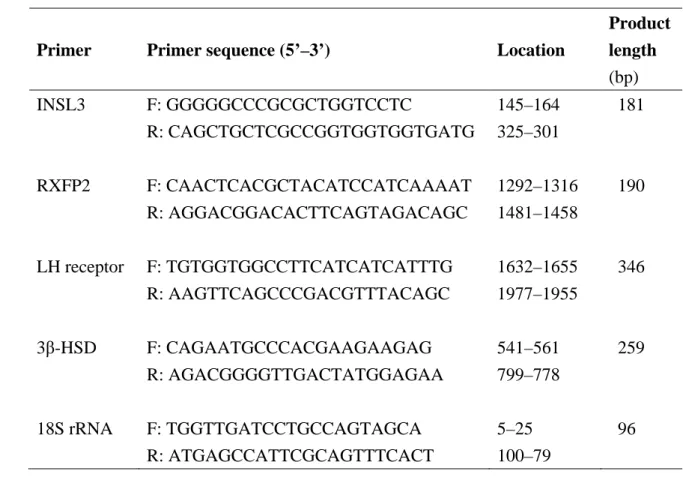
Table 1. Oligonucleotide sequences of the primers used for real-time PCR, their
566
location and the product sizes expected in canines
567
Primer Primer sequence (5’–3’) Location
Product length (bp) INSL3 F: GGGGGCCCGCGCTGGTCCTC 145–164 181 R: CAGCTGCTCGCCGGTGGTGGTGATG 325–301 RXFP2 F: CAACTCACGCTACATCCATCAAAAT 1292–1316 190 R: AGGACGGACACTTCAGTAGACAGC 1481–1458 LH receptor F: TGTGGTGGCCTTCATCATCATTTG 1632–1655 346 R: AAGTTCAGCCCGACGTTTACAGC 1977–1955 3β-HSD F: CAGAATGCCCACGAAGAAGAG 541–561 259 R: AGACGGGGTTGACTATGGAGAA 799–778 18S rRNA F: TGGTTGATCCTGCCAGTAGCA 5–25 96 R: ATGAGCCATTCGCAGTTTCACT 100–79 568
33
Figure legends
569 570
Fig. 1. Changes in testicular concentrations of INSL3 mRNA (A), INSL3 peptide (B),
571
RXFP2 mRNA (C), LH receptor mRNA (D), and total amount per testis of INSL3
572
mRNA (E) and INSL3 peptide (F) in various age groups of normal male dogs. Results
573
are shown for pubertal (6–12 mo, n=19), post-pubertal (1–5 y, n=17) and middle age (5–
574
10 y, n=10). Data are mean ± SEM. a–cValues without a common superscript differed
575
significantly for A and E (P<0.05), and for C and D (P<0.01).
576 577
Fig. 2. Testicular concentrations of INSL3 mRNA (A), INSL3 peptide (B), RXFP2
578
mRNA (C), 3β-HSD mRNA (D), and total amount per testis of INSL3 mRNA (E),
579
INSL3 peptide (F), Rxfp2 mRNA (G) and LH receptor mRNA (H) in normal (n=36),
580
retained (n=19) and scrotal testes (n=11). Data are mean ± SEM. a,bValues without a
581
common superscript differed significantly for B (P<0.05), for A, D, E, F and H (P<0.01)
582
and for C and G (P<0.001).
583 584
Fig. 1 0 0.1 0.2 0.3 0.4 0.5 IN SL3 mRNA/18S rRN A A a b c 0 2 4 6 8 IN SL3 n g/m g p ro tein B 0 2 4 6 8 IN SL3 mRNA/t es tis E a ab b 0 40 80 120 160 200
Pubertal Post-pubertal Middle
IN SL3 n g/te stis F 0 0.4 0.8 1.2 1.6
Pubertal Post-pubertal Middle
RXFP2 m RN A/18 S rRN A C a b ab 0 4 8 12 16 20 LH r ece p to r m RN A/18 S rRN A x 10 6 D a ab b
Fig. 2 Cryptorchid 0 0.2 0.4 0.6 0.8 IN SL3 mRNA/18S rRN A A a b a 0 2 4 6 8 10 IN SL3 n g/m g p ro tein B ab a b 0 0.3 0.6 0.9 1.2 RXFP2 m RN A/18 S rRN A C a b a 0 20 40 60 80
Normal Retained Scrotal
3 ß -H SD m RN A/18S rRNA x 10 6 D a b b 0 2 4 6 IN SL3 mRNA/t es tis E a b ab 0 40 80 120 160 IN SL3 n g/te stis F a b ab 0 4 8 12 16 RXFP2 m RN A/tes tis G a b a 0 0.4 0.8 1.2 1.6 2
Normal Retained Scrotal
LH recep to r mRN A/ tes tis H a b ab Cryptorchid
Supplemental data
Fig. 1. Representative photomicrographs of the immunohistochemical staining of
INSL3 peptide (brown staining) in canine normal (A, pubertal; C, post-pubertal; E, middle age) and retained (G) testes. In both the normal and retained testes, only testicular Leydig cells (black arrows) showed INSL3 immunolabeling. The staining intensity was stronger in the retained testes compared to the normal testes in all age groups. When the primary antibody was omitted, immunolabeling was not observed in the normal (B, pubertal; D, post-pubertal; F, middle age) or retained (H) testes.