IEICE TRANS. ELECTRON., VOL.E100–C, NO.11 NOVEMBER 2017
955
INVITED PAPER
Special Section on Electronic DisplaysColored Magnetic Janus Particles
Hiroshi YABU†a),Nonmember
SUMMARY The aim of this research is realizing a high resolution and a fast color switching of electronic papers. In this report, we realized basis of electric papers comprised on magnetic Janus particles was established.
Colored and magnetic Janus particles were successfully prepared, and mag- netic Janus particles were introduced into honeycomb matrices. Introduced magnetic Janus particles quickly respond to an external magnetic field.
key words: electronic papers, twisting ball, janus particles, magnetic, col- ored
1. Introduction
Electronic paper is one of the thin, flexible and low energy consumption display devises[1]. There are many types of electronic papers depending on their driving systems of im- age displays. For example, one of the main electronic paper devises are using electropholesis of dye pigments charged with negative or positive encapsulated in tens micrometer capsules[2]. Other system, using electrowetting[3], inter- ference controlled by liquid crystals technologies[4] etc.
have been reported. Twisting ball type electronic paper is one of the candidate for future electronic papers[5], in which the pixel color was changed by twisting charged Jauns particles having two aspect hemispheres along to the electric field. This devise requires simple architecture and low energy consumption, however, there are some draw- backs that low resolution due to the size of Jauns particle are in tens micrometer scale, slow response due to the viscosity of dispersion media and monotone color. To improve these problems, Janus particles having small and quick response have been required.
The aim of this research is realizing a high resolution and a fast color switching twisting ball type electronic pa- pers. Strong demand for high-resolution electric papers en- forces developing materials, which allow realizing smaller pixels. The micron-sized Janus particles having reactivity for external stimuli can be applied to the particle rotation type electric paper that is one of the display methods in the electronics.
Many efforts have been done to create small, colorful, and stimuli-responsive Janus particles by using conventional emulsion polymerization and microfluidic devices. For ex- ample, Okubo et. al. have reported that Janus type of phase
Manuscript received February 23, 2017.
Manuscript revised June 9, 2017.
†The author is with WPI-Advanced Institute for Materials Re- search (AIMR), Tohoku University, Sendai-shi, 980–8577 Japan.
a) E-mail: [email protected] DOI: 10.1587/transele.E100.C.955
separation was formed inside of the emulsion of two poly- mer solution and eventually solid Janus particles were ob- tained[6]. Furthermore, mixing two monomers by laminar flows in a microfluidic devise followed by photo polymer- ization results in formation of polymer Jauns particles[7].
We have also reported preparation of polymer Janus parti- cles from two immiscible polymers by precipitating them into poor solvents from their solution[8]. Polystyrene and polyisoprene was dissolved in tetrahydrofrane (THF) and then water was added as a poor solvent. After complete evaporation of THF, particles having PS or PI phase on each hemisphere were obtained. Furthermore, the Janus particles were functionalized polymer-stabilized iron oxide particles and titania particles[9],[10]. However, there is no colored Jauns particles can be applicable to creating colorful dis- plays.
In this report, we show colorful Janus particles re- sponding to magnetic fields prepared from polystyrene (PS), polyisoprene (PI), and polymer-stabilized pigments and in- organic nanoparticles. Magnetic Janus particles were also introduced into micron-sized honeycomb matrices and their rotational motions along to the magnetic fields were demon- strated.
2. Experimental
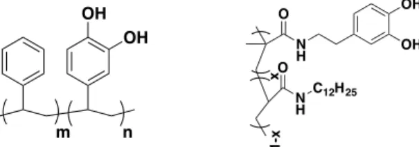
Pigments (Pigment Red 5403, Phthalocyanine Green 3603, Cyanine Blue 6005) were kindly provided from SANYO COLOR WORKS, LTD. Pigments were solubilized in tetrahydrofran (THF) with polymer 14. Fe2O3 nanoparti- cles were also solubilized in THF with polymer 2 (Fig. 1).
The stabilized pigments or Fe2O3 nanoparticles were cor- rected with ultracentrifugation and supernatant was re- moved. The stabilized pigments or Fe2O3 nanoparti- cles were re-dispersed in THF at their concentration of 1.0 mg/mL.
Polystyrene (PS), polyisoprene (PI), were dissolved in
Fig. 1 Chemical Structure of Polymer 1 (left) and 2 (right).
Copyright c2017 The Institute of Electronics, Information and Communication Engineers
956
IEICE TRANS. ELECTRON., VOL.E100–C, NO.11 NOVEMBER 2017
Fig. 2 Photographs of THF dispersion of non-stabilized pigments (left) and polymer-stabilized pigments (right).
THF to prepare 1.0 mg/mL solution and THF dispersion of pigments (red, green and blue) and Fe2O3 nanoparticles were mixed. Then, water was mixed into the THF solution and finally, a water dispersion of Janus particles was ob- tained after evaporation of THF. Three types of Janus parti- cles colored with different pigments were prepared.
Particle size was evaluated by using the dynamic light scattering (DLS) method. The shapes and interior structures of prepared particles were observed by using a transmission electron microscope (TEM).
Honeycomb-structured matrices were prepared from PS by using the breath figure method. Typically, a chlo- roform solution of PS and amphiphilic molecules was cast onto a glass substrate and applied humid air blow. Dur- ing the solvent evaporation, water droplets were condensed from humid air, and then, honeycomb-like porous films were obtained after complete evaporation of solvent. The surface structures of honeycomb matrices were observed by scanning electron microscopy (SEM). A sample cell was prepared by placed honeycomb film and a glass substrate with a small gap.
Magnetic Janus particles were prepared from PS, PI and Fe2O3nanoparticles by same procedures shown in the previously. A water dispersion of micron-sized Janus parti- cles was placed in front of a small inlet (shown in Fig. 2), and then, the ambient atmosphere was evacuated. After pressure recovery to normal atmosphere, the water disper- sion of Janus particles was introduced into the sample cell.
The interior structure of the cell was imaged by optical mi- croscopy.
3. Results and Discussion
Figures 2 (a) and 2 (b) show photographs of THF dispersion of pigments non-stabilized and polymer-stabilized pigments after placed 1 hour, respectively. As shown in Fig. 2 (a), non-stabilized pigments can not be dispersed in THF. On the other hand, polymer-stabilized pigments well dispersed in THF since pigment particles were successfully encapsu- lated in polymer 1.
Figure 3 (a) shows aqueous dispersion of Janus par-
Fig. 3 Photographs of aqueous dispersion of prepared particles incorpo- rated with pigments and iron oxide nanoparticles (a) and after accumulation of particles by using a neodymium magnet (b).
Fig. 4 TEM image of an obtained particle.
Fig. 5 SEM image of a honeycomb matrix (a), schematic illustration of introduction of particles in the honeycomb matrix (b) and optical micro- scope image of Janus particles introduced in honeycomb matrix (c), re- spectively.
ticles. All dispersions were colored with pigments, and opaque due to light scattering of micron-sized particle struc- tures. Figure 3 (b) shows accumulated Janus particles by us- ing a neodymium magnet. The dispersed particles were suc-
YABU: COLORED MAGNETIC JANUS PARTICLES
957
cessfully accumulated at the edge of the vessels by magnetic field. This result indicates prepared particles have magnetic responses.
Figure 4 shows typical TEM image of a prepared Janus particle. Janus type phase separation structure was clearly imaged and pigment particles were introduced into PS phase.
Figure 5 (a) shows typical SEM image of a honeycomb matrix. Uniform-sized porous structure was clearly im- aged. Figure 5 (b) shows preparation procedure of a sample cell. After introduction of aqueous dispersion, the magnetic Janus particles were introduced into each pore (Fig. 5 (c)) and Brownian motion was clearly observed. Furthermore, introduced Janus particles can be rotated very quickly (less than 500 msec.) along to the external magnetic field.
From these results, basis of electric papers comprised on magnetic Janus particles was established.
4. Conclusion
In this report, we realized basis of electric papers comprised on magnetic Janus particles was established. Colored and magnetic Janus particles were successfully prepared, and magnetic Janus particles were introduced into honeycomb matrices. Introduced magnetic Janus particles quickly re- spond to external magnetic field. These results indicate that new type of electric papers, which can be driven by mag- netic fields, can be realized based on these materials.
Acknowledgments
This work has been partially supported by Grant-in-Aid for Young Researchers (A) (No. 26708025) and Grant-in-Aid for Exploratory Research (No. 16K14071), MEXT and Pre- cursory Research for Embryonic Science (PRESTO), Japan Science and Technology Agency (JST).
References
[1] G. Schryen and J. Karla, “Electronic paper - a display technology for a wide range of applications,” Wirtschaftsinf, vol.44, no.6, pp.567–
574, 2002.
[2] T. Bert and H. De Smet, “Dielectrophoresis in electronic paper,” Dis- plays, vol.24, no.4-5, pp.223–230, 2003.
[3] D.Y. Kim and A.J. Steckl, “Electrowetting on paper for elec- tronic paper display,” ACS Appl. Mater. Interfaces, vol.2, no.11, pp.3318–3323, 2010.
[4] T.Z. Kosc, K.L. Marshall, A. Trajkovska-Petkoska, E. Kimball, and S.D. Jacobs, “Progress in the development of polymer cholesteric liquid crystal flakes for display applications,” Displays., vol.25, no.5, pp.171–176, 2004.
[5] R. Ishikawa, S. Maeda, and M. Omodani, “Estimation of Rotation Behavior of Balls for a Twisting Ball Display by Mobility Measure- ments,” Journal of Imaging Science and Technology, vol.50, no.2, pp.168–172, March 2006.
[6] H. Ahmad, N. Saito, Y. Kagawa, and M. Okubo, “Preparation of mi- crometer-sized, monodisperse ‘Janus’ composite polymer particles having temperature-sensitive polymer brushes at half of the surface by seeded atom transfer radical polymerization,” Langmuir., vol.24, no.3, pp.688–691, 2008.
[7] T. Nisisako, T. Torii, T. Takahashi, and Y. Takizawa, “Synthesis of Monodisperse Bicolored Janus Particles with Electrical Anisotropy Using a Microfluidic Co-Flow System,” Adv. Mater., vol.18, no.9, pp.1152–1156, May 2006.
[8] H. Yabu, K. Koike, K. Motoyoshi, T. Higuchi, and M. Shimomura,
“A Novel Route for Fabricating Metal-Polymer Composite Nanopar- ticles with Phase-Separated Structures,” Macromol. Rapid Comm., vol.31, no.14, pp.1267–1271, April 2010.
[9] H. Yabu, H. Ohshima, and Y. Saito, “Double-Phase-Functionalized Magnetic Janus Polymer Microparticles Containing TiO2and Fe2O3
Nanoparticles Encapsulated in Mussel-Inspired Amphiphilic Poly- mers,” ACS Appl. Mater. Interfaces, vol.6, no.20, pp.18122–18128, Oct. 2014.
[10] H. Yabu, M. Kanahara, M. Shimomura, T. Arita, K. Harano, E.
Nakamura, T. Higuchi, and H. Jinnai, “Polymer Janus Particles Con- taining Block-Copolymer Stabilized Magnetic Nanoparticles,” ACS Appl. Mater. Interfaces, vol.5, no.8, pp.3262–3266, April 2013.
Hirosh Yabu rceived the B.S. and M.S. de- grees in Biological Science from Hokkaido Uni- versity in 2000 and 2002, respectively. He re- ceived Ph.D. on Chemistry from Hokkaido Uni- versity in 2004 (early graduation). He served as an assistant professor in RIES, Hokkaido Uni- versity until 2007 and then moved to IMRAM, Tohoku University. He is now Junior Principal Investigator (Associate Prof.) of AIMR, Tohoku University.