1
Running Title: Monogenic mutations and type 1 diabetes
1 2
Correspondence: Maki Fukami, Department of Molecular Endocrinology, National
3
Research Institute for Child Health and Development, 2-10-1 Okura, Setagaya, Tokyo
4
157-8535, Japan. E-mail: [email protected].
5 6
2
Comprehensive screening for monogenic diabetes in 89 Japanese
7
children with insulin-requiring antibody-negative type 1 diabetes
8 9
Kikumi Ushijima
a, Maki Fukami
a, Tadayuki Ayabe
a,b, Satoshi Narumi
a,
10
Misako Okuno
a,c, Akie Nakamura
a, Toshikazu Takahashi
d, Kenji Ihara
e,
11
Kazuhiro Ohkubo
f, Emiko Tachikawa
g, Shoji Nakayama
h, Junichi Arai
i,
12
Nobuyuki Kikuchi
j, Toru Kikuchi
k, Tomoyuki Kawamura
l, Tatsuhiko Urakami
c,
13
Kenichiro Hata
m, Kazuhiko Nakabayashi
m, Yoichi Matsubara
n, Shin Amemiya
k,
14
Tsutomu Ogata
o, Ichiro Yokota
p, Shigetaka Sugihara
q, The Japanese Study Group of
15
Insulin Therapy for Childhood and Adolescent Diabetes
16 17
a
Department of Molecular Endocrinology, National Research Institute for Child Health
18
and Development, Tokyo, Japan
19
b
Department of Pediatrics, Sanaikai General Hospital, Misato, Japan
20
c
Department of Pediatrics and Child Health, Nihon University School of Medicine,
21
Tokyo, Japan
22
d
Takahashi clinic, Kobe, Japan
23
e
Department of Pediatrics, Oita University School of Medicine, Oita, Japan
24
f
Department of Pediatrics, Kyushu University School of Medicine, Fukuoka, Japan
25
g
Department of Pediatrics, Tokyo Women’s Medical University Hospital, Tokyo, Japan
26
h
Department of Pediatrics, Mominoki Hospital, Kochi, Japan
27
i
Department of Pediatrics, Hosogi Hospital, Kochi, Japan
28
j
Department of Pediatrics, Yokohama City Minato Red Cross Hospital, Yokohama,
29
Japan
30
k
Department of Pediatrics, Saitama Medical University Faculty of Medicine, Saitama,
31
Japan
32
l
Department of Pediatrics, Osaka City University School of Medicine, Osaka, Japan
33
3
m
Department of Maternal-Fetal Biology, National Research Institute for Child Health
34
and Development, Tokyo, Japan
35
n
National Research Institute for Child Health and Development, Tokyo, Japan
36
o
Department of Pediatrics, Hamamatsu University School of Medicine, Hamamatsu,
37
Japan
38
p
Department of Pediatrics, Division of Pediatric Endocrinology and Metabolism,
39
Shikoku Medical Center for Children and Adults, Kagawa, Japan
40
q
Department of Pediatrics, Tokyo Women’s Medical University Medical Center East,
41
Tokyo, Japan
42 43
Word count, 2,763; Tables 4; Figure 1
44 45
4
ABSTRACT
46
Background: Mutations in causative genes for neonatal diabetes or maturity-onset
47
diabetes of the young have been identified in multiple patients with autoantibody-
48
negative type 1 diabetes (T1D).
49
Objectives: We aimed to clarify the prevalence and phenotypic characteristics of
50
monogenic abnormalities among 89 children with autoantibody-negative insulin-
51
requiring T1D.
52
Methods: Mutations in 30 genes were screened using next-generation sequencing, and
53
copy-number alterations of four major causative genes were examined using multiplex-
54
ligation dependent probe amplification. We compared the clinical characteristics
55
between mutation carriers and non-carriers.
56
Results: We identified 11 probable pathogenic substitutions (six in INS, two in HNF1A,
57
two in HNF4A, and one in HNF1B) in 11 cases, but no copy-number abnormalities.
58
Only two mutation carriers had affected parents. De novo occurrence was confirmed for
59
three mutations. The non-carrier group, but not the carrier group, was enriched with
60
susceptible HLA alleles. Mutation carriers exhibited comparable phenotypes to those of
61
non-carriers, except for a relatively normal body mass index (BMI) at diagnosis.
62
Conclusions: This study demonstrated significant genetic overlap between
63
autoantibody-negative T1D and monogenic diabetes. Mutations in INS and HNF genes,
64
but not those in GCK and other monogenic diabetes genes, likely play critical roles in
65
children with insulin-requiring T1D. This study also suggests the relatively high de novo
66
rates of INS and HNF mutations, and the etiological link between autoimmune
67
abnormalities and T1D in the non-carrier group. Carriers of monogenic mutations show
68
nonspecific phenotypes among all T1D cases, although they are more likely to have a
69
normal BMI at diagnosis than non-carriers.
70 71
KEY WORDS: INS, HNF, mutation, next-generation sequencing
72
5 73
6
INTRODUCTION
74
Diabetes mellitus is classified into type 1 (T1D), type 2, other specific types, and
75
gestational diabetes (1, 2). T1D is subdivided into type 1A associated with
76
autoantibodies against glutamic acid decarboxylase 65 (GADA), tyrosine phosphatase-
77
like insulinoma antigen 2 (IA2A), insulin (IAA), islet cells (ICA), or β-cell-specific zinc
78
transporter 8 (ZnT8A), and type 1B that occurs independently of these autoantibodies
79
(1, 2). The group of “other specific types of diabetes” includes neonatal diabetes and
80
maturity-onset diabetes of the young (MODY), both of which arise from monogenic
81
mutations. Previous studies have identified 24 genes, including INS, KCNJ11, and
82
ABCC8, that jointly account for most cases of neonatal diabetes, and 13 genes,
83
including GCK, HNF1A, and HNF4A, whose mutations and deletions are present in a
84
substantial fraction of MODY cases (3, 4, 5, 6, 7). Seven genes are involved in both
85
neonatal diabetes and MODY.
86
Patients with monogenic diabetes are usually distinguishable from those with
87
other types of diabetes, because neonatal diabetes is characterized by an early disease
88
onset within the first few months of life, and MODY represents autosomal dominant
89
diabetes with partially preserved insulin secretion (1, 3). Nevertheless, mutations in the
90
monogenic diabetes genes have been identified in multiple patients with childhood-
91
onset autoantibody-negative T1D (8, 9, 10, 11, 12). We, the Japanese Study Group of
92
Insulin Therapy for Childhood and Adolescent Diabetes (JSGIT), previously performed
93
Sanger sequencing-based mutation analysis of INS and KCNJ11 on 34 patients with
94
early onset (≤ 5 years of age) autoantibody-negative diabetes, and identified INS and
95
KCNJ11 mutations in five cases and one case, respectively (13). Similarly, Sanger
96
sequence analysis of INS, KCNJ11, HNF1A, and HNF4A on 32 Japanese patients
97
detected INS and HNF1A mutations in three cases (14).
98
Most recently, Johansson et al. performed the first comprehensive mutation
99
screening of MODY genes on a large cohort of children with autoantibody-negative
100
7
diabetes. The authors studied 469 affected children by next-generation sequencing
101
(NGS) and identified probable damaging variants of GCK, INS, and HNF genes in 6.5%
102
of the cases. These findings provided evidence for a significant association between
103
T1D and monogenic mutations. However, because the subjects of Johansson et al.
104
included patients of various clinical severities, the frequency of monogenic mutations in
105
patients with insulin-requiring T1D remains to be determined. Furthermore, NGS may
106
miss pathogenic copy-number variations (CNVs), although deletions involving HNF1A,
107
HNF1B, and GCK have been identified in a few patients with MODY (15).
108
Here, we conducted a NGS-based mutation screening of 30 monogenic
109
diabetes genes in 89 Japanese children with insulin-requiring autoantibody-negative
110
T1D. We also analyzed CNVs involving GCK, HNF1A, HNF4A, and HNF1B using
111
multiplex-ligation dependent probe amplification (MLPA). The clinical characteristics
112
of mutation carriers were compared to those of non-carriers.
113 114
8
METHODS
115
Participants
116
This study was approved by the Institutional Review Board Committee at the National
117
Center for Child Health and Development and performed in accordance with the
118
Declaration of Helsinki. Written informed consent was obtained from the participants or
119
their parents. We enrolled 89 unrelated Japanese children clinically diagnosed with T1D
120
(Tables 1, 2, and S1). All participants required persistent insulin therapy and satisfied
121
the following criteria: (i) recruited by JSGIT between January 2008 and June 2013; (ii)
122
diagnosed with T1D based on the criteria of the World Health Organization published in
123
1998 (16); (iii) diagnosed between the age of 0.5 and 16.0 years; (iv) had detailed
124
medical records including data of height and weight at diagnosis; and (v) showed
125
negative results for all diabetes-associated autoantibodies examined. In all cases, GADA
126
had been tested at diagnosis, and other autoantibodies were also examined in several
127
cases (Tables 2 and S1). The participants included 47 children who were previously
128
subjected to Sanger sequencing-based mutation analysis (13, 14).
129 130
Molecular analyses
131
Genomic DNA was extracted from peripheral blood samples of the participants. To
132
detect nucleotide substitutions, we designed an NGS panel (HaloplexHS; Agilent
133
Technologies, Santa Clara, USA) targeting the coding- and non-coding exons and their
134
flanking regions of 30 genes known to cause monogenic diabetes (ABCC8, BLK,
135
CDKN1C, CEL, EIF2AK3, FOXP3, GATA4, GATA6, GCK, GLIS3, HNF1A, HNF4A,
136
HNF1B, IER3IP1, INS, KCNJ11, KLF11, MNX1, NEUROD1, NEUROG3, NKX2-2,
137
PAX4, PDX1, PTF1A, RFX6, SIRT1, SLC2A2, SLC19A2, SLC29A3, and ZFP57). The
138
total amplicon number was 11,033 and the target size was 205.8 kb with a theoretical
139
coverage of 98.5% for the targeted regions. According to the manufacture’s protocol,
140
individually indexed HaloplexHS libraries were prepared, and sequenced on a HiSeq
141
9
(Illumina, San Diego, USA). Base calling, read filtering, and demultiplexing were
142
performed with the standard Illumina processing pipeline. We used BWA 0.7.5 to map
143
reads against the human reference genome (build: hg19) with the default settings. Local
144
realignment, quality score recalibration, and variant calling were performed with
145
GATK3.6 using the default setting. We used ANNOVAR for annotation of the called
146
variants.
147
All non-synonymous substitutions and nucleotide changes at a splice site were
148
evaluated by database search [the Exome Aggregation Consortium Browser (ExAC,
149
http://exac.broadinstitute.org); the 1000 Genomes Database
150
(http://www.ncbi.nlm.nih.gov); the Human Genetic Variation Database (HGVD,
151
http://www.hgvd.genome.med.kyoto-u.ac.jp) and the Human Gene Mutation Database
152
(HGMD, http://www.hgmd.cf.ac.uk)]. The functional consequences of missense
153
substitutions were predicted by Sorting Intolerant From Tolerant (SIFT,
154
http://provean.jcvi.org/genome_submit_2.php); PolyPhen-2
155
(http://genetics.bwh.harvard.edu/pph2/); Mutation Taster (http://mutationtaster.org/); the
156
Combined Annotation Dependent Depletion (CADD, http://cadd.gs.washington.edu);
157
and the Mendelian Clinically Applicable Pathogenicity (M-CAP,
158
http://bejerano.stanford.edu/mcap/index.html)]. Substitutions previously identified in
159
patients with diabetes were classified as pathogenic. Nucleotide changes whose
160
frequency in the general population was ≥ 0.001 and were predicted as benign by more
161
than three of the five in silico analysis were excluded as probable benign variants. All
162
variants of interest were confirmed by PCR-based Sanger sequencing. When possible,
163
we analyzed parental DNA samples of mutation-positive participants.
164
To detect CNVs involving GCK, HNF1A, HNF4A, or HNF1B, we performed
165
MLPA analyses using the SALSA MLPA MODY mix-1 probemix (catalog number,
166
P241; MRC-Holland, Amsterdam, The Netherlands).
167 168
10
HLA alleles and clinical characteristics of mutation carriers
169
We genotyped HLA-DRB1 using the Luminex Multi-Analyte Profiling system with the
170
WAKFlow HLAtyping Kit (Wakunaga, Hiroshima, Japan). We determined known
171
diabetes susceptible alleles (*09:01, *04:05, *08:02) and protective alleles (*15:02,
172
*15:01, *08:03, *04:06) in the Japanese population (17). The difference in the
173
frequencies of susceptible and protective alleles among mutation carriers and non-
174
carriers were analyzed. We also compared the frequencies of susceptible and protective
175
alleles between mutation carriers and the Japanese general population [the Database of
176
the HLA laboratory (http://hla.or.jp/)]. In this analysis, the brother of patient 5 who had
177
diabetes and the same mutation as the proband was included in the group of mutation
178
carriers.
179
In addition, we compared phenotypic characteristics between mutation carriers
180
and non-carriers, and between INS mutation carriers and carriers of other mutations. The
181
body mass index (BMI, weight / height
2) SD was calculated based on the data of the
182
Japanese population (18). We examined fasting blood C-peptide values at diagnosis.
183
Cases with C-peptide values less than 0.6 ng/mL were considered as having endogenous
184
insulin deficiency (19). Diabetic ketoacidosis (DKA) was diagnosed according to the
185
Clinical Practice Consensus Guidelines of the International Society for Pediatric and
186
Adolescent Diabetes (ISPAD) (20). In addition, we examined whether mutation carriers
187
had extra-pancreatic complications.
188
The statistical significance in the differences between two participant groups
189
was analyzed using the Fisher’s exact test for categorical variables, and the Mann–
190
Whitney U test for continuous variables. A-two tailed p-value with an alpha level for
191
significance was determined as ≤ 0.05. All statistical analyses were performed using the
192
EZR system (version 1.32, http://www.jichi.ac.jp/saitama-
193
sct/SaitamaHP.files/statmedOSX.html), a graphical user interface for R (21).
194 195
11
RESULTS
196
Molecular analysis
197
Eleven probably pathogenic sequence variants were identified in 11 patients (patients 1–
198
11; Tables 2 and S2 and Figure 1). All variants were present in heterozygous states. The
199
variants consisted of six missense substitutions in INS (p.C31Y, p.V42A, p.G75C,
200
p.R89C, p.C96F, and p.C96R), two in HNF1A (p.R131Q and p.R203S), two in HNF4A
201
(p.Q142H and p.E256A), and one in HNF1B (p.L168P). Of these, p.G75C and p.C96F
202
in INS, p.Q142H and p.E256A in HNF4A, and p.L168P in HNF1B were hitherto
203
unreported, while the remaining six substitutions have previously been identified in
204
patients with diabetes (13, 22, 23, 24, 25). Notably, mutation p.G75C is located in the
205
C-peptide sequence. Nine of 11 variants were not found in the public databases, whereas
206
p.R131Q in HNF1A and p.E256A in HNF4A have been submitted to the ExAC Browser
207
as an extremely rare nucleotide change (allele frequency, 1/121,198 and 1/121,396).
208
Three substitutions in INS (p.C31Y, p.C96F, and p.C96R) affect cysteine residues
209
involved in disulfide bond formation, and p.V42A disrupts an amino acid next to the
210
cysteine residue at the 41st codon (26). In addition, p.R89C is known to cause
211
proinsulin misfolding (26), while the effect of p.G75C remains to be clarified. The
212
variants in HNF1A, HNF4A, and HNF1B are invariably located within functionally
213
important domains (27, 28, 29, 30) (Figure 1). Furthermore, p.Q142H in HNF4A affects
214
the last nucleotide of exon 4. Parental analysis revealed that three INS mutations
215
(p.V42A, p.G75C, and p.R89C) were de novo, while p.C31Y of patient 1 and p.C96F of
216
patient 5 were inherited from their parent with diabetes. Parental samples of other
217
patients were unavailable for genetic analysis.
218
MLPA detected no copy-number alterations of exons of GCK, HNF1A,
219
HNF4A, or HNF1B.
220 221
HLA alleles and clinical characteristics of mutation carriers
222
12
We compared the HLA-DRB1 allele frequencies and clinical information between
223
mutation carriers (n = 12) and non-carriers (n = 78) (Table 3). Susceptible HLA-DRB1
224
alleles were less frequent in the carrier group than the non-carrier group (4/24 vs.
225
96/156, p = 0.00004), while protective HLA-DRB1 alleles were more frequent in the
226
carrier group (6/24 vs. 8/156, p = 0.004). The frequency of susceptible HLA-DRB1
227
alleles in the carrier group was slightly lower than that in the Japanese general
228
population (10,235/31,973, p = 0.01), and the frequency of protective HLA-DRB1
229
alleles were comparable between these two groups (9585/31,973 in the general
230
population, p = 0.43).
231
Clinical features were almost comparable between the two groups, except for
232
the median BMI SD scores at diagnosis, which were normal in mutation carriers and
233
slightly reduced in non-carriers (mean SDS, 0.0 vs. -0.8, p = 0.02). In particular,
234
endogenous insulin deficiency indicated by low C-peptide levels were observed in both
235
carriers and non-carriers (1/5 vs. 25/51, p = 0.36). Patient 11 with p.L168P in HNF1B
236
had end-stage renal failure and was treated with peritoneal dialysis, while the remaining
237
11 carriers had no apparent extra-pancreatic complications.
238
We then compared the clinical characteristics between patients with INS
239
mutations (n = 7) and those with HNF mutations (n = 5) (Table 4). Age at diagnosis was
240
significantly lower in INS mutation carriers than that of HNF mutation carriers (2.3 vs.
241
10.2 years, p = 0.01). DKA was observed in two INS mutation carriers, but was not
242
described in the HNF mutation carriers. Other examined parameters were comparable
243
between the two groups.
244 245
13
DISCUSSION
246
NGS-based mutation screening identified probable pathogenic mutations in 11 of 89
247
patients with autoantibody-negative T1D. Identified mutations consisted of six
248
substitutions in INS, two in HNF1A, two in HNF4A, and one in HNF1B. These results
249
provide further evidence that monogenic mutations account for a small fraction of
250
children with autoantibody-negative T1D. Since MLPA excluded copy-number
251
variations of GCK, HNF1A, HNF4A, and HNF1B in our patients, chromosomal
252
deletions involving these genes appear to be uncommon in this condition. Notably, the
253
results of this study have both similarities and differences to those of the previous
254
mutation screening by Johansson et al (8). Both studies demonstrated significant roles
255
of INS and HNF genes, together with negligible roles of most other monogenic diabetes
256
genes, in the development of autoantibody-negative T1D. However, the frequency of
257
pathogenic INS mutations was much higher in our cohort than that in the cohort of
258
Johansson et al. (6/89 vs. 1/469). In contrast, none of our participants carried GCK
259
mutations which accounted for 6 of 469 patients studied by Johansson et al.
260
Heterozygous GCK mutations are known to represent one of the major causes of
261
MODY (3, 31, 32). These discrepancies between previous studies and ours likely reflect
262
the differences in the clinical severities of the participants. It is known that INS
263
mutations represent the second common cause of permanent neonatal diabetes (
3, 22) 264and often result in insulin-requiring diabetes in early childhood, while heterozygous
265
GCK mutations typically lead to relatively mild diabetes that does not require insulin
266
therapy. Since our study group consisted solely of children with insulin-requiring
267
diabetes, this selection criterion likely contributed to the accumulation of INS mutations
268
and the lack of GCK mutations.
269
Of the 11 mutation carriers, only two had affected parents. These data imply
270
that INS and HNF mutations can be associated with de novo occurrence or incomplete
271
penetrance. We confirmed that at least three of the 11 mutations, i.e., p.V42A, p.G75C
272
14
and p.R89C in INS, were de novo. Although a de novo substitution can be a functionally
273
benign variant (33, 34), pathogenicity of these three INS mutations was supported by
274
multiple in silico programs. In addition, mutations p.V42A and p.R89C have already
275
been reported as causing diabetes in childhood (22, 25, 35). Recently, Stanik et al.
276
revealed that de novo mutations in GCK, HNF1A, and HNF4A are more frequent in
277
MODY cases than previously assumed (36). Our data, in conjunction with those of
278
Stanik et al., suggest that the de novo occurrence of INS or HNF mutations is not rare.
279
Since previous sequence analyses of INS and HNF have focused primarily on patients
280
with a positive family history (15, 37), further studies are necessary to clarify the actual
281
frequency of these mutations among patients with diabetes.
282
The frequencies of susceptible and protective HLA-DRB1 alleles were
283
significantly different between the carrier and non-carrier groups. The frequencies of
284
these alleles in the carrier group were close to those seen in the Japanese general
285
population, whereas the non-carrier group was characterized by relatively high and low
286
frequencies of susceptibility and protective alleles, respectively. These results imply that
287
a substantial fraction of the non-carrier group can be ascribed to autoimmune
288
abnormalities, although we cannot exclude the possibility that some other monogenic
289
diabetes genes remain unidentified. This is consistent with the prior findings by Hameed
290
et al. that a substantial percentage of patients who had no autoantibodies at diagnosis
291
were found to be antibody-positive at retesting. Since previous studies have revealed the
292
ethnic specificity of protective and high risk HLA alleles for T1D (1, 2, 17), the
293
contribution of autoimmune abnormality to T1D in the non-carrier group may differ
294
among ethnic groups. Hameed et al. suggested that repeated measurement of blood C-
295
peptide values provides useful information about the clinical course of T1D; relatively
296
preserved C-peptide levels during the follow-up period are often seen in persistent
297
antibody negative cases. Thus, C-peptide values of our patients need to be carefully
298
monitored, although the values at diagnosis were highly variable and did not reflect the
299
15
presence or absence of monogenic mutations.
300
Clinical examinations revealed three notable findings. First, the clinical
301
features of mutation carriers were comparable to those of non-carriers, except for the
302
median BMI SD scores at diagnosis that were low in the non-carrier group and normal
303
in the carrier group. The normally preserved BMI at diagnosis in the carrier group may
304
reflect the slow progression of the disease, because mutations in HNF1A, HNF4A, and
305
HNF1B are known to cause a gradual impairment of insulin secretion (38, 39). Second,
306
patient 11 with a HNF1B mutation manifested renal failure. This is consistent with
307
previous observations that renal cysts and renal dysplasia are common features of
308
patients with HNF1B mutations (3). The presence of extra-pancreatic lesions appears to
309
be a good marker of monogenic diabetes among patients with autoantibody-negative
310
diabetes. Lastly, compared to carriers of HNF mutations, INS mutation carriers tended
311
to have early disease onset and more frequently experienced DKA. These data are
312
consistent with previous observations that INS mutations are associated with a both
313
severe neonatal insulin deficiency and MODY, while HNF mutations typically lead to
314
late-onset slowly progressive diabetes (33, 38, 39, 40). However, given the small
315
number of participants in this study, further studies are necessary to clarify the
316
frequency and phenotypic characteristics of each monogenic abnormality among
317
autoantibody-negative T1D cases.
318
In conclusion, this study provides further evidence for the significant genetic
319
overlap between autoantibody-negative T1D and monogenic diabetes. Mutations in INS
320
and HNF genes, but not those in GCK and other monogenic diabetes genes, likely play
321
critical roles in childhood-onset insulin-requiring T1D. This study also suggests the
322
relatively high de novo rates of INS and HNF mutations, as well as the etiological link
323
between autoimmune abnormalities and T1D in the non-carrier group. Carriers of
324
monogenic mutations show nonspecific phenotypes among all T1D cases, although they
325
are more likely to have a normal BMI at diagnosis than non-carriers.
326
16 327
17
ACKNOWLEDGEMENTS
328
This work was supported by grant–in–aid for Scientific Research from the Japan
329
Society for the Promotion of Science, by grants from the Manpei Suzuki Diabetes
330
Foundation, the Japan Diabetes Foundation, the Japan Agency for Medical Research
331
and Development, the National Center for Child Health and Development, and the
332
Takeda foundation.
333 334
18
REFERENCES
335
1. Craig ME, Jefferies C, Dabelea D, Balde N, Seth A, Donaghue KC. ISPAD Clinical
336
Practice Consensus Guidelines 2014. Definition, epidemiology, and classification
337
of diabetes in children and adolescents. Pediatr Diabetes. 2014; 15 Suppl 20:4–17.
338
2. Redondo MJ, Eisenbarth GS. Genetic control of autoimmunity in Type I diabetes
339
and associated disorders. Diabetologia. 2002; 45:605–622.
340
3. Rubio-Cabezas O, Hattersley AT, Njolstad PR et al. ISPAD Clinical Practice
341
Consensus Guidelines 2014. The diagnosis and management of monogenic diabetes
342
in children and adolescents. Pediatr Diabetes. 2014; 15 Suppl 20:47–64.
343
4. Slingerland AS. Monogenic diabetes in children and young adults: Challenges for
344
researcher, clinician and patient. Rev Endocr Metab Disord. 2006; 7:171–185.
345
5. Kerns SL, Guevara-Aguirre J, Andrew S et al. A novel variant in CDKN1C is
346
associated with intrauterine growth restriction, short stature, and early-adulthood-
347
onset diabetes. J Clin Endocrinol Metab. 2014; 99:E2117–2122.
348
6. Biason-Lauber A, Boni-Schnetzler M, Hubbard BP et al. Identification of a SIRT1
349
mutation in a family with type 1 diabetes. Cell Metab. 2013; 17:448–455.
350
7. Cliffe ST, Kramer JM, Hussain K et al. SLC29A3 gene is mutated in pigmented
351
hypertrichosis with insulin-dependent diabetes mellitus syndrome and interacts
352
with the insulin signaling pathway. Hum Mol Genet. 2009; 18:2257–2265.
353
8. Johansson BB, Irgens HU, Molnes J et al. Targeted next-generation sequencing
354
reveals MODY in up to 6.5% of antibody-negative diabetes cases listed in the
355
Norwegian Childhood Diabetes Registry. Diabetologia. 2016 Dec 02.
356
9. Brahm AJ, Wang G, Wang J et al. Genetic Confirmation Rate in Clinically
357
Suspected Maturity-Onset Diabetes of the Young. Can J Diabetes. 2016; 40:555–
358
560.
359
10. Shepherd M, Shields B, Hammersley S et al. Systematic Population Screening,
360
Using Biomarkers and Genetic Testing, Identifies 2.5% of the U.K. Pediatric
361
19
Diabetes Population With Monogenic Diabetes. Diabetes Care. 2016; 39:1879-
362
1888.
363
11. Rubio-Cabezas O, Edghill EL, Argente J, Hattersley AT. Testing for monogenic
364
diabetes among children and adolescents with antibody-negative clinically defined
365
Type 1 diabetes. Diabet Med. 2009; 26:1070–1074.
366
12. Hameed S, Ellard S, Woodhead HJ et al. Persistently autoantibody negative (PAN)
367
type 1 diabetes mellitus in children. Pediatr Diabetes. 2011; 12:142–149.
368
13. Moritani M, Yokota I, Tsubouchi K et al. Identification of INS and KCNJ11 gene
369
mutations in type 1B diabetes in Japanese children with onset of diabetes before 5
370
years of age. Pediatr Diabetes. 2013; 14:112–120.
371
14. Moritani M, Yokota I, Horikawa R et al. Identification of monogenic gene
372
mutations in Japanese subjects diagnosed with type 1B diabetes between >5 and
373
15.1 years of age. J Pediatr Endocrinol Metab. 2016; 29:1047–1054.
374
15. Yorifuji T, Fujimaru R, Hosokawa Y et al. Comprehensive molecular analysis of
375
Japanese patients with pediatric-onset MODY-type diabetes mellitus. Pediatr
376
Diabetes. 2012; 13:26–32.
377
16. Alberti KG, Zimmet PZ. Definition, diagnosis and classification of diabetes
378
mellitus and its complications. Part 1: diagnosis and classification of diabetes
379
mellitus provisional report of a WHO consultation. Diabet Med. 1998; 15:539–553.
380
17. Sugihara S, Ogata T, Kawamura T et al. HLA-class II and class I genotypes among
381
Japanese children with Type 1A diabetes and their families. Pediatr Diabetes. 2012;
382
13:33–44.
383
18. Kato N, Takimoto H, Sudo N. The Cubic Functions for Spline Smoothed L, S and
384
M Values for BMI Reference Data of Japanese Children. Clin Pediatr Endocrinol.
385
2011; 20:47–49.
386
19. Kawasaki E, Maruyama T, Imagawa A et al. Diagnostic criteria for acute-onset type
387
1 diabetes mellitus (2012): Report of the Committee of Japan Diabetes Society on
388
20
the Research of Fulminant and Acute-onset Type 1 Diabetes Mellitus. J Diabetes
389
Investig. 2014; 5:115–118.
390
20. Wolfsdorf JI, Allgrove J, Craig ME et al. ISPAD Clinical Practice Consensus
391
Guidelines 2014. Diabetic ketoacidosis and hyperglycemic hyperosmolar state.
392
Pediatr Diabetes. 2014; 15 Suppl 20:154–179.
393
21. Kanda Y. Investigation of the freely available easy-to-use software 'EZR' for
394
medical statistics. Bone Marrow Transplant. 2013; 48:452–458.
395
22. Edghill EL, Flanagan SE, Patch AM et al. Insulin mutation screening in 1,044
396
patients with diabetes: mutations in the INS gene are a common cause of neonatal
397
diabetes but a rare cause of diabetes diagnosed in childhood or adulthood. Diabetes.
398
2008; 57:1034–1042.
399
23. Yamagata K, Oda N, Kaisaki PJ et al. Mutations in the hepatocyte nuclear factor-
400
1alpha gene in maturity-onset diabetes of the young (MODY3). Nature. 1996;
401
384:455–458.
402
24. Colclough K, Bellanne-Chantelot C, Saint-Martin C, Flanagan SE, Ellard S.
403
Mutations in the genes encoding the transcription factors hepatocyte nuclear factor
404
1 alpha and 4 alpha in maturity-onset diabetes of the young and hyperinsulinemic
405
hypoglycemia. Hum Mutat. 2013; 34:669–685.
406
25. Piccini B. Artuso R. Lenzi L et al. Clinical and molecular characterization of a
407
novel INS mutation identified in patients with MODY phenotype. Eur J Med Genet.
408
2016; 59:590–595.
409
26. Liu M, Sun J, Cui J et al. INS-gene mutations: from genetics and beta cell biology
410
to clinical disease. Mol Aspects Med. 2015; 42:3–18.
411
27. Wu G, Bohn S, Ryffel GU. The HNF1beta transcription factor has several domains
412
involved in nephrogenesis and partially rescues Pax8/lim1-induced kidney
413
malformations. Eur J Biochem. 2004; 271:3715–3728.
414
28. Lu P, Rha GB, Chi YI. Structural basis of disease-causing mutations in hepatocyte
415
21
nuclear factor 1beta. Biochemistry. 2007; 46:12071–12080.
416
29. Lu P, Rha GB, Melikishvili M et al. Structural basis of natural promoter recognition
417
by a unique nuclear receptor, HNF4alpha. Diabetes gene product. J Biol Chem.
418
2008; 283:33685–33697.
419
30. Chandra V, Huang P, Potluri N, Wu D, Kim Y, Rastinejad F. Multidomain
420
integration in the structure of the HNF-4alpha nuclear receptor complex. Nature.
421
2013; 495:394–398.
422
31. Fajans SS, Bell GI, Polonsky KS. Molecular mechanisms and clinical
423
pathophysiology of maturity-onset diabetes of the young. N Engl J Med. 2001;
424
345:971–980.
425
32. Kawakita R, Hosokawa Y, Fujimaru R et al. Molecular and clinical characterization
426
of glucokinase maturity-onset diabetes of the young (GCK-MODY) in Japanese
427
patients. Diabet Med. 2014; 31:1357–1362.
428
33. Bonfanti R, Colombo C, Nocerino V et al. Insulin gene mutations as cause of
429
diabetes in children negative for five type 1 diabetes autoantibodies. Diabetes Care.
430
2009; 32:123–125.
431
34. Liu M, Lara-Lemus R, Shan SO et al. Impaired cleavage of preproinsulin signal
432
peptide linked to autosomal-dominant diabetes. Diabetes. 2012; 61:828–837.
433
35. Colombo C, Porzio O, Liu M et al. Seven mutations in the human insulin gene
434
linked to permanent neonatal/infancy-onset diabetes mellitus. J Clin Invest. 2008;
435
118:2148–2156.
436
36. Stanik J, Dusatkova P, Cinek O et al. De novo mutations of GCK, HNF1A and
437
HNF4A may be more frequent in MODY than previously assumed. Diabetologia.
438
2014; 57:480–484.
439
37. Irgens HU, Molnes J, Johansson BB et al. Prevalence of monogenic diabetes in the
440
population-based Norwegian Childhood Diabetes Registry. Diabetologia. 2013;
441
56:1512–1519.
442
22
38. Amed S, Oram R. Maturity-Onset Diabetes of the Young (MODY): Making the
443
Right Diagnosis to Optimize Treatment. Can J diabetes. 2016; 40:449–454.
444
39. Timsit J, Saint-Martin C, Dubois-Laforgue D, Bellanne-Chantelot C. Searching for
445
Maturity-Onset Diabetes of the Young (MODY): When and What for? Can J
446
diabetes. 2016; 40:455–461.
447
40. Molven A, Ringdal M, Nordbo AM et al. Mutations in the insulin gene can cause
448
MODY and autoantibody-negative type 1 diabetes. Diabetes. 2008; 57:1131–1135.
449 450
23
Table 1. Characteristics of participants.
451
Total number 89
Male : Female 34 : 55
Age at diagnosis (yr) 8.2 (3.6–11.0)
Diabetes duration (yr) 3.3 (1.7–7.2)
Parental history of diabetes 8 / 75
BMI SD score at diagnosis -1.0 ± 1.2
HbA1c at diagnosis (NGSP, %) 11.3 (9.5–13.4) HbA1c at diagnosis (IFCC, mmol/mol) 99.6 (80.3–122.9) C-peptide negative at diagnosis
#26 / 56
DKA at diagnosis* 24 / 69
Birth weight (g) 3,056 ± 404.0
Gestational age (weeks) 39.3 ± 1.5
Susceptible HLA-DRB1 alleles† 100 / 178 Protective HLA-DRB1 alleles† 13 / 178
BMI, Body Mass Index; DKA, diabetic ketoacidosis; IFCC, International Federation of
452
Clinical Chemistry; NGSP, National Glycohemoglobin Standardization Program. Data
453
are represented as median (interquartile range) or mean ± SD.
454
#
Patients with a fasting C-peptide <0.6 ng/ml were assessed as C-peptide negative (19)
455
* DKA was diagnosed according to the International Society for Pediatric and
456
Adolescent Diabetes (ISPAD) Clinical Practice Consensus Guidelines 2014 (20).
457
† Susceptible and protective HLA-DRB1 alleles are defined based on the report of
458
Sugihara et al (17).
459 460
24
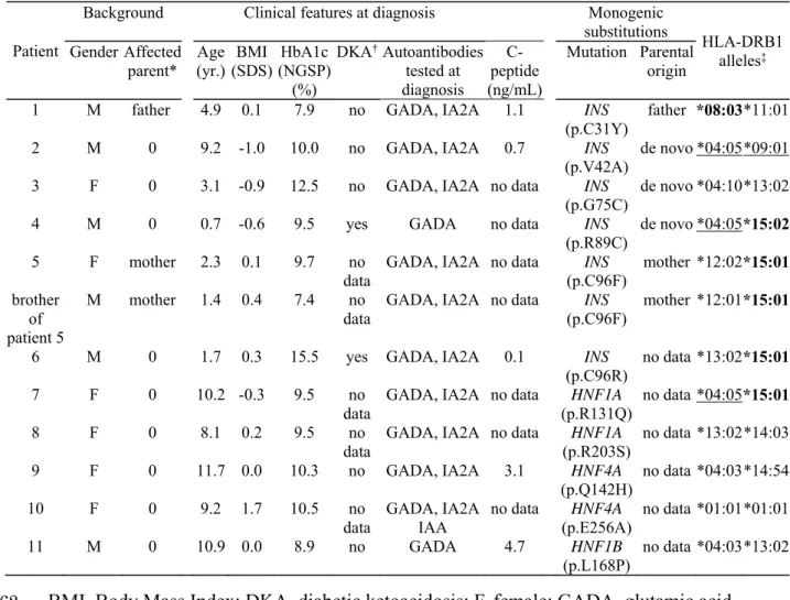
Table 2. Clinical and molecular findings of mutation carriers.
461
BMI, Body Mass Index; DKA, diabetic ketoacidosis; F, female; GADA, glutamic acid
462
decarboxylase 65 antibody; IA2A tyrosine phosphatase-like insulinoma antigen 2
463
antibody; IAA, insulin autoantibody; M, male; NGSP, National Glycohemoglobin
464
Standardization Program.
465
* The number of parents with history of diabetes.
466
† DKA was diagnosed according to the International Society for Pediatric and
467
Adolescent Diabetes (ISPAD) Clinical Practice Consensus Guidelines 2014 (20).
468
‡ Susceptible HLA-DRB1 alleles are underlined, and protective alleles are boldfaced.
469
These alleles were defined based on the report of Sugihara et al (17).
470
# Variants are described according to the HGMD reference sequences: HNF1A
471
NM_000545.6; HNF4A NM_175914.4; HNF1B NM_000458.3; INS NM_001185098.1.
472 Patient
Background Clinical features at diagnosis Monogenic
substitutions HLA-DRB1 alleles‡ Gender Affected
parent* Age (yr.) BMI
(SDS) HbA1c (NGSP)
(%)
DKA† Autoantibodies tested at diagnosis
C- peptide (ng/mL)
Mutation Parental origin 1 M father 4.9 0.1 7.9 no GADA, IA2A 1.1 INS
(p.C31Y) father *08:03 *11:01 2 M 0 9.2 -1.0 10.0 no GADA, IA2A 0.7 INS
(p.V42A) de novo *04:05 *09:01 3 F 0 3.1 -0.9 12.5 no GADA, IA2A no data INS
(p.G75C) de novo *04:10 *13:02 4 M 0 0.7 -0.6 9.5 yes GADA no data INS
(p.R89C)
de novo *04:05 *15:02 5 F mother 2.3 0.1 9.7 no
data GADA, IA2A no data INS
(p.C96F) mother *12:02 *15:01 brother
of patient 5
M mother 1.4 0.4 7.4 no data
GADA, IA2A no data INS (p.C96F)
mother *12:01 *15:01
6 M 0 1.7 0.3 15.5 yes GADA, IA2A 0.1 INS (p.C96R)
no data *13:02 *15:01 7 F 0 10.2 -0.3 9.5 no
data GADA, IA2A no data HNF1A
(p.R131Q) no data *04:05 *15:01 8 F 0 8.1 0.2 9.5 no
data GADA, IA2A no data HNF1A
(p.R203S) no data *13:02 *14:03 9 F 0 11.7 0.0 10.3 no GADA, IA2A 3.1 HNF4A
(p.Q142H) no data *04:03 *14:54 10 F 0 9.2 1.7 10.5 no
data GADA, IA2A
IAA no data HNF4A
(p.E256A) no data *01:01 *01:01
11 M 0 10.9 0.0 8.9 no GADA 4.7 HNF1B
(p.L168P) no data *04:03 *13:02
25 473
26
Table 3. Comparison between mutation carriers and non-carriers.
474
Mutation
carriers Non-carriers p-value (n = 12) (n = 78)
Male : Female 6 : 6 29 : 49 0.53
Age at diagnosis (yr) 6.5 (2.1–9.5) 8.3 (3.8–11.5) 0.12 Diabetes duration (yr) 2.9 (1.4–3.1) 3.7 (1.8–8.1) 0.14
Parental history of diabetes 3 / 12 6 / 64 0.15
BMI SD score at diagnosis 0 ± 0.7 -0.8 ± 1.2 0.02
HbA1c at diagnosis (NGSP, %) 9.6 (9.4–10.4) 11.7 (9.6–13.4) 0.08 C-peptide negative at diagnosis
#1 / 5 25 / 51 0.36
DKA at diagnosis
*2 / 7 22 / 62 1.00
Birth weight (g) 3,027 ± 404.5 3,058 ± 409.9 0.82 Gestational age (weeks) 38.8 ± 1.6 39.3 ± 1.5 0.33 Susceptible HLA-DRB1 alleles
†4 / 24 96 / 156 0.00004 Protective HLA-DRB1 alleles
†6 / 24 8 / 156 0.004 BMI, Body Mass Index; DKA, diabetic ketoacidosis; NGSP, National Glycohemoglobin
475
Standardization Program. Data are represented as median (interquartile range) or mean
476
± SD.
477
#
Patients with a fasting C-peptide <0.6 ng/ml were assessed as C-peptide negative (19).
478
*
DKA was diagnosed according to the International Society for Pediatric and
479
Adolescent Diabetes (ISPAD) Clinical Practice Consensus Guidelines 2014 (20).
480
†
Susceptible and protective HLA-DRB1 alleles are defined based on the report of
481
Sugihara et al (17).
482 483
27
Table 4. Comparison between INS mutation carriers and carriers of HNF1A, HNF4A,
484
and HNF1B mutations.
485
INS mutation carriers
HNF mutation
carriers p-value
(n = 7) (n = 5)
Male : Female 5 : 2 1 : 4 0.24
Age at diagnosis (yr) 2.3 (1.5–4.0) 10.2 (9.2–10.9) 0.01 Diabetes duration (yr) 3.0 (1.5–5.2) 2.9 (0.7–2.9) 0.34
Parental history of diabetes 3 / 7 0 / 5 0.21
DKA at diagnosis
*2 / 5 0 / 2 1.00
BMI SD score at diagnosis -0.2 ± 0.6 0.32 ± 0.8 0.20 HbA1c at diagnosis
(NGSP, %) 9.7 (8.7–11.3) 9.5 (9.5–10.3) 1.00
Birth weight (g) 3,045 ± 361.0 2,995 ± 531.7 0.86
BMI, Body Mass Index; DKA, diabetic ketoacidosis; NGSP, National Glycohemoglobin
486
Standardization Program. Data are represented as median (interquartile range) or mean
487
± SD.
488
*
DKA was diagnosed according to the International Society for Pediatric and
489
Adolescent Diabetes (ISPAD) Clinical Practice Consensus Guidelines 2014 (20).
490 491
28
FIGURE LEGENDS
492
Figure 1. Monogenic mutations identified in the present study. Chromatographs of
493
patients 1–11 are shown. Arrows indicate mutated nucleotides. The S symbols depict
494
disulfide bonds. DNA binding domains of HNF1A and HNF1B consist of the POU
495
specific domain (POU
S) and the POU homeodomain (POU
H).
496
1
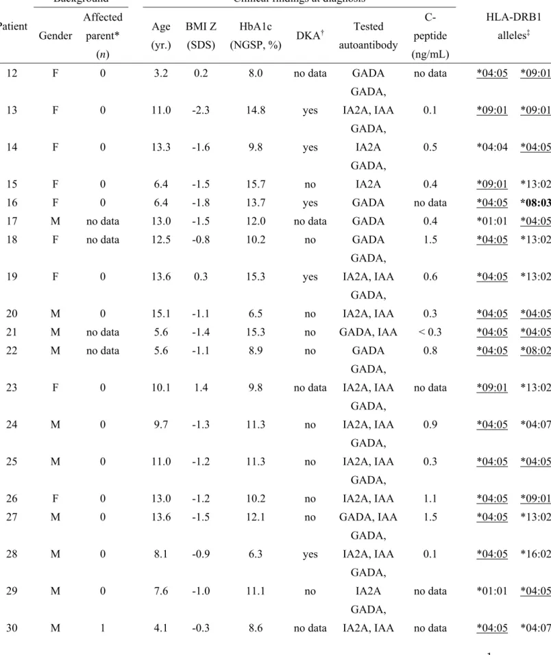
Table S1. Clinical findings of mutation non-carriers.
Patient
Background Clinical findings at diagnosis
HLA-DRB1 alleles‡ Gender
Affected parent*
(n)
Age (yr.)
BMI Z (SDS)
HbA1c
(NGSP, %) DKA† Tested autoantibody
C- peptide (ng/mL)
12 F 0 3.2 0.2 8.0 no data GADA no data *04:05 *09:01
13 F 0 11.0 -2.3 14.8 yes
GADA,
IA2A, IAA 0.1 *09:01 *09:01
14 F 0 13.3 -1.6 9.8 yes
GADA,
IA2A 0.5 *04:04 *04:05
15 F 0 6.4 -1.5 15.7 no
GADA,
IA2A 0.4 *09:01 *13:02
16 F 0 6.4 -1.8 13.7 yes GADA no data *04:05 *08:03
17 M no data 13.0 -1.5 12.0 no data GADA 0.4 *01:01 *04:05
18 F no data 12.5 -0.8 10.2 no GADA 1.5 *04:05 *13:02
19 F 0 13.6 0.3 15.3 yes
GADA,
IA2A, IAA 0.6 *04:05 *13:02
20 M 0 15.1 -1.1 6.5 no
GADA,
IA2A, IAA 0.3 *04:05 *04:05 21 M no data 5.6 -1.4 15.3 no GADA, IAA < 0.3 *04:05 *04:05
22 M no data 5.6 -1.1 8.9 no GADA 0.8 *04:05 *08:02
23 F 0 10.1 1.4 9.8 no data
GADA,
IA2A, IAA no data *09:01 *13:02
24 M 0 9.7 -1.3 11.3 no
GADA,
IA2A, IAA 0.9 *04:05 *04:07
25 M 0 11.0 -1.2 11.3 no
GADA,
IA2A, IAA 0.3 *04:05 *04:05
26 F 0 13.0 -1.2 10.2 no
GADA,
IA2A, IAA 1.1 *04:05 *09:01
27 M 0 13.6 -1.5 12.1 no GADA, IAA 1.5 *04:05 *13:02
28 M 0 8.1 -0.9 6.3 yes
GADA,
IA2A, IAA 0.1 *04:05 *16:02
29 M 0 7.6 -1.0 11.1 no
GADA,
IA2A no data *01:01 *04:05
30 M 1 4.1 -0.3 8.6 no data
GADA,
IA2A, IAA no data *04:05 *04:07
2
31 F 0 2.5 -2.7 13.4 no data GADA, IAA no data *09:01 *09:01 32 F 0 12.2 -2.1 15.6 no GADA, IAA no data *01:01 *04:05
33 M 0 3.6 0.9 7.6 no GADA no data *01:01 *09:01
34 F 0 2.3 -1.7 10.0 yes
GADA,
IA2A no data *09:01 *09:01
35 M 1 10.7 1.1 11.7 no
GADA,
IA2A no data *09:01 *13:02
36 F 0 6.1 -2.4 9.5 no GADA, IAA no data *01:01 *04:05
37 F 0 4.9 -0.2 14.5 yes
GADA,
IA2A 0.5 *09:01 *12:01
38 M 0 12.1 -1.3 5.6 yes GADA no data *01:01 *09:01
39 F 1 8.6 2.5 12.9 no data GADA, IAA < 0.3 *04:03 *15:02
40 F 0 14.4 -0.9 5.6 yes GADA 0.1 *04:05 *08:02
41 F no data 2.9 -3.1 12.8 yes
GADA,
IA2A no data *04:05 *08:02
42 F 0 2.4 -1.6 14.7 no GADA < 0.3 *04:05 *13:02
43 F 0 4.3 0.5 13.2 yes GADA, IAA 0.1 *04:05 *13:02
44 F no data 11.5 -1.0 13.0 yes GADA no data *04:05 *04:07
45 M 0 3.4 -3.1 12.3 no GADA, IAA 0.1 *04:05 *08:02
46 F no data 10.3 -2.1 13.3 yes GADA 0.4 *09:01 *13:02
47 M 0 2.6 0.8 9.6 no GADA 0.4 *01:01 *04:05
48 F 0 10.5 -0.9 10.4 no
GADA,
IA2A 0.7 *04:10 *09:01
49 M 0 2.6 -0.3 12.3 no GADA 0.1 *04:05 *13:02
50 F 0 5.3 0.6 6.2 no data
GADA,
IA2A 0.7 *04:05 *11:01
51 F 0 9.0 -2.1 15.0 yes
GADA,
IA2A, IAA 0.1 *04:05 *13:02
52 F 0 9.0 -2.1 11.2 no
GADA,
IA2A, IAA 0.6 *09:01 *14:06
53 F 0 5.3 -0.6 8.7 no data GADA no data *09:01 *13:02
54 M 0 2.6 -2.2 11.0 no data GADA, ICA no data *04:05 *09:01
55 M 0 2.7 -0.4 13.4 yes
GADA,
IA2A no data *09:01 *11:05
56 F 0 3.8 -2.3 13.2 no GADA 0.5 *04:05 *09:01
57 M 0 1.1 0.6 11.5 no data GADA 0.7 *04:05 *09:01
3
58 F 0 1.5 0.0 9.8 yes
GADA,
IA2A, ICA no data *09:01 *13:02
59 F 0 4.9 0.0 11.9 no GADA 0.5 *04:05 *12:01
60 M 0 10.7 -0.7 8.3 no
GADA,
IA2A 1.1 *09:01 *09:01
61 M 0 11.8 -0.1 11.9 no GADA 0.8 *01:01 *04:05
62 F 0 13.1 -0.7 12.4 no data
GADA,
IA2A, ICA 0.9 *09:01 *15:01
63 M 0 3.5 -0.4 11.3 no
GADA,
IA2A, ICA 0.3 *04:05 *04:07
64 M 0 3.8 -0.9 14.5 no GADA 0.6 *09:01 *13:02
65 M 0 14.7 -2.3 15.6 no data
GADA,
IA2A 1.3 *08:02 *12:01
66 F 0 10.1 0.0 13.6 no
GADA,
IA2A 1.2 *01:01 *09:01
67 F 0 7.5 -1.4 10.4 no
GADA,
IA2A 0.8 *01:01 *04:05
68 F 1 1.7 -0.9 9.3 no data GADA 0.1 *04:05 *08:02
69 F 1 3.6 0.1 11.7 yes GADA 0.9 *09:01 *09:01
70 F 0 14.2 -1.4 9.6 yes
GADA,
IA2A 1.0 *04:05 *11:01
71 F 0 3.4 -1.5 9.1 no GADA no data *09:01 *13:02
72 M 0 0.9 0.0 6.8 no
GADA,
IA2A no data *01:01 *04:05
73 F 0 5.2 -2.8 16.6 yes
GADA,
IA2A no data *04:05 *09:01
74 F 0 8.6 0.4 13.6 no GADA 0.8 *04:05 *13:02
75 F 0 8.4 0.9 5.8 no GADA 2.2 *03:01 *04:05
76 F 0 13.3 1.2 13.4 yes GADA, ICA no data *11:01 *15:01
77 F no data 15.1 -0.3 9.9 no
GADA,
IA2A 1.2 *14:06 *15:02
78 F 1 9.0 -0.7 14.8 no data GADA no data *09:01 *13:02
79 M no data 10.9 -0.9 7.1 no data GADA no data *13:02 *13:02 80 M no data 13.3 1.2 11.2 no data GADA no data *09:01 *13:02
81 M 0 8.2 -1.1 15.7 yes
GADA,
IA2A 0.2 *04:05 *08:02
4
82 F 0 11.5 -1.8 6.6 no
GADA,
IA2A, ICA 3.6 *04:05 *09:01
83 F no data 5.5 -2.6 17.0 yes GADA 0.2 *04:05 *08:02
84 F 0 14.0 1.2 14.5 no GADA 1.0 *04:05 *13:02
85 F no data 11.5 -1.2 14.9 no GADA, ICA 1.2 *08:03 *08:03
86 M no data 10.6 -1.6 6.9 no
GADA,
IA2A 0.7 *04:05 *09:01 87 F no data 2.4 -0.1 13.0 no GADA no data *09:01 *13:02
88 F 0 10.3 0.9 12.0 no
GADA,
IA2A no data *09:01 *15:02
89 F 0 4.3 -1.7 12.4 no
GADA,
IA2A, IAA 0.3 *09:01 *16:02
BMI, Body Mass Index; DKA, diabetic ketoacidosis; F, female; GADA, glutamic acid
decarboxylase 65 antibody; IA2A, tyrosine phosphatase-like insulinoma antigen 2 antibody; IAA, insulin autoantibody; ICA, islet cell antibody; M, male; NGSP, National Glycohemoglobin Standardization Program.
Patients 1-11 had monogenic mutations (see Table 2).
* The number of parents with history of diabetes.
† DKA was diagnosed according to the International Society for Pediatric and Adolescent Diabetes (ISPAD) Clinical Practice Consensus Guidelines 2014 (20).
‡ Susceptible HLA-DRB1 alleles are underlined, and protective alleles are boldfaced.
These alleles were defined based on the report of Sugihara et al (17).
# Variants are described according to the HGMD reference sequences: HNF1A
NM_000545.6; HNF4A NM_175914.4; HNF1B NM_000458.3; INS NM_001185098.1.
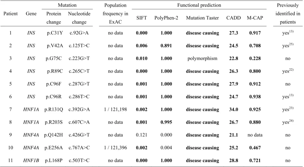
1
Table S2. Mutations identified in present study.
Patient Gene
Mutation Population
frequency in ExAC
Functional prediction Previously
identified in patients Protein
change
Nucleotide
change SIFT PolyPhen-2 Mutation Taster CADD M-CAP
1 INS p.C31Y c.92G>A no data 0.000 1.000 disease causing 27.3 0.917 yes13) 2 INS p.V42A c.125T>C no data 0.006 0.891 disease causing 24.5 0.708 yes25) 3 INS p.G75C c.223G>T no data 0.010 1.000 polymorphism 22.8 0.228 no 4 INS p.R89C c.265C>T no data 0.000 1.000 disease causing 26.3 0.800 yes22) 5 INS p.C96F c.287G>T no data 0.001 1.000 disease causing 27.9 0.912 no 6 INS p.C96R c.286T>C no data 0.001 1.000 disease causing 24.7 0.938 yes13) 7 HNF1A p.R131Q c.392G>A 1 / 121,198 0.002 1.000 disease causing 34.0 0.925 yes23) 8 HNF1A p.R203S c.607C>A no data 0.001 0.995 disease causing 26.7 0.880 yes24) 9 HNF4A p.Q142H c.426G>T no data 0.121 0.000 disease causing 21.1 no data no 10 HNF4A p.E256A c.767A>C 1 / 121,396 0.002 0.004 disease causing 25.2 0.467 no 11 HNF1B p.L168P c.503T>C no data 0.000 1.000 disease causing 28.8 0.721 no CADD, the Combined Annotation Dependent Depletion; ExAC, the Exome Aggregation Consortium Browser; M-CAP, the Mendelian
2
Clinically Applicable Pathogenicity; SIFT, Sorting Intolerant From Tolerant.
Scores classified as pathogenic by in silico analysis (SIFT scores < 0.05; PolyPhen-2 scores > 0.8; CADD scores > 20; and M-CAP scores > 0.025) are boldfaced.