Histological and Histochemical Studies of
Gonads of Nautilus pompilius from Fiji
著者
TSUKAHARA Junzo
journal or
publication title
南方海域調査研究報告=Occasional Papers
volume
4
page range
50-60
URL
http://hdl.handle.net/10232/15859
Kagoshima Univ. Res. Center S. Pac. Occasional Papers, No. 4, p. 50-60, 1985
4.
Histological and Histochemical Studies of
Gonads of Nautilus pompilius from Fiji
by
Junzo TSUKAHARA"
Introduction
For the investigation of the reproduction of Nautilus pompilius, living specimens collected in Fiji were dissected to take out the gonads and other reproductive organs. These organs were immediately weighed and fixed for histologcal and histochemical studies. Results of these studies through the optical and electron microscopic observations are discussed in this article in compari son with those of Nautilus in the Philippines and a few other kinds of cephalopod.
Materials and Methods
21 specimens of Nautilus pompilius captured off Suva and 15 specimens off Pacific Harbour in Fiji late in August and in September, 1983, were used for these studies. The soft parts of these 36 specimens were dissected and the gonads and other reproductive organs were taken out. After weighing, these organs were immediately fixed. The gonad index {%) defined as follows was used in the present study.
gonad index =
gonad weight
° soft part weight
A single large testis is located near the siphancle in the posterior position of soft body of male
(PL 10, fig. 1, PI. II. figs. 1, 2). The other reproductive organs such as an accessory organ, a
spermatophore sac and a penis lie successively on the anterior position (PI. 11, fig. 1). The spermatophore is formed in the accessory organ and stored in the spermatophore sac (PI. 11. fig. 3). It is coiled and surrounded by jelly-like viscous fluid in the sac.A single ovary is located also in the posterior position of the soft body of female. It is covered
with almost transparent thin membrane. Various sizes of the oocytes were found in clusters
attached to the ovarian wall (PI. 11, fig. 4). Growing pear shaped oocytes are covered with a meshwork of follicle epithelium and contain amber-colored fluid of yolks.
To weigh the growing oocytes, clusters of oocytes were fixed with 3 % glutaraldehyde for
5 or 10 hr and dissected into individual oocytes.
Preparation of specimens for optical microscopy
Fixations were taken for 24 or 48 hr at room temperature with 5 %neutral formaldehyde in 1) Department of Biology, Faculty of Science, Kagoshima University, Kagoshima 890, Japan.
51 Tsukahara : Gonads of Nautilus pompilius
90% sea water or 2.5 %glutaraldehyde in 90%sea water. Most specimens were dehydrated and
embedded in paraffin, while the rest of them were transferred into 30% sucrose solution for freezing sections.
Histochemical studies were performed for detecting the chemical natures of yolk substances in oocytes. Paraffin sections were stained with periodic acid Schiff (PAS) staining reagent (Mc-Mannls, 1946) or 0.02% toluidine blue buffered with 0.1 M phosphate buffer solution at pH 4.0. Frozen sections were stained with 0.5 %Sudan black B. Salivary ^-amylase was used to digest the
glycogen. The enzyme solution was prepared by adding 10ml of saliva to 50 ml of Tris-HCl buffer solution (pH 7.2), and filtering. Samples fixed with glutaraldehyde were incubated in the enzyme solution for 30 min or 3 hr at 35° C, followed by washing with 0.1 M buffer solution.
Preparation of specimens for electron microscopy
Small pieces of gonads and spermatophore were prefixed with 2.5 %glutaraldehyde in 90% sea water adjusted at pH 7.4 with 0.05 M cacodylate buffer solution. The fixation was performed at room temperature for 5 or10 hr. After rinsed three times with buffered sea water, post fixation was carried out for 2 hr with 1%Os04 in 90%sea water buffered with 0.05 M cacodylate solution (pH 7.4) at 0° C. The tissues were then dehydrated and embedded in Spurr resin. Ultra-thin sections were stained with uranyl acetate and lead citrate. These sections were observed by JEM 100 C transmission electron microscope.
Scanning electron microscopic observation was undertaken with the specimens, that were fixed with 2.5 %glutaraldehyde, dehydrated and dried by crytical point dryer. These specimens were coated with gold and observed by H 450 scanning electron microscope.
Observations and Discussions
Gonad weight and index
Table 1 shows weights of reproductive organs of males and ovaries of females. Gonad indexes
are also shown. The largest two testes among those of the specimens at hand are both 26.0 g in weight, about 3cmX4.5cm in diameters, and have a highest value of gonad index (see SV-13-9, SV-13-13). Almost all the males heavier than 350 g in soft part weight have a developing spermatophore in a spermatophore sac. The most grown spermatophore is 2.8 g in weight and measured approximately 16mmXl8mm in diameters. This specimen has another mature
spermatophore, which is holded by labial tentacles. When the semen is taken out from well
developed spermatophore and mixed by stirring with sea water, many spermatozoa move
vigorously in sea water.
The present writer preliminarily reported that some male specimens captured in summer season (early December) in Fiji, had large testes of about 16 g in weight and spermatophore of approximately 15 mm X 18 mm in diameters (Tsukahara and Ono, 1983). Haven (1977) found the coiled spermatophore, measuring approximately 6 mm X 25 mm, that was removed from the spermatophore sac of a male collected on October 1st in the Philippines. The writer also had a chance to weigh and measure the specimens collected by Hayasaka et al. (1982) from the Tafion area, the Philippines. They captured the specimens during September 7th to 19th, 1981. The result is shown in Table 2. Most of the male and female specimens in the Philippines have bigger
Kagoshima Univ. Res. Center S. Pac, Occasional Papers, No. 4, 1985 52
Table. 1. Weights of reproductive organs and gonad index of N. pompilius from Fiji. Male
Body No. S. P. W.(g) R.O.W.(g) T. W. (g) A. 0. W. (g) S. S. W. (g) G. I. (%)
SV 7- 1 276 8.2 6.8 0.9 0.4 2.5 SV12- 4 425 23.0 17.0 3.0 2.0 4.0 SV13- 2 371 12.3 9.3 2.1 1.1 2.5 SV13- 6 400 17.9 14.4 2.5 0.4 3.6 SV13- 9 410 37.0 26.0 7.0 3.0 6.3 SVI3-I0 355 18.8 14.9 2.0 0.7 4.2 SV13-13 413 35.0 26.0 6.0 2.5 6.3 SVI3-I6 230 1.0 0.2 0.5 0.25 0.1 SVI3-18 245 10.7 8.1 0.4 0.3 3.3 SVI3-I9 304 10.5 8.2 1.2 0.3 2.7 SV14- 2 348 10.0 7.0 2.0 0.5 2.0 SVI4- 3 408 15.7 11.9 2.2 0.7 2.9 SVI4- 5 394 29.3 20.2 5.4 2.3 5.1 PH 3-13 371 15.5 11.5 2.5 1.0 3.1 PH 5- 4 440 18.2 13.6 3.5 0.9 3.1 PH 5- 9 429 17.5 13.7 2.6 0.5 3.2 PH 6- 2 339 16.3 12.4 2.6 0.8 3.7 PH 6- 3 331 5.7 4.2 0.8 0.15 1.3 PH 6- 6 410 17.8 13.8 3.0 0.7 3.4 PH 6-11 390 10.1 7.5 1.7 0.2 1.9 PH 6-15 430 14.2 11.1 2.2 0.7 2.6 PH 6-18 443 18.7 14.7 2.7 0.5 3.3 PH 6-19 388 18.1 15.0 1.9 0.4 3.9 Female
Body No. S. P.W.i(g) O. W. (g)• G.I.(%)
SV 5- 3 276 3.3 1.2 SV12- 1 313 7.0 2.2 SV12- 3 197 1.0 0.5 SV12- 7 176 0.3 0.2 SV13- 3 323 11.3 3.5 SV13- 8 364 15.3 4.2 SV13-14 270 7.3 2.7 SV14- 1 298 9.9 3.3 PH 2- 1 247 3.2 1.3 PH 4- 9 440 11.9 2.7 PH 4-16 254 0.6 0.2 PH 6- 5 306 10.9 3.6 PH 6- 7 355 11.8 3.3 Sex unknown Body No. S. P . w. (g) G. W.(g) SV10- 6 125 0.15
S. P. W. Soft Part Weight
R. O. W. Reproductive Organ Weight T. W. Testis Weight
A. O. W. Accessory Organ Weight S. S. W. Spermatophore Sac Weight
O. W. Ovary Weight G.W. Gonad Weight
53 Tsukahara : Gonads of Nautilus pompilius
Male
Table 2. Weights of reproductive organs and gonad index of N pompilius from the Tafion area, the Philippines.
Body No. S. P. W. (g) R. 0. W. (g) T. W. (g) A. O. W. (g) S. S. W. (g) G. I. (%) B-10 536 19.84 14.830 4.065 0.768 2.68 B-24 445 30.03 24.289 4.541 0.729 5.46 B-29 543 33.66 27.15 5.001 0.910 5.00 No- 1 451 29.63 22.22 5.993 0.786 4.93 Female Body No. S. P. W. (g) O. W. (g) G. I. (%) B- 2 B- 3 B- 6 B-ll B-13 B-14 B-17 B-18* B-19 B-23 B-30 B-33 No- 2 No- 3 219 9.757 4.46 415 30.769 7.41 365 24.576 6.73 395 20.964 5.31 408 17.200 4.22 535 23.530 4.40 380 25.326 6.66 445 129.441 29.09 446 26.202 5.87 370 18.151 4.91 383 19.603 5.12 468 31.523 6.74 437 29.822 6.82 400 11.322 2.83-*B-18 Ovarian Tumor
S. P. W. Soft Part Weight
R. O. W. Reproductive Organ Weight T. W. Testis Weight
A. O. W. Accessory Organ Weight S. S. W. Spermatophore Sac Weight
O. W. Ovary Weight
Kagoshima Univ. Res. Center S. Pac., Occasional Papers, No. 4, 1985 54 reproductive organs than those in Fiji. Gonad indexes also show somewhat larger values in the
Philippines.
The largest ovary among 13 females has approximately 15.3 g in weight and 4.2 %as gonad index (SV 13-8). In the Philippines the largest ovary was found in late July (Haven, 1977). It is 25.50 g in weight and has the biggest oocyte of 3.6 g in weight. Table 2 shows that the largest ovary in September in the Philippines is 31.523 g (B-33). Although the body numbered B-18 has
the biggest ovary, a large ovarian tumor is increased in it.
The distribution of the oocyte weight is shown in Fig. 1. Although the number of oocytes in
(9) 2.5 2JD-•c 1.5 Z 5 o O 1.0 0.5-Sp. No. Ov. W. (g) SV 13- 8 15.25 SV 13- 3 11.35 SV 14- 1 9.86 SV 13-14 7.30 SV 5- 3 3.35 » - < 5 10
Ranking of Oocyte Weight
15
Fig. I. Distribution of the weight of oocytes more than 0.01 g of N. pompilius collected from Fiji.
55 Tsukahara : Gonads of Nautilus pompilius
the figure comprises only those heavier than 0.01 g, amber yolk fluid can be found in the oocyte
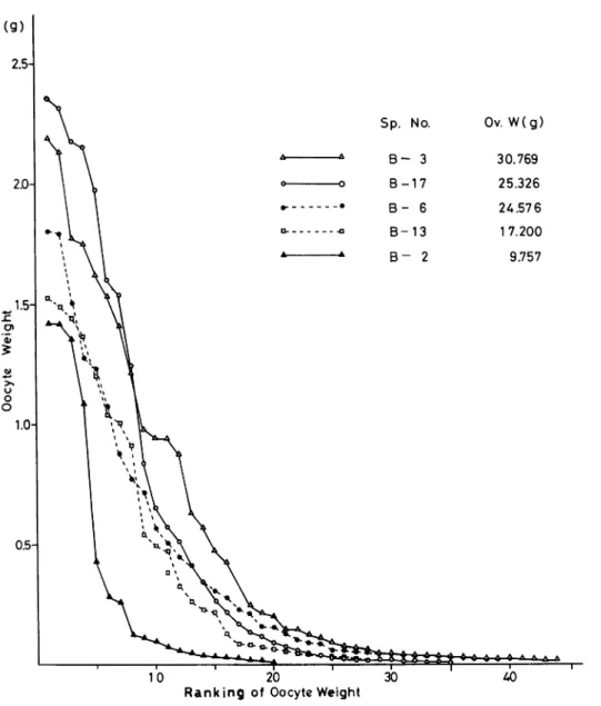
heavier than 0.05 g. When the oocyte weight attains to 0.3 g, rapid deposition of yolks may be occurred. Fig. 2 also shows the weight distribution of oocytes of the specimens collected in the
Philippines (Hayasaka et al., 1982). The biggest oocyte among them is 2.35 g in weight and
slightly smaller than those in Fiji. The numbers ofoocyte heavier than 0.01 g, however, are almost
twice or thrice as much as those shown in Fig. 1.
Haven (1977) carried out trapping in Tafion strait for about one year and captured the mature females within the period from August through March. She reported that the higher average
Sp. No. Ov. W(g) B - 3 30.769 B-17 25.326 B - 6 24.576 B-13 1 7.200 B - 2 9.757 10 20 30
Ranking of Oocyte Weight
Fig. 2. Distribution of the weight of oocytes more than 0.0I g of N. pompilius collected from
Kagoshima Univ. Res. Center S. Pac, Occasional Papers, No.4, 1985 56
values of oocytes weight more than lg are 3.15 g in late July and 2.18 g in mid-August in the Philippines. In Fiji, the largest oocyte of 2.35 g was taken in early December (Tsukahara and Ono, 1983). From the records in Fiji and in the Philippines, it may be suggested that
the reproductive season of Nautilus in Fiji seems to be in summer or in winter. It must be necessary to try the collection of Nautilus on the other seasons in Fiji.
Histological observations
Spermatogenesis and spermiogenesis
There are numerous seminiferous tubules in the testis. Many spermatogonia can be seen near the basal lamina. Meiotic division of primary or secondary spermatocytes is found on slightly
inner side of tubules (PI. 12, fig. 1). Single spermatocyte remains connected to each other by
cytoplasmic bridges throughout their differentiation into mature sperm. Many mitochondria are scattered over the cytoplasm of spermatocyte.
When the youngest spermatid starts on its spermiogenesis, many mitochondria are crowded near one side of nucleus (PL 12, fig. 2) and fused gradually with one another to make two larger mitochondria (PI. 13, fig. 1). A pair of centrioles is found near the peripheral cytoplasm at the beginning of spermiogenesis (PI. 12, fig. 2) and then moves toward the nuclear envelope. One of them changes into distal basal body forming a flagellum (PI. 13, fig. 1). Proximal basal body gets closely into the pit of nuclear envelope. These two basal bodies are set on a slightly acute angle each other. Many microtubules were extended into the cytoplasm and beside the nuclear envelope from the periphery of one of the basal bodies (PI. 13, fig. 2). Arnold and Arnold (1978) reported that microtubules appeared first at right angles to the distal centriole of the spermatid of
Nautilus pompilius collected in the Philippines.
Chromatin condensation begins to appear in the uncleus near the mitochondria (PI. 13, fig. 1), and slightly later in development many clusters of dense granules are found in the
nucleoplasm (PI. 14, fig. 1).
Spherical nucleus elongates gradually to pear shape as subsequent development progress (PI. 14, fig. 1) and later becomes to spindle shape (PI. 14, fig. 2). Proximal and distal basal bodies are almost parallel with each other in a implantation fossa of the nuclear envelope. Two mitochon dria are elongated along the envelope for opposite side to the fossa. Many parallel microtubules surround nuclear envelope and mitochondria elongate from the fossa side to the top of nucleus as the manchette structure (PI. 15, fig. 1). Two elongated mitochondria are situated just on the opposite side of the nucleus to each other in the cross section (PI. 15, fig. 1, PI. 17, figs. 1, 2)
Near the top of spindle shaped nucleus dense amorphous materials formed in the Golgi apparatus are crowded (PI. 14, fig. 1), and later may be set on the top of the nucleus as the
acrosomal granule (PI. 14, fig. 2). Many nuclear pores are found clearly in the nuclear envelope,
where the acrosomal granule is settled on (PI. 14, figs. 1, 2).
The nucleus elongates gradually to long cylindrical shape (PI. 15, fig. 2). The acrosomal granule is surrounded by many microtubules and nuclear pores (PI. 16, fig. 1). At the posterior side of the nucleus, two basal bodies with electron dense cap are embedded entirely in the fossa (PI. 16, fig. 2). These are a set of nine doublets on each basal body in the cross section
57 0) E o E Z
Tsukahara : Gonads of Nautilus pompilii
5 A 3 2 1 Diameter of nucleus (pm) 0.75 xCU 0.5 x0.3
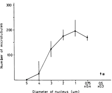
Fig. 3. Variation of the number of microtubules surrounding the nucleus of spermatid during the spermiogenesis. asterisk ; the nucleus of mature spermatozoon in the spermatophore.
As shown In the cross sections of nucleus (PI. 15, fig. 1, PI. 17, figs. 1, 2, PI. 18, fig. 1) and
summarized in Fig. 3, the number of microtubules in the manchette structure changes in accor dance with the diameter of nucleus. When the nuclear diameter diminishes just under 2//m, however, the number of microtubules may be almost invariable. Mature sperm nucleus in the
spermatophore was surrounded by only 20-30 microtubules (PI. 19, fig. 2).
Chromatine substances become gradually condensed in the peripheral regions of nucleus (PI. 17, figs. 1, 2). On the almost matured spermatozoon in the testis the rod shaped nucleus includes a mass of dense and fine threads of chromatin substances (PL 18, fig. 1).
Mature sperm in the spermatophore
Numberless mature spermatozoa are arranged their head toward the outer jelly-like substance
of spermatophore tubules (PL 18, fig. 2). Their nuclear length is reached approximately 37 ^m. In the cross section the nucleus is found slightly ellipsoidal and approximately 0.5 X 0.3//m in diameters, although the area near the top of the nucleus is more flat and 0.7 X 0.15//m in
diameters (PL 19, fig. 1). Nautilus pompilius captured in the Philippines has a mature spermatozoa
with a length approximately 35 ptm and a diameter approximately 0.3jxm (Arnold and Arnold,
1978). Chromatin substance in the nucleus is shown as many fine amorphous masses (PL 19, fig. 2). On the acrosomal region of sperm anterior rod structure and a posterior clear vesicle can be detected in the longitudinal section (PL 20, fig. 1). Many fine granules are found in the space
between nucleus and plasma membrane. They were shown as glycogen granules by Arnold and Arnold (1978).
Oogenesis
Kagoshima Univ. Res. Center S.Pac, Occasional Papers. No. 4, 1985 58
with a developing nucleolus is situated near the center ofthe ooplasm (PL 10, fig. 2). The ooplasm is stained almost uniformly with neutral toluidine blue. Thin follicle epithelium is surrounded entirely by the oocyte. On the electron microscopic observation there are many mitochondria and vesicles scattered in the cytoplasm with numerous ribosomal granules (PL 20, fig. 2). Electron dense nucleolus of approximately 30 pm in diameter is located near the nuclear envelope, and has
many small electron light spots.
When the diameter of oocyte grows up to approximately 500 /*m, the oocyte alters its shape
into pear-like. Nucleus gradually moves for opposite side to the stalk extended to the ovary wall
(PL 10, fig. 3). A protoplasmic mass surrounding the nucleus is better stained with neutral toluidine blue than the others. Some parts of the surface of the oocyte near the stalk invaginate and follicle epithelium develops to a vast array of interdigitation.
Electron microscopic observations show that the surface of the oocyte near nucleus is almost smooth and covered with about 5 layers of flat follicle cells (PL 21, fig. 1). On the opposite side, however, some clusters of microvilli grow on the surface of the oocyte near the stalk (PL 21, fig. 2). Follicle cells arranged on the oocyte become gradually bottle shaped. The surface of the follicle cell faced on the oocyte makes also some clusters of microvilli. When the oocyte reaches
approximately 800 ^m in diameter, the follicular epithelium complex folding into the oocyte
grows just like the meshwork (PL 10, fig. 4). Nucleus in the oocyte is more flat and may be pressed on one side of the surface. Most parts of cytoplasm are transparent.
Electron micrographs show that a lot of well extended microvilli are found on both surfaces of the oocyte and follicle cells (PL 22, fig. 1). The space between them is almost 3//m in width,, and the longest villus in the figure reaches approximately 3//m (PL 22, fig. 2). Cortical layer of the oocyte possesses numerous electron light vesicles, and they may be fused with each other to make larger yolk vesicles in the inner ooplasm. There are no typical pinocytotic coated vesicles among them. Follicle cells arrange nearly at right angle against the surface of the oocyte. They have many endoplasmic reticulum, mitochondria and electron dense granules in the cytoplasm.
Yung (1930) reported that in the cuttle-fish Sepia officinalis the proliferation of the follicle epithelium is strongest on the attached vegetative side; here the folding of the granulosa is most pronounced. In Loligo bravis the folliclar epithelium also invades the oocyte as fold of tissue with a high mitotic in the vegetative side (Cowden, 1968). Aizenshtadt (1969) reported through electron microscopy that when the follicle epithelium of Rossia pacifera begins to flatten out, the
interdigitations start to withdraw and become microvillous, but close contact with the oocyte is
still maintained. In Octopus vulgaris, however, Wells (1960) showed that the germinal vesicle still remains in almost central position during oogenesis. From these observations it may be suggested that the oogenesis of Nautilius pompilius grows as the same manner as that of the cuttle-fish. The pole of the oocyte where the nucleus moves toward may represent as the animal pole, where the futured blastodisc is formed; the opposite side with a stalk may be vegetal pole. PL 10, fig. 5 shows that there is a micropyle-like pore on the follicle epithelium near the animal
pole of the oocyte with 3 mm in diameter.
Scanning electron microscopic observations of oocytes
Animal pole of the oocyte with the diameter of about 3 mm is almost flat (PL 23, fig. 1), but has a slightly concave place (PL 23, fig. 2). However, it may not be a typical micropyle, which is
59 TSUKAHARA : Gonads of Nautilus pompilius
it is shown that the follicle epithelium is infolded inside of the oocyte cytoplasm. Numerous yolk granules of various sizes are formed at the periphery of the ooplasm (PL 23, figs. 3, 4).
Histochemistry of yolks
PAS staining
Yolk granules in the oocyte of more than approximately 1 mm in diameter are dyed magenta
with PAS reagent in paraffin or freezing section (PL 10, fig. 6). When these sections are treated
with salivary ^-amylase, the yolks still remain PAS positive. From these results yolks in the oocyte contain a lot of carbohydrates except glycogen-like substances. In the cytoplasm of the
follicle epithelium numerous PAS positive fine granules can be detected, and are located near the oocyte surface. PAS positive yolk granules may be prepared in the follicle cells and transferred into the oocyte through microvilli of both cells.
Acidic toluidine blue staining
Acidic toluidine blue (pH 4.0) stainsyolk substances in metachromatic violet-blue (PL 10, fig.
7). They may contain acid mucopolysaccharide. The cytoplasm of the follicle cell, however,
shows scarecely metachromatic reactions.
Sudan black B staining
Freeze sections stained with Sudan black B show various deep blue-black stained granules located at the periphery of the elder oocyte (PL 10, fig. 8). According to the result, yolk granules may contain also a lot of the fatty substances. Follicle cell cytoplasm is also dyed deep blue-black.
Summary
In the present article, anatomical and histological observations were given on the reproductive organs of 36 specimens of N. pompilius collected in Fiji late in August and in September, 1983.
A mature spermatophore in the spermatophore sac is observed to develop in most of the adult
males. A developing ovary including oocytes of various sizes and a few full-grown oocytes are in
most of adult females.
The nucleus of the early spermatid begins to elongate remarkably. Crowds of mitochondria fuse one another to make two large mitochondria, and extremely elongate along the cylindrical nucleus. Numbers of microtubules surround the elongating nucleus and mitochondria resulting in a manchette structure. The acrosomal rods and vesicle are formed on the apical area of the cylindrical nucleus. Condensation of the chromatin occurrs during the elongation of nucleus. Two basal bodies lie in parallel to each other in a deep implantaion fossa of the nuclear envelope. The nucleus of mature spermatozoa in the spermatophore is extremely elongated with a length approximately 37 pm and diameters approximately 0.5 X 0.3 pm.
Young oocytes have a germinal vesicle with a spherical nucleolus. They are surrounded by a flat follicle epithelium. According to the growth of the oocyte, the nucleus moves to one side of the oocyte surface, where the futured blastodisc is formed. On the opposite side follicle epithelium develops to a vast array of interdigitaion. A lot of microvilli are extended on the
Kagoshima Univ. Res. Center S. Pac, Occasional Papers. No. 4. 1985 60
surface of the oocyte and the follicle cells. The follicle epithelium complex folding into the elder oocyte grows just like a meshwork. Yolk substance in the growing oocytes includes a lot of
carbohydrate, especially acid mucopolysaccharide. They also include a lot of fatty substances.
The biggest oocyte in the ovary has approximately 2.35 g in weight.
Acknowledgements
The writer wishes to express his appreciation to Professor Shozo Hayasaka of Kagoshima
University for his kind criticisms and improvement of the manuscript. The writer also wishes to
express his deep gratitude to Professor Uday Raj and the staff of the Institute of Marine Resources,
the University of the South Pacific for their help in preparation of samples.
References
Aizenshtadt, J. B., 1969 : Cytomorphological study of egg follicles in the cuttlefish. 1. Fine structure of cells of the follicular epithelium. Tsitologia, 11, 401-409.
Arnold, J. M. and Williams-Arnold, L. D., 1978: Spermiogenesis of Nautilus pompilius. 1. General survay. J. Exp. Zool., 205, 13-26.
Cowden, R. R., 1968 : Cytological and cytochemical studies of oocyte development and develop ment of the follicular epithelium in the squid Loligo brevis. Acta Embryol. Morphol., 10, 160-173.
Haven, N., 1977 : The reproductive biology of Nautilus pompilius in the Philippines. Mar. Biol., 42, 177-184.
Hayasaka, S., Saisho, T., Kakinuma, Y., Shinomiya, A., Oki, K., Hamada, T., Tanabe, K., Kanie, Y., Hattori, M, Vande Vusse, F., Alcala, L., Cordero, P. A.Jr ., Cabrera, J.
J. and Garcia, R.G., 1982 : Field study on the Nautilus in the environs of Cebu and Negros
Islands, the Philippines. Mem. Kagoshima Univ. Res. Center S. Pac, 3, 67-115.
McManus, J.F.A., 1946 : Histological demonstration of mucin after periodic acid. Nature, 158,
202.
Tsukahara, J. and Ono, S., 1983 : Preliminary study on the reproductive biology of Nautilus
pompilius in Fiji. Promp. Rep. Sci. Sun: S. Pac, Kagoshima Univ. Res. Center S. Pac,
2, 19-23.
Wells, M.J., 1960 : Optic glands and the ovary of Octopus. Symp. Zool. Soc. London, 2, 87-101.
Yung, K. C. M., 1930 : Contribution a I'etude cytologique de l'ovogenese, du developpement et
Abbreviations
A Accessory Organ AG Acrosomal Granule
AS Acrosomal Substance AV Acrosomal Vesicle
Ch Chromosome DB Distal Basal Body (Centriole)
F Flagellum (DC in Plate 16)
G Golgi Apparatus FC Follicle Cell
Mv Microvillus M Mitochondrion
No Nucleolus N Nucleus
OC Oocyte Cytoplasm NP Nuclear Pore PB Proximal Basal Body (Centricle) P Penis
(PC in Plate 16) S Spermatophore Sac
Explanation of Plate 10
Fig. 1. Soft body of a male of N. pompilius.
Fig. 2. Young oocyte stained with neutral toluidine blue.
Fig. 3. Growing oocyte of approximately 500 pm in diameter stained with neutral toluidine blue. Fig. 4. Growing oocyte of approximately 800 pm in diameter stained with alcian blue and PAS. Fig. 5. Micropyle-like pore(arrow)on the follicle epithelium near the animal pole of the oocyte. Fig. 6. PAS positive yolk substances in the ooplasm and follicle cell cytoplasm.
Fig. 7. Yolk substances showing metachromatic violet-blue by staining with acidic toluidine blue.
Explanation of Plate 11
Fig. 1. Soft body of a male of N. pompilius.
Fig. 2. Testis.
Fig. 3. Spermatophore. Fig. 4. Cluster of oocytes.
Tsukahara: Gonads of Nautilus pompilius Plate
* .
Explanation of Plate 12
Fig. 1. Meiosis of the spermatocytes linked with a cytoplasmic bridge. X 6000.
Fig. 2. Young spermatid in which the mitochondria crowded at one side ofthe nucleus, arrow;
Tsukahara : Gonads of Nautilus pompilius
>
**'
'31ft m m 'Ts -V ,*§; -i> . ... ' . , ' . • :-v- • • • • / ;€N
mmCh
-4f»."'•^'"^<
m
*'.««;: •-Plate 12 ; . ^ ***'?'#!
v, , J Jgfs • / :m
••? " "Explanation of Plate 13
Fig. 1. Spermatid in which two large mitochondria located at the posterior side of the nucleus. arrow ; chromatin condensation. X 10000.
Tsukahara : Gonads of Nautilus pompilius Plate 13
I
•
*•%•....;•
Explanation of Plate 14
Fig. 1. Elongation of the spermatid nucleus, arrow; nuclear pore. X 12500.
Fig. 2. Continued elongation of nucleus with acrosomal-like granules, arrow ; nuclear pore.
Tsukahara : Gonads of Nautilus pompilius Plate 14
>:•
Explanation of Plate 15
Fig. 1. Cross section of the elongating nucleus with a manchette. arrow ; microtubule.
X 25000.
Explanation of Plate 16
Fig. 1. Anterior region of the sylindrical nucleus, arrow; microtubule. X 10000.
Fig. 2. Posterior region of the sylindrical nucleus. X 30000. inset; a set of nine doublets in the
Tsukahara : Gonads of Nautilus pompilius Plate 16 -• ; ••» ;
MP
M ?J; IjMIHI m*PC
*
},fjDC
?:
,Explanation of Plate 17
Fig. 1. Cross section of the sylindrical nucleus with approximately 2pm in diameter. X 45000.
Fig. 2. Cross section of the sylindrical nucleus with approximately 1pm in diameter. X45000.
Tsukahara : Gonads of Nautilus pompilius Plate 17 »f.»-. - . ' » •
I-•*»:••
>
t-M
-M
I *
. N
/ %.«V1
%*, * "Explanation of Plate 18
Fig. 1. Fully growing spermatozoa in the testis. X 45000.
Tsukahara : Gonads of Nautilus pompilius HBl i ' , : Plate 18 i
Explanation of Plate 19
Fig. 1. Cross section of mature nucleus of spermatozoa, arrow; acrosomal region of the
spermatozoon. X 25000.
Tsukahara : Gonads of Nautilus pompilius Plate 19 m :,:«» II
ill
Jmmm#
it*
''""• ''-i-'-'iL ' ' '".a*,^ =1'.? liifliir ^
i ««iiSpill*
•-...' •j#w '.i-tf*Explanation of Plate 20
Fig. 1. Anterior region of the mature nucleus, arrow ; acrosomal rod. X 50000. Fig. 2. Young oocyte nucleus with a spherical nucleolus. X 6000.
Tsukahara : Gonads of Nautilus pompilius , „ r « : . .,..,.„ .* •.•.'•••.• . .• . . . --;;«; '48 llliili •
i§M|iliiiit,
Plate 20 Ills r \ V ! -»i =:: iSffi;: y-ms'SiiExplanation of Plate 21
Fig. 1. Oocyte surface of animal pole region surrounded by flat follicle epithelium. X7500.
Fig. 2. Oocyte surface of vegetal pole region surrounded by bottle-shaped follicle cells, arrow;
Tsukahara : Gonads of Nautilus pompilius
1 : *
>MF
in* ...« jfcjaQ |
j ^
%•ow
i. . V* •i. ) « . . , > . . . . Plate 21P
,->.;
Explanation of Plate 22
Fig. 1. Surface of the growing oocyte with well extended microvilli. X 7500.
Tsukahara : Gonads of Nautilus pompilius Plate 22
*
-^.M *
Explanation of Plate 23
Fig. 1. Animal pole of the growing oocyte with approximately 3 mm in diameter. Fig. 2. Slightly concave place on the animal pole of growing oocyte.
Fig. 3. Fractural surface of the oocyte with interdigitation of follicle epithelium. Fig. 4. Yolk granules located near the surface of the oocyte.
Tsukahara : Gonads of Nautilus pompilius Plate 23 .