https://doi.org/10.1177/2324709620953283 Journal of Investigative Medicine High Impact Case Reports
Volume 8: 1–10
© 2020 American Federation for Medical Research
DOI: 10.1177/2324709620953283 journals.sagepub.com/home/hic
Creative Commons Non Commercial CC BY-NC: This article is distributed under the terms of the Creative Commons Attribution-NonCommercial 4.0 License (https://creativecommons.org/licenses/by-nc/4.0/) which permits non-commercial use, reproduction and distribution of the work without further permission provided the original work is attributed as specified on the SAGE and Open Access pages (https://us.sagepub.com/en-us/nam/open-access-at-sage).
Case Report
Background
Juvenile idiopathic arthritis is relatively common in the Caucasian population but appears to be rare in the Japanese
population.1-3 Caucasian ethnicity is a risk factor for
develop-ing juvenile idiopathic arthritis,1 but the incidence of juvenile
idiopathic arthritis in different populations is yet to be deter-mined. To establish the common ground for the diagnosis, the International League of Associations for Rheumatology (ILAR) has published classification criteria for juvenile
idiopathic arthritis.4 Recently, the Pediatric Rheumatology
Association of Japan (PRAJ) has surveyed the prevalence of uveitis as 6.1% in Japanese patients with the diagnosis of juvenile idiopathic arthritis, based on the ILAR classification
criteria.5 The prevalence of juvenile idiopathic arthritis–
associated uveitis in the Japanese population is at the lower
end of the prevalence in the other populations (4.7% to 20.5%).5
In the field of ophthalmology, band keratopathy, posterior iris synechiae, and complicated cataract in both eyes have
been described in textbooks as hallmark features of uveitis in
children with juvenile idiopathic arthritis.6-8 Indeed, these
clinical features are sequelae to persistent iridocyclitis. Recent advances in clinical imaging such as optical coherence tomography have demonstrated macular edema as
1Okayama University Graduate School of Interdisciplinary Science and
Engineering in Health Systems, Okayama City, Japan
2Ophthalmology, Okayama University Hospital and Okayama University
Medical School, Okayama City, Japan
3Pediatrics, Okayama University Hospital and Okayama University
Graduate School of Medicine, Dentistry, and Pharmaceutical Sciences, Okayama City, Japan
Received June 7, 2020. Revised June 28, 2020. Accepted August 2, 2020.
Corresponding Author:
Toshihiko Matsuo, Regenerative and Reconstructive Medicine (Ophthalmology), Okayama University Graduate School of
Interdisciplinary Science and Engineering in Health Systems, Shikata-cho 2-5-1, Okayama City 700-8558, Japan.
Email: [email protected]
Long-Term Control of Macular Edema
With Adalimumab After Cataract
Surgery in a Japanese Child With Juvenile
Idiopathic Arthritis: Case Report and
Review of 26 Japanese Patients
Toshihiko Matsuo, MD, PhD
1,2and Masato Yashiro, MD, PhD
3Abstract
Juvenile idiopathic arthritis–associated uveitis is rare in the Japanese population. In this article, we report a child whose macular edema was controlled for years after cataract surgery with adalimumab, and reviewed 26 Japanese patients in the literature. In this case report, a 4-year-old boy developed band keratopathy, posterior iris synechiae, and complicated cataract in both eyes. Oral prednisolone prescribed at another hospital was discontinued due to high intraocular pressure in both eyes as a steroid responder. At the age of 5 years, he started oral methotrexate 8 mg weekly for recurrent bilateral iridocyclitis and then underwent lensectomy with core vitrectomy in both eyes. Planned intraocular lens implantation was cancelled at surgery because the anterior vitreous had severe inflammatory opacity with diffuse retinal edema in both eyes. Due to persistent macular edema in both eyes 5 months postoperatively, at the age of 6 years, he began to use adalimumab injection 20 mg every 2 weeks. The macular structure depicted by optical coherence tomography became normal in 2 months. At final visit at the age of 11 years, he had the best-corrected visual acuity of 0.8 in the right eye and 0.4 in the left eye, with adalimumab 40 mg every 2 weeks and methotrexate 8 mg weekly. In conclusion, macular edema persistent despite oral methotrexate after cataract surgery could be controlled for long term by adalimumab in a child with juvenile idiopathic arthritis. In the Japanese literature, only 26 additional cases with juvenile idiopathic arthritis–associated uveitis have been reported so far.
Keywords
juvenile idiopathic arthritis, JIA, macular edema, uveitis, adalimumab, steroid responder, cataract surgery, methotrexate, literature review, Japanese
2 Journal of Investigative Medicine High Impact Case Reports a vision-threatening feature of uveitis in children with
juvenile idiopathic arthritis.9-15 Uveitis is more dominant
at younger ages and often associated with oligoarticular (pauciarticular) juvenile idiopathic arthritis that tends to lack
apparent systemic manifestations such as fever.5 Arthritis
may appear after the onset of uveitis. Therefore, children with juvenile idiopathic arthritis–associated uveitis have more chance to be presented first to ophthalmologists and to be referred later to pediatricians.
In this study, we report a Japanese child with oligoarticu-lar juvenile idiopathic arthritis who underwent cataract surgery during the course of oral methotrexate. Persistent macular edema was dramatically controlled with the induc-tion of adalimumab for the following years. We also reviewed Japanese patients with juvenile idiopathic arthritis–associ-ated uveitis in the literature.
Case Report
A 4-year and 8-month boy was referred from a local eye doctor to our hospital for band keratopathy, posterior iris synechiae, and complicated cataract in both eyes. The best-corrected visual acuity was 0.3 with +1.75 diopters and cylindrical −1.0 diopters at axis of 30° in the right eye and 0.1 with +3.75 diopters and cylindrical −2.25 diopters at axis of 170°, determined by cycloplegic refraction with topi-cal 1% cyclopentolate. The intraocular pressure was 10 mm Hg in both eyes. He had no active inflammation in both eyes as evidenced by neither aqueous cells nor keratic precipi-tates. The retina in both eyes could be visualized as normal through the pupil with synechiae. The patient was referred to a pediatric rheumatologist and was diagnosed as juvenile idiopathic arthritis because he had intermittent joint symp-toms. At the time of the diagnosis, he had no arthritis, and thus was followed with no medication. Antinuclear antibody, rheumatoid factor, and anti–cyclic citrullinated peptide (anti-CCP) antibody were negative throughout the course.
He was delivered with the birthweight of 2420 g by emer-gency Cesarean section at 41 weeks of gestation because of a reduced heart rate to 50 per minute when the mother suffered from the exacerbation of pregnancy-induced hypertension, proteinuria, and lower limb edema. He had been well except for hand, foot, and mouth disease at the age of 1 year and 9 months, and varicella at the age of 3 years and 8 months. Family history had nothing particular except for the mother who later developed immunoglobulin A (IgA) nephropathy at the age of 40 years.
At the age of 5 years and 1 month, oral prednisolone at daily dose of 20 mg (1 mg/kg of body weight) for 2 weeks, followed by 15 mg daily, was prescribed at another hospital where the mother sought the consultation at her own discre-tion. At the visit back to our hospital in 3 weeks after the start of oral prednisolone, the intraocular pressure was 40 mmHg in the right eye and 30 mmHg in the left eye. No iridocyclitis was noted in both eyes. With the diagnosis of a steroid responder who showed the elevation of intraocular pressure
in response to corticosteroid administration, the dose of oral prednisolone was reduced to 5 mg daily and discontinued in a week. Topical 0.1% fluorometholone 4 times daily was replaced by topical bromfenac twice daily. The intraocular pressure became 9 mmHg in both eyes. Oral ibuprofen 600 mg daily was prescribed by the pediatrician.
At the age of 5 years and 6 months (Figure 1A and B), oral
methotrexate 10 mg/m2 (8 mg) weekly was initiated by the
pediatrician for recurrent bilateral iridocyclitis in both eyes, and 2 months later (Figure 1C and D), he underwent poste-rior synechialysis, anteposte-rior capsulotomy, and lens cortex aspiration with a 25-gauge vitrectomy cutter under the irriga-tion with a 25-gauge irrigating cannula, both of which were
inserted through 2 ports at the corneal limbus.16 Planned
intraocular lens implantation was cancelled at this time point of surgery because the anterior vitreous was visualized through the posterior capsule to have severe inflammatory opacity with diffuse retinal edema in both eyes. Total poste-rior capsulectomy and core vitrectomy was done through the limbal side ports (Figure 1E and F).
Postoperative visual acuity by correction with glasses (+14.75 diopters in the right eye and +14.25 diopters in the left eye on the focus of 50 cm from the eyes) remained at 0.04 in both eyes due to macular edema (Figure 2A and B) when he used oral methotrexate 10 mg in a week and topical 0.1% fluorometholone 4 times daily. At the age of 6 years, 5 months postoperatively, he began to use subcutaneous injec-tion of adalimumab 20 mg every 2 weeks. In 2 months with scheduled injection of adalimumab, the macular retina was demonstrated to have normal structure by optical coherence tomography (Figure 2C and D). He gained the best-corrected visual acuity of 0.5 in the right eye and 0.2 in the left eye at the age of 6 years and 4 months when he was admitted to a local elementary school. The intraocular pressure in both eyes was kept in the normal range by occasional switching of topical bromfenac to topical 0.1% fluorometholone when keratic precipitates as a sign of iritis appeared.
At the age of 7 years and 4 months, he started topical latano-prost once daily when the intraocular pressure in the right eye had tendency to show a higher value of 25 mmHg. One month later, 0.5% timolol gel–forming solution once daily was pre-scribed in addition to latanoprost but was discontinued in the event of an asthma attack. At the final visit at the age of 11 years and 6 months, he had the best-corrected visual acuity of 0.8 in the right eye and 0.4 in the left eye (Figure 2E and F), with adalimumab 40 mg every 2 weeks and methotrexate 8 mg in a week. He had no intraocular inflammation with no topical steroid and maintained normal intraocular pressure in the range of 10 to 15 mmHg in both eyes with topical latanoprost and brinzolamide (Figure 1G and H). He had experienced no sys-temic adverse event in the course of adalimumab.
Methods
To analyze historical cases from the literature, the Japanese literature was searched for the keywords
“juvenile idiopathic arthritis (in Japanese)” or “juvenile rheumatoid arthritis (in Japanese)” and “uveitis (in Japanese)” in the bibliographic database of medical
literature in Japanese (Igaku Chuo Zasshi, Japana Centra Revuo Medicina, Ichushi-Web), published by the Japan Medical Abstracts Society (JAMAS). Old literatures were Figure 1. Slit-lamp biomicroscopic photographs. At the age of 5 years and 6 months (A, right eye; B, left eye) when oral methotrexate
was started. At the age of 5 years and 8 months just before cataract surgery (C, right eye; D, left eye) and 5 days after the surgery (E, right eye; F, left eye). At the age of 7 years (G, right eye; H, left eye), 1 year after the start of adalimumab. Note that dense band keratopathy in both eyes (C, D) has been scaled off and that inflammation has subsided in aphakic eyes (E, F).
4 Journal of Investigative Medicine High Impact Case Reports
further collected from references cited in the articles iden-tified during the literature search. PubMed was also searched for the keywords “juvenile idiopathic arthritis” and “uveitis” or “juvenile rheumatoid arthritis” and
“uveitis.” The sufficient description was found in 26 Japanese patients with juvenile idiopathic arthritis–asso-ciated uveitis in 22 case reports written in Japanese except
for one in English (Table 1).17-38
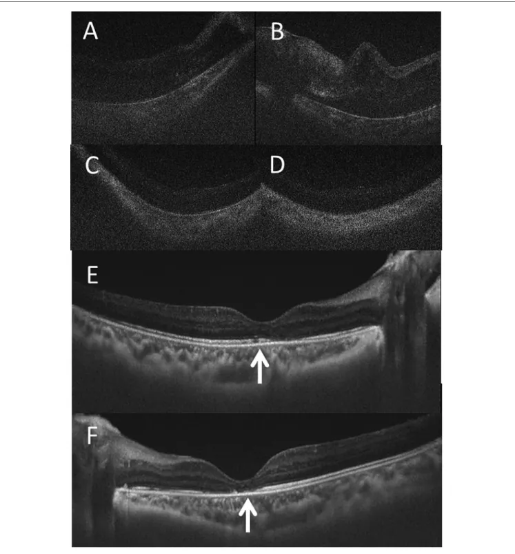
Figure 2. Optical coherence tomography. At the age of 5 years of 11 months (A, right eye; B, left eye), 1 month before the start of
adalimumab. At the age of 6 years and 2 months (C, right eye; D, left eye), 2 months after the start of adalimumab. At the age of 8 years and 1 month (E, right eye; F, left eye). Note that macular edema in both eyes (A, B) has been resolved in 2 months (C, D) after the start of adalimumab. Note also that ellipsoid zone at the fovea (arrows in E, F), indicative of photoreceptor outer segments, is more well visualized in the right eye (E) compared with the left eye (F), explaining better visual acuity in the right eye.
5
Table 1.
Review of 27 Japanese Patients With Juvenile Idiopathic Arthritis–Associated Uveitis Including the Present Patient.
Case no./ gender
Age at onset Category ANA RF Preceding treatment before uveitis Age at uveitis Eye manifestations Eye(s) involved Systemic treatment for uveitis a Eye surgery
Visual acuity at final visit, right/Left
Author 1/Male 3 years Systemic ND ND ND 7 years
Band keratopathy Iridocyclitis Subretinal exudates Optic papillitis
Bilateral
Prednisolone
Left iridectomy
0.9/0.1
Urayama and Sakuragi
17 2/Female 3 years Oligo ND No Simultaneous onset
Band keratopathy Iridocyclitis Cataract Optic papillitis
Right
No
No
0.03/1.5
Urayama and Sakuragi
17 3/Female 7 years Systemic Yes No Simultaneous onset
Iridocyclitis Retinal vasculitis Optic papillitis
Bilateral Betamethasone No 0.9/0.9 Tabuchi et al 4/Female 4 years 1 month Systemic ND ND Simultaneous onset Iridocyclitis Bilateral Prednisolone No 1.2/0.4 Fujiwara et al 5/Female 6 years Oligo Yes No Simultaneous onset
Iridocyclitis Retinal vasculitis
Bilateral Prednisolone Aspirin Bilateral trabeculectomy 0.1/0.7 Takano et al 6/Female 2 years Systemic No No Aspirin 4 years
Iridocyclitis Retinal vasculitis Optic papillitis
Bilateral No No 1.5/1.5 Takano et al 7/Female 2 years Oligo Yes No Aspirin 5 years
Band keratopathy Iridocyclitis Cataract Retinal vasculitis
Bilateral
Aspirin
Left cataract surgery
1.2/0.1 Takano et al 8/Female 3 years Poly ND ND Prednisolone 15 years
Band keratopathy Iridocyclitis Cataract Retinal vasculitis
Bilateral
ND
Left trabeculectomy Bilateral cataract surgery
0.02/0.04 Sakai et al 21 9/Female 9 years Systemic Yes No Simultaneous onset
Iridocyclitis Retinal vasculitis Optic papillitis
Right Dexamethasone No 1.5/1.5 Uezato et al 10/Male 12 years Oligo No No ND 13 years
Iridocyclitis Retinal vasculitis
Bilateral ND No 1.0/1.2 Akaki et al 23 11/Male 17 years Systemic No No Methylprednisolone pulse 18 years Retinal vasculitis Bilateral Prednisolone No 1.0/1.0 Iizuka et al 24 12/Female 3 years Systemic No No Simultaneous onset
Iridocyclitis Retinal vasculitis Subretinal exudates Optic papillitis
Bilateral Prednisolone Aspirin No 0.8/0.8 Nakayama et al 13/Female 20 years Systemic Yes No Simultaneous onset
Iridocyclitis Optic papillitis
Bilateral
Methylprednisolone pulse Aspirin
No 1.2/1.5 Igari et al 26 14/Female 2 years Oligo Yes No ND 7 years Iridocyclitis Cataract Bilateral
Levothyroxine for Hashimoto disease Bilateral cataract surgery
1.5/1.5
Honda et al
6
Case no./ gender
Age at onset Category ANA RF Preceding treatment before uveitis Age at uveitis Eye manifestations Eye(s) involved Systemic treatment for uveitis a Eye surgery
Visual acuity at final visit, right/Left
Author 15/Male 12 years Poly No No Simultaneous onset
Iridocyclitis Retinal vasculitis Optic papillitis
Bilateral
Prednisolone
No
1.2/1.2
Amano and Mochizuki
28 16/Female 10 years Systemic No No Simultaneous onset Subretinal exudates Bilateral Aspirin No 1.0/1.2 Higuchi et al 29 17/Female 5 years Systemic No No Simultaneous onset
Iridocyclitis Retinal vasculitis Optic papillitis
Bilateral Aspirin Prednisolone No 1.5/1.2 Sakaguchi et al 30 18/Male 2 years Systemic Yes No Aspirin 3 years
Subretinal exudates (brain basal ganglia mass)
Right Prednisolone No ND/ND Hirabayashi et al 31 19/Female 12 years Systemic Yes No ND 15 years
Iridocyclitis Subretinal exudates Optic papillitis
Bilateral No No 1.0/1.0 Ishihara et al 32 20/Female 5 years Systemic No No ND 11 years
Iridocyclitis Retinal vasculitis Subretinal exudates Optic papillitis
Bilateral Prednisolone Aspirin No 0.8/0.6 Sekine et al 33 21/Male 19 years Systemic No No Simultaneous onset
Iridocyclitis Retinal vasculitis Optic papillitis
Bilateral Prednisolone Methotrexate No 1.0/1.0 Namba et al 34 22/Female 4 years Oligo Yes ND Simultaneous onset
Iridocyclitis Subretinal exudates Vitreous opacity
Bilateral Prednisolone Cyclosporine No 2.0/2.0 Matsushita et al 35 23/Female 1 year 7 months Oligo Yes No
Ibuprofen Prednisolone Methotrexate
11 years
Iridocyclitis Retinal vasculitis Macular edema Optic papillitis
Bilateral Etanercept No 1.0/1.0 Kinouchi et al 36 24/Female 4 years Poly Yes ND Simultaneous onset
Band keratopathy Iridocyclitis
Bilateral
Prednisolone
Bilateral cataract surgery with IOL Bilateral PTK
0.09/0.1 Ishikura et al 37 25/Female 2 years 9 months Oligo Yes No Simultaneous onset 5 years
Iridocyclitis Cataract Retinal vasculitis Macular edema
Right
Ibuprofen Prednisolone Adalimumab Right cataract surgery
0.9/ND Shimizu 38 26/Male 5 years 1 month Oligo Yes No Simultaneous onset
Band keratopathy Iridocyclitis Macular edema
Left
Ibuprofen Methotrexate Adalimumab Left therapeutic keratectomy
ND/0.4 Shimizu et al 38 27/Male 4 years 8 months Oligo No No Simultaneous onset
Band keratopathy Iridocyclitis Cataract Retinal vasculitis Macular edema
Bilateral
Methotrexate Adalimumab Bilateral cataract surgery Bilateral therapeutic keratectomy
0.8/0.4
Matsuo and Yashiro (this case)
Abbreviations: ANA, antinuclear antibody; RF, rheumatoid factor; ND, not described; systemic
, systemic arthritis; oligo, oligoarthritis; poly, polyarthritis; IOL, intraocular lens implantation; PTK, phototherapeutic ke
ratectomy.
a“No” in systemic treatment for uveitis indicates topical medication only.
Tab
le 1. (contin
ue
Results
In review of the literature, 27 patients with juvenile idio-pathic arthritis–associated uveitis, including the present patient, were 8 male and 19 female, with the age at the diag-nosis of juvenile idiopathic arthritis, ranging from 1 year and 7 months to 20 years (median = 5 years). The timing at the diagnosis of uveitis was the same as the onset of systemic manifestations and, hence, as the diagnosis of juvenile idio-pathic arthritis in 15 patients, while the diagnosis of uveitis was delayed in 12 patients by the range from 1 year to 12 years (median = 2.5 years) after the onset of systemic mani-festations. The category of juvenile idiopathic arthritis was systemic arthritis in 14 patients, oligoarthritis in 10 patients, and polyarthritis (rheumatoid factor–negative) in 3 patients. Antinuclear antibody was positive in 13 patients and nega-tive in 10 patients, while the remaining 4 patients had no description. Rheumatoid factor was negative in all 22 patients with its description.
All 27 patients, including the present patient, showed iridocyclitis manifested as keratic precipitates and aqueous cells sometimes with synechia iris posterior. Both eyes were involved in 22 patients, only the right eye in 4 patients, and only the left eye in 1 patient. Persistent irido-cyclitis was associated with band keratopathy in 7 patients and complicated cataract in 6 patients. Only 3 patients had iridocyclitis with no description of fundus (posterior seg-ment) manifestations. Lens extraction with no intraocular lens implantation as cataract surgery was done in 4 patients while intraocular lens implantation was done in 1 patient (Case 24). Phototherapeutic keratectomy using laser was done in both eyes of 1 patient with dense band keratopathy (Case 24).
The other 24 patients had fundus manifestations: 2 of these 24 patients had fundus manifestations only and did not have iridocyclitis, one with retinal vasculitis (Case 11) and another with subretinal exudates (Case 16) in both eyes. As for fundus (posterior segment) manifestations, retinal vascu-litis was described in 16 patients, optic papilvascu-litis in 12 patients, subretinal exudates in 7 patients, and macular edema in 3 patients. The retinal vasculitis manifested as vas-cular leakage on fluorescein angiography, diffuse retinal edema, and vascular sheathing. The optic papillitis mani-fested as blurred hyperemic swollen optic disc and occasion-ally as exudates around the optic disc. The subretinal exudates manifested usually as multiple spotty white lesions and occasionally as diffuse large exudation. The macular edema in recent patients was detected by optical coherence tomography.
In treatment for uveitis, adalimumab was prescribed in 3 most recent patients including the present patient. Oral ste-roids were given in 16 patients, cyclosporine combined with prednisolone in 1 patient (Case 22), and etanercept only in another patient (Case 23). Aspirin (salicylate) was given in 7 patients: independently in 2 patients and combined with
prednisolone in 5 patients. Ibuprofen was used in 2 most recent patients.
Discussion
The present patient was diagnosed as antinuclear antibody– negative oligoarticular juvenile idiopathic arthritis, based on intermittent joint symptoms and typical eye signs after the exclusion of infectious uveitis and other types of noninfec-tious uveitis. In the field of ophthalmology, pediatric uveitis is rare, and it is mandatory to make differential diagnosis of other types of uveitis from juvenile idiopathic arthritis–asso-ciated uveitis. Bilateral iridocyclitis with retinal capillaritis
in juveniles39 and tubulointerstitial nephritis and uveitis
syndrome40 are 2 entities of pediatric uveitis that must be
considered at this setting. In contrast with juvenile idiopathic arthritis–associated uveitis, these 2 types of uveitis have ten-dency not to cause severe complications of band keratopathy and cataract.
The present patient developed retinal vasculitis and mac-ular edema, concurrently with iridocyclitis that resulted in band keratopathy and complicated cataract in both eyes. The retinal involvement was noted at the time of cataract surgery in which diffuse retinal edema, together with severe anterior vitreous inflammation, was visualized. Male gender has been shown as a risk factor for developing complicated
cata-ract and optic papillitis associated with uveitis.41 Intraocular
lens implantation was cancelled in the present patient with severe anterior vitreous inflammation involving the poste-rior capsule of the lens. Intraocular lens implantation at cataract surgery might not be a risk factor for macular edema and optic papillitis as well as glaucoma under the maximum
control of inflammation,42 but we avoided taking a risk in
the present patient under the circumstances of severe vitre-ous inflammation.
After cataract surgery to remove the entire lens in addition to core vitrectomy in both eyes, the intraocular inflammation became calm during the course of oral methotrexate. However, the patient had poor visual acuity in both eyes due to persistent macular edema revealed by optical coherence tomography. After 2 months of adalimumab therapy, the structure of the macula in both eyes had dramatically nor-malized, leading to good visual acuity that satisfied the stan-dard for the elementary school admission. As shown in the present patient, adalimumab is now the standard therapy for juvenile idiopathic arthritis–associated uveitis with retinal
complications such as macular edema.43-49 In this context,
liaison between ophthalmologists and pediatricians is crucial not to miss a chance to rescue the vision by starting adalim-umab. Concurrent with the present report, case series of Japanese patients with juvenile idiopathic
arthritis–associ-ated uveitis were reported by pediatricians.50 Those 9 patients
were all in the category of oligoarthritis, and 8 of them received biologics such as adalimumab. The authors empha-sized the planned transitional care for children with juvenile
8 Journal of Investigative Medicine High Impact Case Reports idiopathic arthritis to avoid the blindness caused by sequelae
to uveitis.
In review of the literature17-38 in the present study, most
Japanese patients with juvenile idiopathic arthritis–associ-ated uveitis had fundus manifestations such as retinal vascu-litis, optic papilvascu-litis, and subretinal exudates. A high rate of fundus manifestations in juvenile idiopathic arthritis–associ-ated uveitis might be attributed to reporting bias to describe rarer presentations. Indeed until now, fundus manifestations in juvenile idiopathic arthritis–associated uveitis have not been well described in the textbooks. Optical coherence tomography in widespread use at ophthalmology clinics would help accurate diagnosis of macular edema, which is a vision-threatening condition in uveitis associated with juve-nile idiopathic arthritis, as shown in the present patient.
The present patient had 2 risk factors for developing juve-nile idiopathic arthritis–associated uveitis: the younger age
of onset at 4 years and the category of oligoarthritis.5 It
should be noted in review of the literature that about a half of the historical Japanese patients with juvenile idiopathic arthritis–associated uveitis belonged to the category of
sys-temic arthritis.17-38 This finding is in marked contrast with
the recent reports that the category of oligoarthritis was pre-dominant also in Japanese patients with juvenile idiopathic
arthritis–associated uveitis,5,50 in parallel with the Caucasian
population.1 The reason for the categorical difference
between the historical cohort and the recent cohort5 in the
Japanese population remains unknown. As described above, the historical cohort in the present study is indeed the collec-tion of case reports that tended to describe fundus manifesta-tions as a rare event, and thus would have reporting bias from the viewpoint of the disease category.
In conclusion, the present study focused on the Japanese patients with juvenile idiopathic arthritis–associated uveitis. In the historical Japanese patients, the category of systemic arthritis was dominant and was followed by the category of oligoarthritis. The fundus manifestations, such as retinal vas-culitis, subretinal exudates, and optic papillitis, were common in addition to well-recognized features of iridocyclitis, band keratopathy, and complicated cataract in the younger ages.
Authors’ Note
Data are available on reasonable request to the corresponding author.
Author Contributions
TM, as an ophthalmologist, followed the patient and did the surgery. MY, as a pediatrician, introduced methotrexate and adalimumab and followed the patient. TM wrote the manuscript, and MY did critical review of the manuscript. Both authors read and approved the manuscript.
Declaration of Conflicting Interests
The author(s) declared no potential conflicts of interest with respect to the research, authorship, and/or publication of this article.
Funding
The author(s) received no financial support for the research, author-ship, and/or publication of this article.
Ethics Approval
Ethics committee review was not applicable to case reports, based on the Ethical Guidelines for Medical and Health Research Involving Human Subjects, issued by the Government of Japan.
Informed Consent
Oral informed consent was obtained from the patient and the mother.
ORCID iD
Toshihiko Matsuo https://orcid.org/0000-0001-6570-0030
References
1. Saurenmann RK, Rose JB, Tyrrell P, et al. Epidemiology of juvenile idiopathic arthritis in a multiethnic cohort: ethnicity as a risk factor. Arthritis Rheum. 2007;56:1974-1984.
2. Kobayashi T, Tanaka S, Maeda M, Okubo H, Matsuyama T, Watanabe N. A study of prognosis in 52 cases with juvenile rheumatoid arthritis. Acta Pediatr Jpn. 1993;35:439-446. 3. Fujikawa S, Okuni M. Clinical analysis of 570 cases with
juve-nile rheumatoid arthritis: results of a nationwide retrospective survey in Japan. Acta Pediatr Jpn. 1997;39:245-249.
4. Petty RE, Southwood TR, Manners P, et al. International League of Associations for Rheumatology classification of juvenile idiopathic arthritis: second revision, Edmonton, 2001.
J Rheumatol. 2004;31:390-392.
5. Yasumura J, Yashiro M, Okamoto N, et al. Clinical features and characteristics of uveitis associated with juvenile idiopathic arthritis in Japan: first report of the Pediatric Rheumatology Association of Japan (PRAJ). Pediatr Rheumatol Online J. 2019;17:15.
6. Rosenberg AM. Uveitis associated with juvenile rheumatoid arthritis. Semin Arthritis Rheum. 1987;16:158-173.
7. Kanski JJ. Juvenile arthritis and uveitis. Surv Ophthalmol. 1990;34:253-267.
8. Kump LI, Castañeda RAC, Androudi SN, Reed GF, Foster CS. Visual outcomes in children with juvenile idiopathic arthritis-associated uveitis. Ophthalmology. 2006;113:1874-1877. 9. Woreta F, Thorne JE, Jabs DA, Kedhar SR, Dunn JP.
Risk factors for ocular complications and poor visual acu-ity at presentation among patients with uveitis associated with juvenile idiopathic arthritis (JIA). Am J Ophthalmol. 2007;143:647-655.
10. de Lahitte GD, Terrada C, Tran THC, et al. Maculopathy in uveitis of juvenile idiopathic arthritis: an optical coherence tomography study. Br J Ophthalmol. 2008;92:64-69.
11. Sabri K, Saurenmann RK, Silverman ED, Levin AV. Course, complications, and outcome of juvenile arthritis-related uve-itis. J AAPOS. 2008;12:539-545.
12. Paroli MP, Spinucci G, Fabiani C, Pivetti-Pezzi P. Retinal complications of juvenile idiopathic arthritis-related uveitis: a microperimetry and optical coherence tomography study. Ocul
13. Vitale AT, Graham E, de Boer JH. Juvenile idiopathic arthri-tis-associated uveitis: clinical features and complications, risk factors for severe course, and visual outcome. Ocul Immunol
Inflamm. 2013;21:478-485.
14. Paroli MP, Fabiani C, Spinucci G, Abicca I, Sapia A, Spadea L. Severe macular edema in patients with juvenile idiopathic arthritis-related uveitis. Case Rep Ophthalmol Med. 2013;2013: 803989.
15. de Boer J, Steijaert A, van den Bor R, Stellato R, van Norel JO. Development of macular edema and impact on visual acu-ity in uveitis associated with juvenile idiopathic arthritis. Ocul
Immunol Inflamm. 2015;23:67-73.
16. Matsuo T. Intraocular lens implantation in unilateral congenital cataract with minimal levels of persistent fetal vasculature in the first 18 months of life. Springerplus. 2014;3:361.
17. Urayama A, Sakuragi S. Pediatric uveitis: especially regarding Still’s disease [in Japanese]. Ganka. 1969;11:358-363. 18. Tabuchi A, Takeda J, Takagi K, et al. A case of juvenile
rheumatoid arthritis with papillitis and retinal vasculitis [in Japanese]. Nippon Ganka Kiyo (Folia Ophthalmol Jpn). 1979;30:497-502.
19. Fujiwara H, Ota T, Okamoto S. Ocular complications in the cases with juvenile rheumatoid arthritis [in Japanese]. Nippon
Ganka Kiyo (Folia Ophthalmol Jpn). 1980;31:333-337.
20. Takano S, Ohnishi Y, Mitsuhashi M, et al. Three cases of juvenile rheumatoid arthritis-associated uveitis [in Japanese].
Ganka. 1984;26:77-84.
21. Sakai M, Kanari T, Yaginuma Y, et al. Sister cases of arthropa-thy and uveitis associated either with sarcoidosis or juvenile rheumatoid arthritis [in Japanese]. Ganka Rinsho Iho (Jpn Rev
Clin Ophthalmol). 1986;80:1077-1081.
22. Uezato T, Kuwae Y, Uehara M, et al. A case of juvenile rheu-matoid arthritis with fundus involvement [in Japanese]. Rinsho
Ganka (Jpn J Clin Ophthalmol). 1986;40:378-379.
23. Akaki Y, Haruta Y, Nakagawa Y, et al. Eight cases of chronic recurrent uveitis with hypopyon [in Japanese]. Nippon Ganka
Kiyo (Folia Ophthalmol Jpn). 1986;37:201-206.
24. Iizuka M, Hayakawa M, Yoshiike A, et al. A case of retinal vasculitis associated with juvenile rheumatoid arthritis [in Japanese]. Ganka Rinsho Iho (Jpn Rev Clin Ophthalmol). 1989;83:84-87.
25. Nakayama T, Otaki C, Makino S. Posterior uveitis in juvenile rheumatoid arthritis [in Japanese]. Ganka Rinsho Iho (Jpn Rev
Clin Ophthalmol). 1989;83:284-287.
26. Igari S, Onizawa N, Yoshida T, et al. A case of adult-onset Still’s disease with iridocyclitis and papillitis [in Japanese].
Ryumachika (Rheumatology, Tokyo). 1989;1:527-533.
27. Honda T, Ishibashi T, Inomata H, et al. A case of juvenile rheumatoid arthritis associated with iridocyclitis, cataract and Hashimoto’s disease [in Japanese]. Nippon Ganka Kiyo (Folia
Ophthalmol Jpn). 1989;40:663-667.
28. Amano S, Mochizuki M. A case of juvenile rheumatoid arthri-tis with panuveiarthri-tis [in Japanese]. Rinsho Ganka (Jpn J Clin
Ophthalmol). 1990;44:205-208.
29. Higuchi H, Kimura S, Mukuno K, et al. A case of juvenile rheu-matoid arthritis manifested isolated retinochoroidal lesions.
Rinsho Ganka (Jpn J Clin Ophthalmol). 1990;44:1969-1973.
30. Sakaguchi S, Hara T, Saishin M, et al. A case of juvenile rheu-matoid arthritis with papilledema and retinal vasculitis. Rinsho
Ganka (Jpn J Clin Ophthalmol). 1991;45:723-726.
31. Hirabayashi S, Kasai S, Tsukahara T, Fujisawa N, Watanabe T. Basal ganglia mass lesions in juvenile rheumatoid arthritis.
Pediatr Neurol. 1991;7:141-143.
32. Ishihara M, Nakamura S, Okada K, et al. A case of juvenile rheu-matoid arthritis with panuveitis [in Japanese]. Nippon Ganka
Gakkai Zasshi (Acta Soc Ophthalmol Jpn). 1992;96:109-113.
33. Sekine N, Takeda M, Suzuki J. Iridocyclitis with subreti-nal flecked lesions in a case of juvenile rheumatoid arthritis [in Japanese]. Rinsho Ganka (Jpn J Clin Ophthalmol). 1993; 47:131-134.
34. Namba K, Tsuda K, Ohnishi K, et al. Iridocyclitis, papill-edema and retinal phlebitis in a case of adult-onset Still’s dis-ease [in Japanese]. Rinsho Ganka (Jpn J Clin Ophthalmol). 1996;50:1687-1690.
35. Matsushita E, Fushishima A, Hayashi N, et al. Panuveitis in a case of juvenile rheumatoid arthritis [in Japanese]. Rinsho
Ganka (Jpn J Clin Ophthalmol). 2002;56;617-620.
36. Kinouchi R, Hirokawa H, Igarashi S, et al. A case of panuve-itis with optic disc neovascularization associated with juvenile idiopathic arthritis which progressed during a clinical trial of etanercept. Nippon Ganka Gakkai Zasshi (J Jpn Ophthalmol
Soc). 2007;111:970-975.
37. Ishikura R, Miyazaki D, Inoue Y, et al. A case of juvenile rheumatoid arthritis with cataract surgery after phototherapeu-tic keratectomy [in Japanese]. Ganka Rinsho Kiyo (Folia Jpn
Ophthalmol Clin). 2008;1:1163-1167.
38. Shimizu M, Takemoto Y, Takakura M, et al. Usefulness of adalimumab in the treatment of refractory uveitis associated with juvenile idiopathic arthritis [in Japanese]. Shoni Ryumachi
(J Clin Pediatr Rheumatol). 2015;6:33-38.
39. Matsuo T, Matsuo N. Bilateral iridocyclitis with retinal capil-laritis in juveniles. Ophthalmology. 1997;104:939-944. 40. Matsuo T. Fluorescein angiographic features of tubulointerstitial
nephritis and uveitis syndrome. Ophthalmology. 2002;109:132-136.
41. Ayuso VK, Cate HATT, van der Does P, Rothova A, de Boer JK. Male gender as a risk factor for complications in uveitis associated with juvenile idiopathic arthritis. Am J Ophthalmol. 2010;149:994-999.e5.
42. Sijssens KM, Los LI, Rothova A, et al. Long-term ocular complications in aphakic versus pseudophakic eyes of chil-dren with juvenile idiopathic arthritis-associated uveitis. Br J
Ophthalmol. 2010;94:1145-1149.
43. Samra KA, Maghsoudlou A, Roohipoor R, Valdes-Navarro M, Lee S, Foster CS. Current treatment modalities of JIA-associated uveitis and its complications: literature review. Ocul
Immunol Inflamm. 2016;24:431-439.
44. Asproudis I, Katsanos A, Kozeis N, Tantou A, Konstas AG. Update on the treatment of uveitis in patients with juvenile idiopathic arthritis: a review. Adv Ther. 2017;34:2558-2565. 45. Tynjala P, Kotaniemi K, Lindahl P, et al. Adalimumab in juve-nile idiopathic arthritis-associated chronic anterior uveitis.
Rheumatology (Oxford). 2008;47:339-344.
46. Díaz-Llopis M, Salom D, Garcia-de-Vicuña C, et al. Treatment of refractory uveitis with adalimumab: a prospective multicenter study of 131 patients. Ophthalmology. 2012;119:1575-1581. 47. Zannin ME, Birolo C, Gerloni VM, et al. Safety and efficacy
of infliximab and adalimumab for refractory uveitis in juvenile idiopathic arthritis: 1-year follow-up data from the Italian reg-istry. J Rheumatol. 2013;40:74-79.
10 Journal of Investigative Medicine High Impact Case Reports 48. Muñoz-Gallego A, Barral E, Enríquez E, Tejada P, Barceló
A, de Inocencio J. Adalimumab for the treatment of refrac-tory noninfectious pediatric uveitis. Int Ophthalmol. 2017;37: 719-725.
49. Zierhut M, Heiligenhaus A, de Boer J, Cunningham ET, Tugal-Tutkan I. Controversies in juvenile idiopathic
arthritis-associated uveitis. Ocul Immunol Inflamm 2013;21: 167-179.
50. Yamazaki S, Shimbo A, Akutsu Y, Takase H, Morio T, Mori M. Importance of pediatric rheumatologists and transitional care for juvenile idiopathic arthritis-associated uveitis: a retrospec-tive series of 9 cases. Pediatr Rheumatol Online J. 2020;18:26.